
Journal of Southern Medical University ›› 2026, Vol. 46 ›› Issue (10): 2309-2324.doi: 10.12122/j.issn.1673-4254.2026.10.03
Xiaoyu LU1( ), Tianxiao PANG2, Ye LIU2, Xuehong HE2(), Zhiming LI2()
Received:2026-03-17
Online:2026-10-20
Published:2026-10-26
Contact:
Xuehong HE, Zhiming LI
E-mail:dfs1107@163.com;lnzysnk@163.com;zglzmzg@163.com
Xiaoyu LU, Tianxiao PANG, Ye LIU, Xuehong HE, Zhiming LI. Shenqi Xiezhuo Decoction ameliorates renal fibrosis in rats by inhibiting excessive autophagy via modulating the ERK1/2-mTOR signaling pathway[J]. Journal of Southern Medical University, 2026, 46(10): 2309-2324.
Add to citation manager EndNote|Ris|BibTeX
URL: https://www.j-smu.com/EN/10.12122/j.issn.1673-4254.2026.10.03
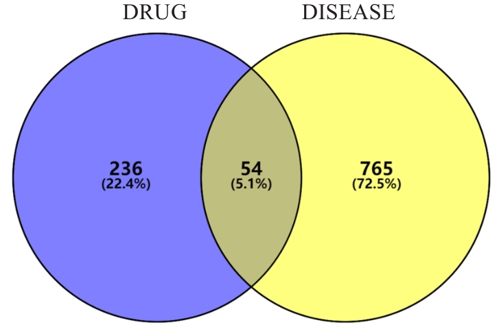
Fig.1 Venn diagram of the action targets of SQXZD for delaying progression of renal fibrosis.
| Components | Degree | Components | Degree |
|---|---|---|---|
| benzoyl paeoniflorin | 201 | Digallic acid | 131 |
| Diisooctyl phthalate | 161 | Physciondiglucoside | 129 |
| 4-O-methylpaeoniflorin_qt | 153 | Acanthoside B | 128 |
| Procyanidin B-5,3'-O-gallate | 136 | Poricoic acid B | 128 |
| Poricoic acid A | 135 | Sennoside D_qt | 125 |
Tab.1 Top 10 key components of SQXZD for delaying progression of renal fibrosis and their degree values
| Components | Degree | Components | Degree |
|---|---|---|---|
| benzoyl paeoniflorin | 201 | Digallic acid | 131 |
| Diisooctyl phthalate | 161 | Physciondiglucoside | 129 |
| 4-O-methylpaeoniflorin_qt | 153 | Acanthoside B | 128 |
| Procyanidin B-5,3'-O-gallate | 136 | Poricoic acid B | 128 |
| Poricoic acid A | 135 | Sennoside D_qt | 125 |
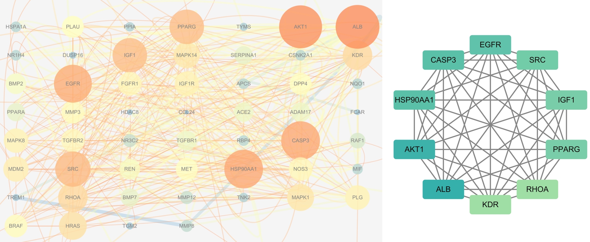
Fig.2 Key targets of SQXZD for delaying progression of renal fibrosis (Color intensity and node area are based on degree values).
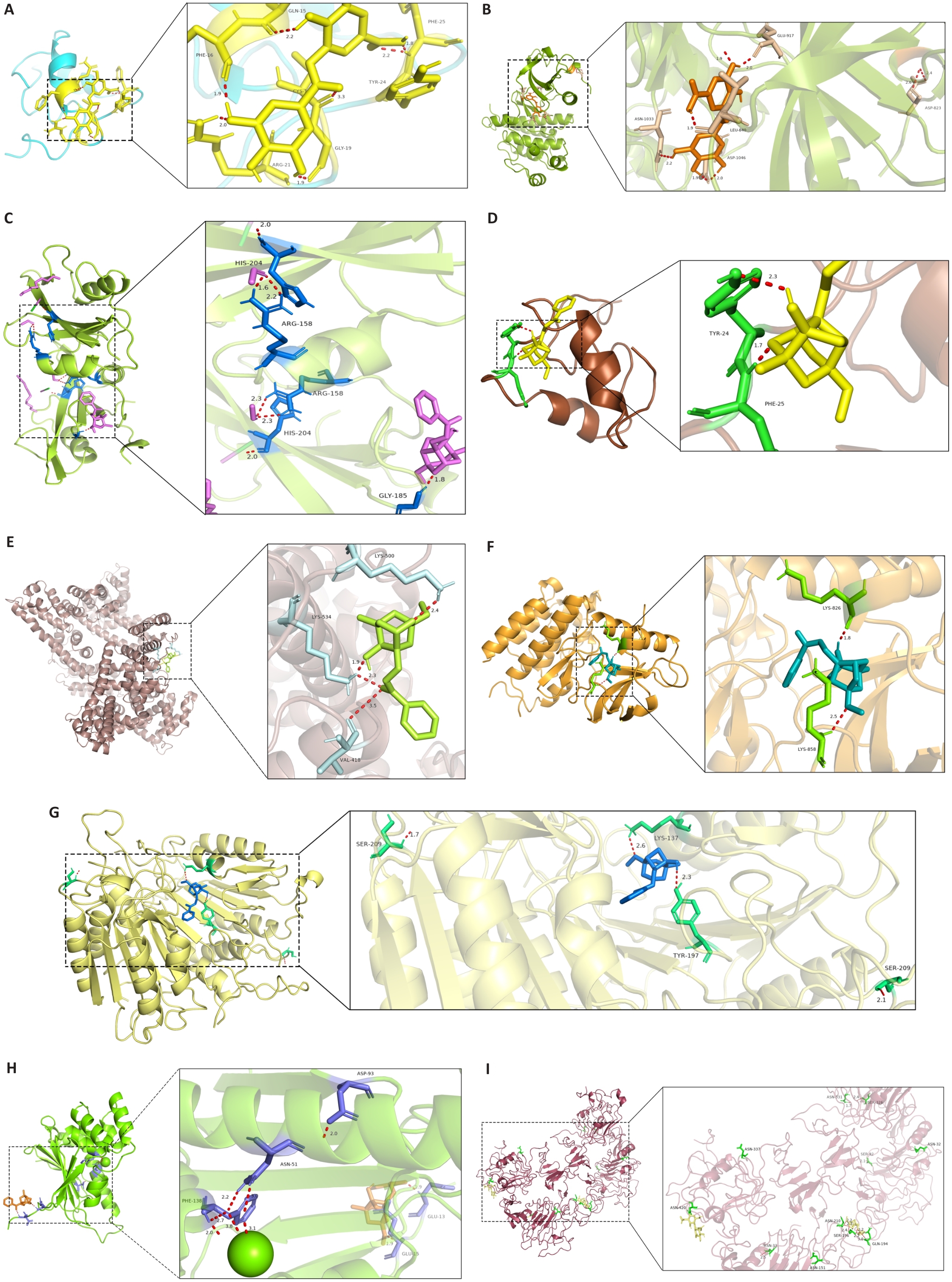
Fig.3 Molecular docking of the core active components of SQXZD with their core targets. A: DGA-IGF1. B: DGA-KDR. C: 4-O-methylpaeoniflorin-SRC. D: 4-O-methylpaeoniflorin-IGF1. E: 4-O-methylpaeoniflorin-ALB. F: 4-O-methylpaeoniflorin-KDR. G: 4-O-methylpaeoniflorin-CASP3. H: 4-O-methylpaeoniflorin-HSP90AA1. I: 4-O-methylpaeoniflorin-EGFR. DGA: Digallic acid.
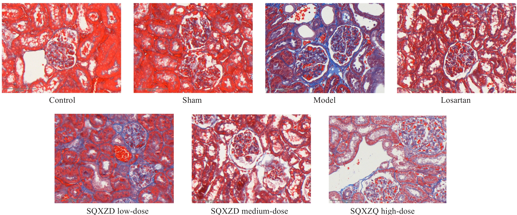
Fig.4 Effect of SQXZD on collagen deposition in the surgical-side kidney in UUO rats (Original magnification: ×400).
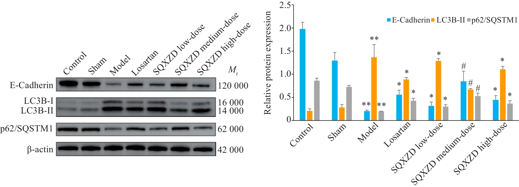
Fig.6 Effect of SQXZD on expression of EMT and autophagy-related proteins in the surgical-side kidney in UUO rats. **P<0.01 vs Sham group; *P<0.05, #P<0.01 vs Model group, n=3.
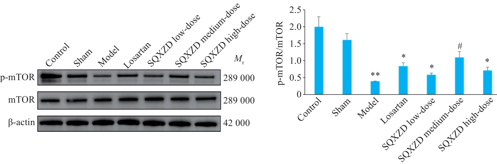
Fig.7 Effect of SQXZD on the expression of p-mTOR and mTOR in the surgical-side kidney in UUO rats. **P<0.01 vs Sham group; *P<0.05, #P<0.01 vs Model group, n=3.
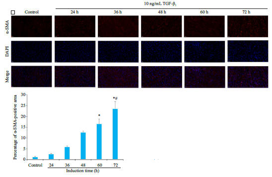
Fig.9 Effects of different concentrations of TGF-β1 on α-SMA expression in NRK-52E cells (×400). Induction time: A, 12 h; B, 24 h; C, 36 h; D, 48 h. *P<0.05, #P<0.01 vs Control group; △P<0.01 vs 12 h-induced group by 10 ng/mL TGF-β1; ▲P<0.05 vs 12 h-induced group by 12.5 ng/mL TGF-β1 and 24 h-induced group by 10 ng/mL TGF-β1. n=3.
| Item | Blank group | Control group | Dilutions of SQXZD-medicated serum groups (%) | |||||||
|---|---|---|---|---|---|---|---|---|---|---|
| 2.5 | 5 | 7.5 | 10 | 12.5 | 15 | 17.5 | 20 | |||
| NRK-49F | 0.189±0.006 | 2.062±0.116 | 1.831±0.291 | 2.300±0.120 | 2.257±0.123 | 2.282±0.088 | 2.294±0.095 | 2.332±0.112* | 2.164±0.180 | 2.117±0.087 |
| NRK-52E | 0.207±0.001 | 1.256±0.051 | 1.862±0.424 | 1.702±0.151 | 1.992±0.271 | 1.937±0.224 | 2.017±0.050* | 2.284±0.146# | 2.238±0.153* | 0.826±0.065 |
Tab.2 Effect of different Dilutions of SQXZD-medicated serum on viability of NRK-49F and NRK-52E cells (Mean±SD, n=3)
| Item | Blank group | Control group | Dilutions of SQXZD-medicated serum groups (%) | |||||||
|---|---|---|---|---|---|---|---|---|---|---|
| 2.5 | 5 | 7.5 | 10 | 12.5 | 15 | 17.5 | 20 | |||
| NRK-49F | 0.189±0.006 | 2.062±0.116 | 1.831±0.291 | 2.300±0.120 | 2.257±0.123 | 2.282±0.088 | 2.294±0.095 | 2.332±0.112* | 2.164±0.180 | 2.117±0.087 |
| NRK-52E | 0.207±0.001 | 1.256±0.051 | 1.862±0.424 | 1.702±0.151 | 1.992±0.271 | 1.937±0.224 | 2.017±0.050* | 2.284±0.146# | 2.238±0.153* | 0.826±0.065 |
| Group | Induction time NRK-49F | Induction time NRK-52E | ||
|---|---|---|---|---|
| Blank | 1.711±0.053 | 1.830±0.018 | ||
| Control | 2.041±0.061 | 2.612±0.427 | ||
| SQXZD-medicated serum | 0 h | 1.999±0.029 | 0 h | 2.052±0.044 |
| 24 h | 2.014±0.104 | 12 h | 2.093±0.039 | |
| 48 h | 1.915±0.059* | 24 h | 2.049±0.038* | |
Tab.3 Effect of different treatment durations with SQXZD-medicated serum on viability of TGF-β1-induced NRK-49F and NRK-52E cells (Mean±SD, n=3)
| Group | Induction time NRK-49F | Induction time NRK-52E | ||
|---|---|---|---|---|
| Blank | 1.711±0.053 | 1.830±0.018 | ||
| Control | 2.041±0.061 | 2.612±0.427 | ||
| SQXZD-medicated serum | 0 h | 1.999±0.029 | 0 h | 2.052±0.044 |
| 24 h | 2.014±0.104 | 12 h | 2.093±0.039 | |
| 48 h | 1.915±0.059* | 24 h | 2.049±0.038* | |
| Cell | Blank group | Control group | Concentration of DGA (ng/mL) | ||||
|---|---|---|---|---|---|---|---|
| 44.5 | 89 | 178 | 356 | 712 | |||
| NRK-49F | 1.927±0.046 | 1.808±0.015 | 2.259±0.150 | 0.980±0.080 | 0.273±0.009 | 0.611±0.019 | 0.921±0.016 |
| NRK-52E | 1.256±0.051 | 1.267±0.110 | 1.818±0.172 | 1.018±0.090 | 0.848±0.066 | 0.634±0.150 | 0.862±0.122 |
Tab.4 Effect of different concentrations of DGA on viability of NRK-49F and NRK-52E cells (Mean±SD, n=3)
| Cell | Blank group | Control group | Concentration of DGA (ng/mL) | ||||
|---|---|---|---|---|---|---|---|
| 44.5 | 89 | 178 | 356 | 712 | |||
| NRK-49F | 1.927±0.046 | 1.808±0.015 | 2.259±0.150 | 0.980±0.080 | 0.273±0.009 | 0.611±0.019 | 0.921±0.016 |
| NRK-52E | 1.256±0.051 | 1.267±0.110 | 1.818±0.172 | 1.018±0.090 | 0.848±0.066 | 0.634±0.150 | 0.862±0.122 |
| Group | Induction time NRK-49F | Induction time NRK-52E | ||
|---|---|---|---|---|
| Blank | 1.711±0.053 | 1.819±0.031 | ||
| Control | 2.093±0.063 | 2.219±0.041 | ||
| DGA | 0 h | 2.101±0.032 | 0 h | 2.108±0.027* |
| 24 h | 2.082±0.113 | 12 h | 2.052±0.084* | |
| 48 h | 1.974±0.049* | 24 h | 1.943±0.039# | |
Tab.5 Effect of different treatment durations with DGA on viability of TGF-β1-induced NRK-49F and NRK-52E (Mean±SD, n=3)
| Group | Induction time NRK-49F | Induction time NRK-52E | ||
|---|---|---|---|---|
| Blank | 1.711±0.053 | 1.819±0.031 | ||
| Control | 2.093±0.063 | 2.219±0.041 | ||
| DGA | 0 h | 2.101±0.032 | 0 h | 2.108±0.027* |
| 24 h | 2.082±0.113 | 12 h | 2.052±0.084* | |
| 48 h | 1.974±0.049* | 24 h | 1.943±0.039# | |
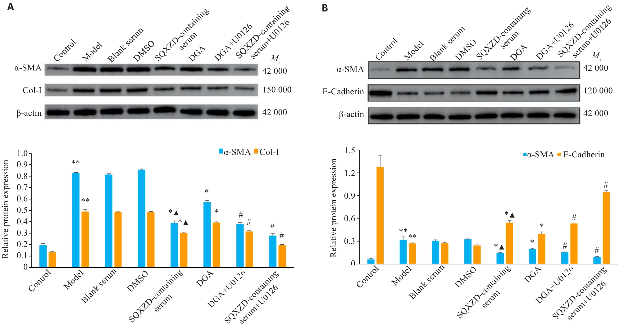
Fig.10 Effects of SQXZD-medicated serum and DGA on expression of α-SMA, Col-I, and E-cadherin in TGF-β1-induced NRK-49F (A) and NRK-52E cells (B). **P<0.01 vs Control group; *P<0.05, #P<0.01 vs Model group; ▲P<0.05 vs DGA group. n=3.
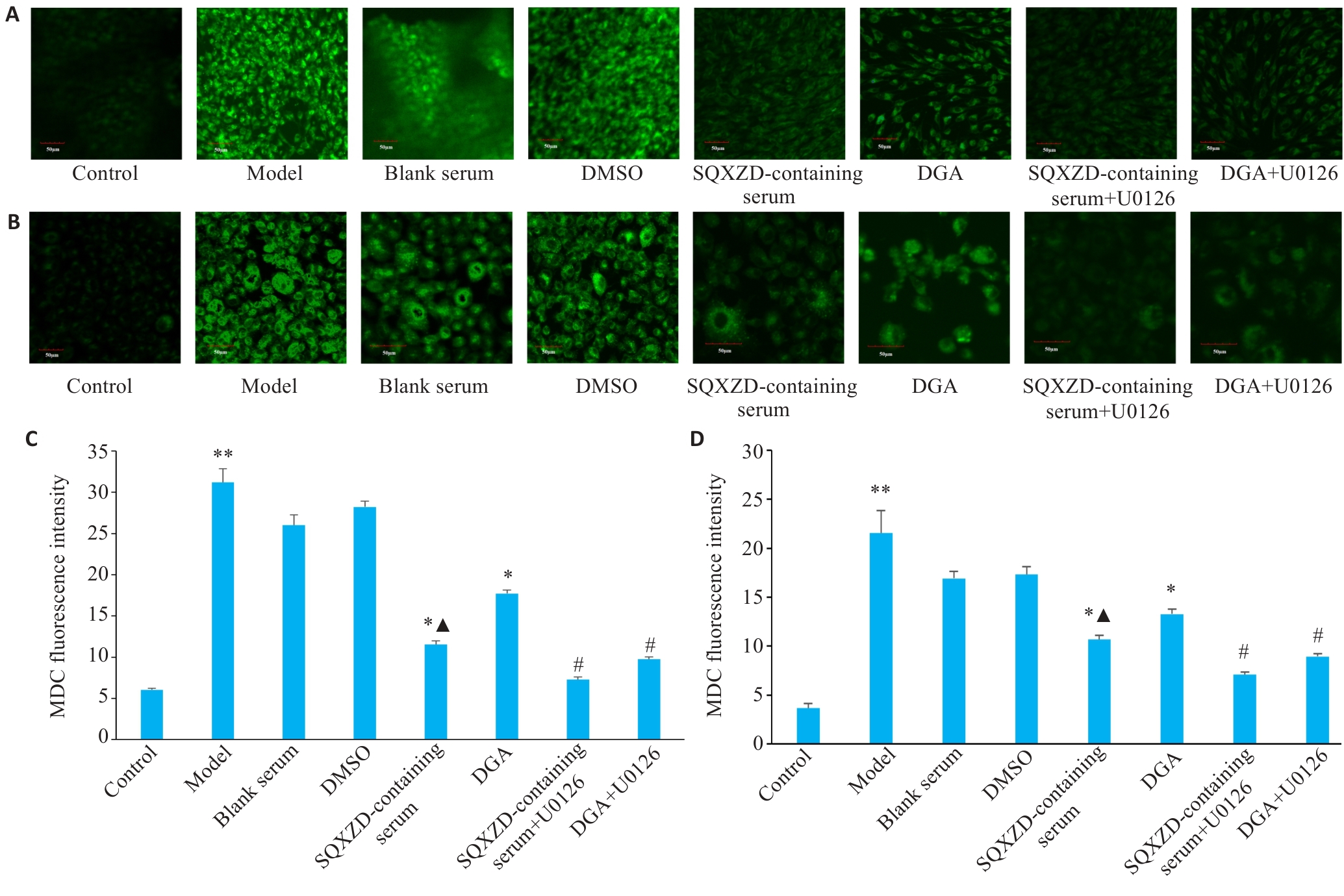
Fig.11 Effects of SQXZD-medicated serum and DGA on autophagic fluorescence in TGF-β1-induced NRK-49F cells (A, C) and NRK-52E cells (B, D) (×400). **P<0.01 vs Control group; *P<0.05, #P<0.01 vs Model group; ▲P<0.05 vs DGA group. n=3.
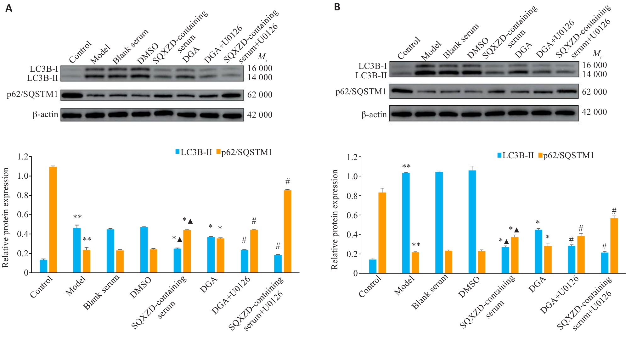
Fig.12 Effects of SQXZD-medicated serum and DGA on the expression of LC3B-II and p62/SQSTM1 in TGF-β1-induced NRK-49F (A) and NRK-52E cells (B). **P<0.01 vs Control group; *P<0.05, #P<0.01 vs Model group; ▲P<0.05 vs DGA group. n=3.
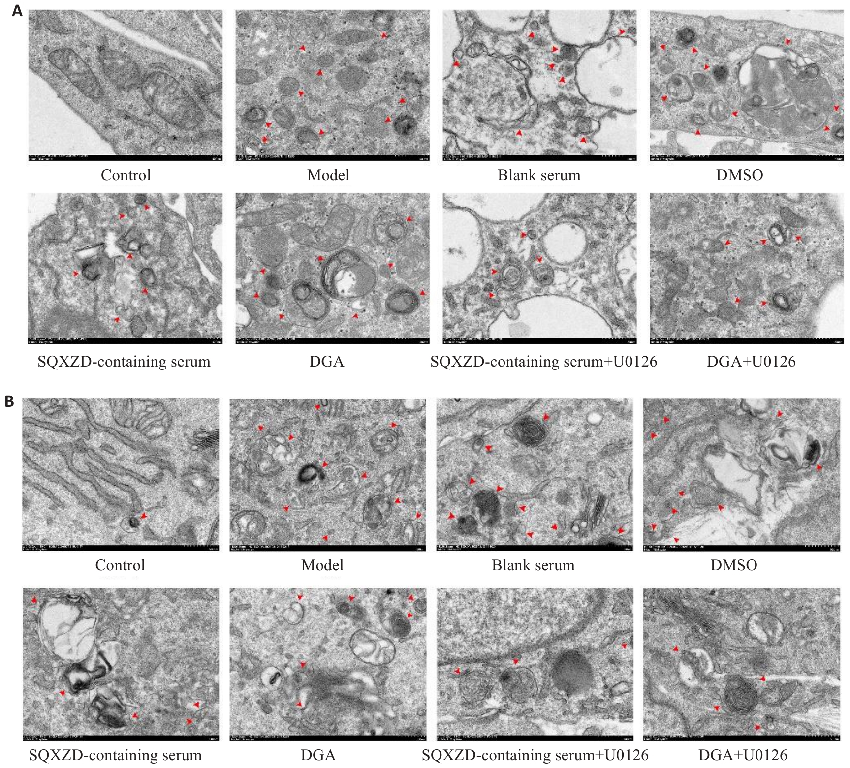
Fig.13 Effects of SQXZD-medicated serum and DGA on ultrastructure of TGF-β1-induced NRK-49F (A) and NRK-52E cells (B) (Transmission electron microscopy, ×12 000), red arrows indicate autophagosomes.
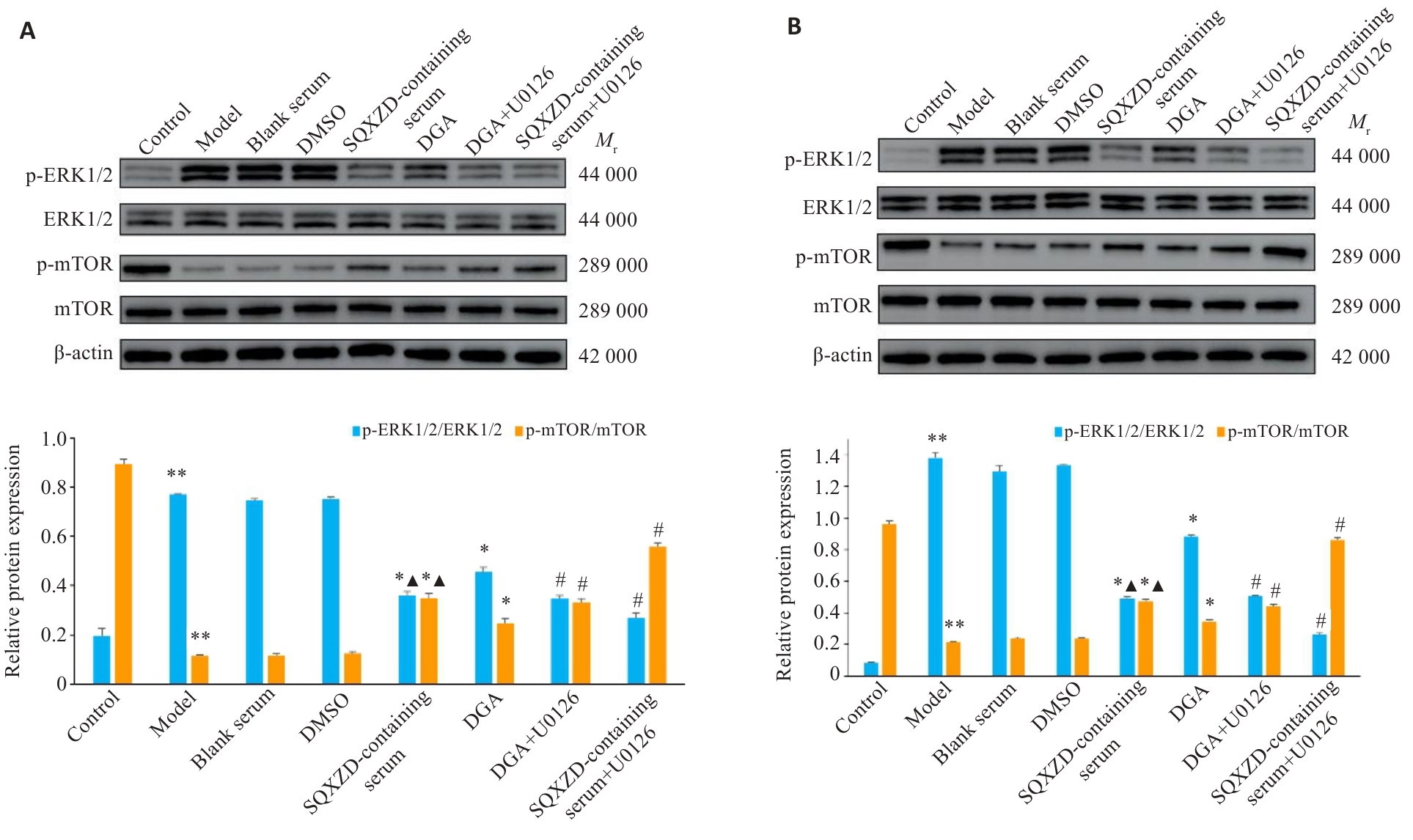
Fig.14 Effects of SQXZD-medicated serum and DGA on expression of p-ERK1/2/ERK1/2 and p-mTOR/mTOR in TGF-β1-induced NRK-49F (A) and NRK-52E cells (B). **P<0.01 vs Control group; *P<0.05, #P<0.01 vs Model group; ▲P<0.05 vs DGA group. n=3.
| [1] | Liu YH. Cellular and molecular mechanisms of renal fibrosis[J]. Nat Rev Nephrol, 2011, 7(12): 684-96. doi:10.1038/nrneph.2011.149 |
| [2] | Zeisberg M, Neilson EG. Mechanisms of tubulointerstitial fibrosis[J]. J Am Soc Nephrol, 2010, 21(11): 1819-34. doi:10.1681/asn.2010080793 |
| [3] | GBD 2023 Chronic Kidney Disease Collaborators. Global, regional, and national burden of chronic kidney disease in adults, 1990-2023, and its attributable risk factors: a systematic analysis for the Global Burden of Disease Study 2023[J]. Lancet, 2025,406(10518):2461-82. |
| [4] | Lovisa S, Zeisberg M, Kalluri R. Partial epithelial-to-mesenchymal transition and other new mechanisms of kidney fibrosis[J]. Trends Endocrinol Metab, 2016, 27(10): 681-95. doi:10.1016/j.tem.2016.06.004 |
| [5] | Dai R, Zhang L, Jin H, et al. Autophagy in renal fibrosis: Protection or promotion[J]? Front Pharmacol, 2022, 13: 963920. doi:10.3389/fphar.2022.963920 |
| [6] | Mizushima N, Komatsu M. Autophagy: renovation of cells and tissues[J]. Cell, 2011, 147(4): 728-41. doi:10.1016/j.cell.2011.10.026 |
| [7] | Kaushal GP, Chandrashekar K, Juncos LA, et al. Autophagy function and regulation in kidney disease[J]. Biomolecules, 2020, 10(1): 100. doi:10.3390/biom10010100 |
| [8] | Takemura K, Nishi H, Inagi R. Mitochondrial dysfunction in kidney disease and uremic sarcopenia[J]. Front Physiol, 2020, 11: 565023. doi:10.3389/fphys.2020.565023 |
| [9] | Bai L, Wu Q, Zhang X, Z, et al. Autosis as a selective type of cell death[J]. Front Cell Dev Biol, 2023, 11:1164681-93. doi:10.3389/fcell.2023.1164681 |
| [10] | Inoki K. mTOR signaling in autophagy regulation in the kidney[J]. Semin Nephrol, 2014, 34(1): 2-8. doi:10.1016/j.semnephrol.2013.11.002 |
| [11] | Xue X, Ren JF, Sun XL, et al. Protein kinase Cα drives fibroblast activation and kidney fibrosis by stimulating autophagic flux[J]. J Biol Chem, 2018, 293(28): 11119-30. doi:10.1074/jbc.ra118.002191 |
| [12] | Nam SA, Kim WY, Kim JW, et al. Autophagy attenuates tubulointerstital fibrosis through regulating transforming growth factor‑β and NLRP3 inflammasome signaling pathway[J]. Cell Death Dis, 2019, 10(2): 78. doi:10.1038/s41419-019-1356-0 |
| [13] | Wu YR, Wang LY, Deng D, et al. Renalase protects against renal fibrosis by inhibiting the activation of the ERK signaling pathways[J]. Int J Mol Sci, 2017, 18(5): 855. doi:10.3390/ijms18050855 |
| [14] | Zhu LN, Yang T, Li LJ, et al. TSC1 controls macrophage polarization to prevent inflammatory disease[J]. Nat Commun, 2014, 5: 4696. doi:10.1038/ncomms5696 |
| [15] | Bermudez O, Marchetti S, Pagès G, et al. Post-translational regulation of the ERK phosphatase DUSP6/MKP3 by the mTOR pathway[J]. Oncogene, 2008, 27(26): 3685-91. doi:10.1038/sj.onc.1211040 |
| [16] | Gao Y, Li H, Han Q, et al. Overexpression of DUSP6 enhances chemotherapy-resistance of ovarian epithelial cancer by regulating the ERK signaling pathway[J]. J Cancer, 2020, 11(11): 3151-64. doi:10.7150/jca.37267 |
| [17] | Ponnusamy M, Liu N, Sellamuthu R, et al. Autophagy protects against necrotic renal epithelial cell-induced death of renal interstitial fibroblasts[J]. Am J Physiol Ren Physiol, 2012, 303(1): F83-91. doi:10.1152/ajprenal.00027.2012 |
| [18] | 卢晓宇, 刘智慧, 刘 烨, 等. 参芪泄浊饮通过调控Rap1/MAPK/FoxO3a信号通路改善氧化应激及炎症反应延缓大鼠肾纤维化[J].南方医科大学学报, 2025, 45(12): 2585-97. |
| [19] | 中华中医药学会. T/ ,中药汤剂煎煮规范[S]. 北京: 中国标准出版社, 2021. |
| [20] | 马园园, 刘成海, 陶艳艳. 肾纤维化动物模型特点与研究进展[J]. 中国实验动物学报, 2018, 26(3): 398-403. |
| [21] | Wang SY, Sun ZQ, Yang SX, et al. CTRP6 inhibits cell proliferation and ECM expression in rat mesangial cells cultured under TGF-β1[J]. Biomed Pharmacother, 2018, 97: 280-5. doi:10.1016/j.biopha.2017.10.091 |
| [22] | Bhouri W, Derbel S, Skandrani I, et al. Study of genotoxic, antigenotoxic and antioxidant activities of the digallic acid isolated from Pistacia lentiscus fruits[J]. Toxicol Vitro, 2010, 24(2): 509-15. doi:10.1016/j.tiv.2009.06.024 |
| [23] | 李建省, 王英明, 闫燕顺, 等. “肾虚络瘀,肾络癥瘕”病机观与肾间质纤维化中自噬不足的相关性[J]. 中国实验方剂学杂志, 2023, 29(2):186-94. |
| [24] | Papaconstantinou J. The role of signaling pathways of inflammation and oxidative stress in development of senescence and aging phenotypes in cardiovascular disease[J]. Cells, 2019, 8(11): 1383. doi:10.3390/cells8111383 |
| [25] | Savill J. Apoptosis and the kidney[J]. J Am Soc Nephrol, 1994, 5(1): 12-21. doi:10.1681/asn.v5112 |
| [26] | Bi XX, Han L, Qu TG, et al. Anti-inflammatory effects, SAR, and action mechanism of monoterpenoids from Radix Paeoniae alba on LPS-stimulated RAW 264.7 cells[J]. Molecules, 2017, 22(5): 715. doi:10.3390/molecules22050715 |
| [27] | Kopp JB, Factor VM, Mozes M, et al. Transgenic mice with increased plasma levels of TGF-beta 1 develop progressive renal disease[J]. Lab Invest, 1996, 74(6):991-1003. |
| [28] | Li YS, Yuan Y, Huang ZX, et al. GSDME-mediated pyroptosis promotes inflammation and fibrosis in obstructive nephropathy[J]. Cell Death Differ, 2021, 28(8): 2333-50. doi:10.1038/s41418-021-00755-6 |
| [29] | Wang Z, Zhou ZK, Zhao ZH, et al. A network toxicology and machine learning approach to investigate the mechanism of kidney injury from melamine and cyanuric acid co-exposure[J]. Ecotoxicol Environ Saf, 2025, 293: 118029. doi:10.1016/j.ecoenv.2025.118029 |
| [30] | Yang SB, Zhong S, Deng ZJ, et al. Hyperforin regulates renal fibrosis via targeting the PI3K-AKT/ICAM1 axis[J]. Cell Signal, 2023, 108: 110691. doi:10.1016/j.cellsig.2023.110691 |
| [31] | Yao WJ, Liu MH, Li ZH, et al. CD38hi macrophages promote fibrotic transition following acute kidney injury by modulating NAD+ metabolism[J]. Mol Ther, 2025, 33(7): 3434-52. doi:10.1016/j.ymthe.2025.04.039 |
| [32] | Chen JC, Peng FF, Jiang MD, et al. EGF receptor activation stimulates SOX9 expression in injured renal proximal tubule epithelial cells[J]. J Am Soc Nephrol, 2026, 37(2): 232-42. doi:10.1681/asn.0000000830 |
| [33] | 周 琪, 钱光荣, 汪凤英, 等. 雷帕霉素对阿霉素肾损害大鼠肾组织的保护作用及机制[J]. 山东医药, 2013, 53(41): 23-6. doi:10.3969/j.issn.1002-266X.2013.41.008 |
| [34] | Li XZ, Pan J, Li HL, et al. DsbA-L mediated renal tubulointerstitial fibrosis in UUO mice[J]. Nat Commun, 2020, 11: 4467. doi:10.1038/s41467-020-18304-z |
| [35] | Wang J, Zhuang SG. Src family kinases in chronic kidney disease[J]. Am J Physiol Ren Physiol, 2017, 313(3): F721-8. doi:10.1152/ajprenal.00141.2017 |
| [36] | Genovese F, Manresa AA, Leeming DJ, et al. The extracellular matrix in the kidney: a source of novel non-invasive biomarkers of kidney fibrosis [J]. Fibrogenesis Tissue Repair, 2014, 7(1): 4. doi:10.1186/1755-1536-7-4 |
| [37] | Ong CH, Tham CL, Harith HH, et al. TGF‑β‑induced fibrosis: a review on the underlying mechanism and potential therapeutic strategies[J]. Eur J Pharmacol, 2021, 911: 174510. doi:10.1016/j.ejphar.2021.174510 |
| [38] | Sharma AK, Mauer SM, Kim Y, et al. Interstitial fibrosis in obstructive nephropathy[J]. Kidney Int, 1993, 44(4): 774-88. doi:10.1038/ki.1993.312 |
| [39] | Onikanni SA, Yang CY, Noriega L, et al. U0126 compound triggers thermogenic differentiation in preadipocytes via ERK-AMPK signaling axis[J]. Int J Mol Sci, 2023, 24(9): 7987. doi:10.3390/ijms24097987 |
| [40] | Mateus A, Gordon LJ, Wayne GJ, et al. Prediction of intracellular exposure bridges the gap between target- and cell-based drug discovery[J]. Proc Natl Acad Sci U S A, 2017, 114(30): 6231-9. doi:10.1073/pnas.1701848114 |
| [41] | Liu XR, Wright M, Hop CECA. Rational use of plasma protein and tissue binding data in drug design: miniperspective[J]. J Med Chem, 2014, 57(20): 8238-48. doi:10.1021/jm5007935 |
| [42] | Tian XY, Teng JL, Chen JG. New insights regarding SNARE proteins in autophagosome-lysosome fusion[J]. Autophagy, 2021, 17(10): 2680-8. doi:10.1080/15548627.2020.1823124 |
| [43] | Li JX, Yan Q, Liu N, et al. The prognostic value of autophagy-related markers bclin-1 and LC-3 in colorectal cancers: a systematic review and meta-analysis[J]. Evid Based Complementary Altern Med, 2020, 2020: 8475840. doi:10.1155/2020/8475840 |
| [44] | Lamark T, Svenning S, Johansen T. Regulation of selective autophagy: the p62/SQSTM1 paradigm[J]. Essays Biochem, 2017, 61(6): 609-24. doi:10.1042/ebc20170035 |
| [45] | Zhao YG, Codogno P, Zhang H. Machinery, regulation and pathophysiological implications of autophagosome maturation[J]. Nat Rev Mol Cell Biol, 2021, 22(11): 733-50. doi:10.1038/s41580-021-00392-4 |
| [46] | 魏 升, 钟光辉, 曹晓丹, 等. 温阳消癥方通过ERK2/TFEB信号通路调控肾小管上皮细胞自噬抑制肾间质纤维化的机制研究[J]. 中药材, 2025, (9): 2346-51. |
| [47] | Liu DD, Wang H, Li JL, et al. Non-lethal sonodynamic therapy mitigates hypertensive renal fibrosis through the PI3K/AKT/mTORC1-autophagy pathway[J]. Sci Rep, 2025, 15: 4534. doi:10.1038/s41598-025-86973-1 |
| [48] | Livingston MJ, Ding HF, Huang S, et al. Persistent activation of autophagy in kidney tubular cells promotes renal interstitial fibrosis during unilateral ureteral obstruction[J]. Autophagy, 2016, 12(6): 976-98. doi:10.1080/15548627.2016.1166317 |
| [49] | Li HY, Peng X, Wang YT, et al. Atg5-mediated autophagy deficiency in proximal tubules promotes cell cycle G2/M arrest and renal fibrosis[J]. Autophagy, 2016, 12(9): 1472-86. doi:10.1080/15548627.2016.1190071 |
| [50] | Ganley IG, Lam DH, Wang JR, et al. ULK1·ATG13·FIP200 complex mediates mTOR signaling and is essential for autophagy[J]. J Biol Chem, 2009, 284(18): 12297-305. doi:10.1074/jbc.m900573200 |
| [51] | Yuan HX, Russell RC, Guan KL. Regulation of PIK3C3/VPS34 complexes by MTOR in nutrient stress-induced autophagy[J]. Autophagy, 2013, 9(12): 1983-95. doi:10.4161/auto.26058 |
| [52] | Martina JA, Chen Y, Gucek M, et al. MTORC1 functions as a transcriptional regulator of autophagy by preventing nuclear transport of TFEB[J]. Autophagy, 2012, 8(6): 903-14. doi:10.4161/auto.19653 |
| [53] | Kim J, Kundu M, Viollet B, et al. AMPK and mTOR regulate autophagy through direct phosphorylation of Ulk1[J]. Nat Cell Biol, 2011, 13(2): 132-41. doi:10.1038/ncb2152 |
| [54] | Joshi JN, Lerner AD, Scallo F, et al. mTORC1 activity oscillates throughout the cell cycle, promoting mitotic entry and differentially influencing autophagy induction[J]. Cell Rep, 2024, 43(8): 114543. doi:10.1016/j.celrep.2024.114543 |
| [55] | Wang XY, Zhang Y, Ding ZH, et al. Cross-talk between the RAS-ERK and mTOR signalings-associated autophagy contributes to Tripterygium glycosides tablet-induced liver injury[J]. Biomed Pharmacother, 2023, 160: 114325. doi:10.1016/j.biopha.2023.114325 |
| [56] | 赵 阳, 郭献炳, 许藏藏, 等. 基于PI3K/Akt/mTOR和ERK/mTOR信号通路探究肾消通络方对糖尿病肾病大鼠自噬的调节作用[J]. 中华中医药杂志, 2025, 40(8): 4255-9. |
| [1] | Zhanfa XU, Zhenyue CHEN, Liping XIE, Xinyi LI, Genggeng QIN, Jie GAO. Jianwei Shoutai Pill reduces spontaneous abortion in mice by inhibiting Exo-miR-410-3p uptake and promoting trophoblast epithelial-mesenchymal transition, migration, and invasion [J]. Journal of Southern Medical University, 2026, 46(9): 2006-2022. |
| [2] | Yuanhuan CHEN, Bin YUE, Quansheng WU, Haiyan MAO, Xiujia JI, Cuijun CHU, Lin CHEN, Yuzhi ZHANG, Bingxia LI, Xiaohua ZHANG, Peng FENG. Activation of the CaMKKβ/AMPK/HIF-1α signaling pathway promotes angiogenesis in endometriosis by regulating mitophagy [J]. Journal of Southern Medical University, 2026, 46(9): 2046-2055. |
| [3] | Liping HUANG, Yu SHAO, Wanbing ZHOU, Jingping HE. Hepatocellular carcinoma-derived lactate-mediated histone H3K18 lactylation modifies S100A2 transcription to promote proliferation, migration, invasion and epithelial-mesenchymal transition of hepatocellular carcinoma cells [J]. Journal of Southern Medical University, 2026, 46(9): 2056-2068. |
| [4] | Baofeng ZHAO, Xueying TAN, Xuewen LIANG, Yuyao PAN, Jiangshui YUAN, Weiqing SONG. MIS18BP1 regulates epithelial-mesenchymal transition, proliferation, and migration of renal cancer cells via the PI3K/AKT pathway [J]. Journal of Southern Medical University, 2026, 46(9): 2159-2172. |
| [5] | Baichao LI, Bingzhe WANG, Xianghui MEI, Ye TIAN, Liang HAN, Jiuna ZHANG. Phosphatidylinositol glycan anchor biosynthesis class T promotes malignant phenotype of colon cancer through the NF-κB signaling pathway [J]. Journal of Southern Medical University, 2026, 46(9): 2173-2184. |
| [6] | Xinyue LIU, Keni ZHANG, Tong QIAO, Lin YIN, Longtao ZHANG, Xinke CHENG, Ben WANG, Jinlin CAO, Zhijun GENG, Hao ZHAO. Anwulignan inhibits malignant phenotypes of gastric cancer cells by antagonizing activation of the JAK1/STAT3 signaling pathway [J]. Journal of Southern Medical University, 2026, 46(9): 2242-2257. |
| [7] | Weili WANG, Yilin GAO, Xin HONG, Yizhen CHEN, Meng CHENG, Lei ZHANG, Rong DAI, Yiping WANG. Qingshen Granules alleviate renal fibrosis in mice with unilateral ureteral obstruction by regulating Akt3-mediated M1 macrophage polarization [J]. Journal of Southern Medical University, 2026, 46(6): 1216-1227. |
| [8] | Minglang SONG, Hai SU, Hongxing XIA, Shiyu CHEN, Lili SHAO, Zhibin JIANG. Mechanism of Tongxie Yaofang for reducing 5-HT-induced oxidative stress injury in BRL 3A cells: the mediating role of the SREBP-1/CD1 pathway [J]. Journal of Southern Medical University, 2026, 46(6): 1385-1394. |
| [9] | Ziyuan LI, Xiaoyu SU, Zhihan TIAN, Xingliang CHAO, Yong WANG, Dufang MA. Glutamine-induced autophagy exacerbates muscle atrophy in cachectic nude mice: a multi-omics analysis [J]. Journal of Southern Medical University, 2026, 46(4): 816-824. |
| [10] | Caixia LI, Lihua CUI, Jie CAO, Yuxing FAN, Xueying ZHOU, Shukun ZHANG, Yanjie ZUO. Huoxue Qingjie Ling alleviates bile duct ligation-induced hepatic fibrosis in rats by regulating the Sirt1-autophagy signaling pathway [J]. Journal of Southern Medical University, 2026, 46(3): 513-522. |
| [11] | Yizhen CHEN, Weili WANG, Meng CHENG, Wei ZHANG, Yilin GAO, Xin HONG, Lei ZHANG, Rong DAI, Yiping WANG. Qingshen Granules inhibits renal fibrosis in mice by regulating glycolytic reprogramming and H3K18 lactylation [J]. Journal of Southern Medical University, 2026, 46(3): 582-591. |
| [12] | Yuyang ZHANG, Ying SHEN, Xinyu TONG, Yukui DUAN, Yunna LUO, Wenqi GUO. Silybin inhibits fibrosis after glaucoma filtration surgery in rabbits by promoting fibroblast autophagy [J]. Journal of Southern Medical University, 2026, 46(3): 666-674. |
| [13] | Fang TANG, Yongyi WANG, Tingting LIANG, Weiying HE, Huilv LAO, Jiale SONG. Folic acid supplementation alleviates high-salt diet-induced renal fibrosis in mice [J]. Journal of Southern Medical University, 2026, 46(3): 707-714. |
| [14] | Tong QIAO, Lin YIN, Keni ZHANG, Minzhu NIU, Ju HUANG, Zhijun Geng, Jing LI, Jianguo HU. Poricoic acid A alleviates dextran sulfate sodium-induced colitis in mice by regulating AMPK/mTOR-mediated autophagy and inhibiting intestinal epithelial cell apoptosis [J]. Journal of Southern Medical University, 2026, 46(1): 131-140. |
| [15] | Siyuan MA, Bochao ZHANG, Chun PU. Circ_0000437 promotes proliferation, invasion, migration and epithelial-mesenchymal transition of breast cancer cells by targeting the let-7b-5p/CTPS1 axis [J]. Journal of Southern Medical University, 2025, 45(8): 1682-1696. |
| Viewed | ||||||
|
Full text |
|
|||||
|
Abstract |
|
|||||
