
Journal of Southern Medical University ›› 2026, Vol. 46 ›› Issue (10): 2424-2433.doi: 10.12122/j.issn.1673-4254.2026.10.12
Yingying HUANG1,2( ), Wei WU1,2(), Xiaoying HONG1,2, Xiandong JIANG1,2, Yan XUE1,2(), Donghong LIN1,2()
Received:2026-04-24
Online:2026-10-20
Published:2026-10-26
Contact:
Yan XUE, Donghong LIN
E-mail:xxx810310@163.com;914525752@qq.com;XY0804@139.com;lindh65@163.com
Supported by:Yingying HUANG, Wei WU, Xiaoying HONG, Xiandong JIANG, Yan XUE, Donghong LIN. Sphingomyelin phosphodiesterase 1 knockdown promotes exogenous dihomo-γ-linolenic acid-induced ferroptosis in acute myeloid leukemia cells[J]. Journal of Southern Medical University, 2026, 46(10): 2424-2433.
Add to citation manager EndNote|Ris|BibTeX
URL: https://www.j-smu.com/EN/10.12122/j.issn.1673-4254.2026.10.12
| Primer name | Sequences(5'-3') |
|---|---|
| β-actin-F | TGACGTGGACATCCGCAAAG |
| β-actin-R | CTGGAAGGTGGACAGCGAGG |
| TfR1-F | GGCTACTTGGGCTATTGTAAAGG |
| TfR1-R | CAGTTTCTCCGACAACTTTCTCT |
| PTGS2-F | CTGGCGCTCAGCCATACAG |
| PTGS2-R | CGCACTTATACTGGTCAAATCCC |
| SMPD1-F | CAGGTATGAGAACACCCTGGC |
| SMPD1-R | CCGATGTAGGTAGTTGCACTG |
Tab.1 Primer sequences
| Primer name | Sequences(5'-3') |
|---|---|
| β-actin-F | TGACGTGGACATCCGCAAAG |
| β-actin-R | CTGGAAGGTGGACAGCGAGG |
| TfR1-F | GGCTACTTGGGCTATTGTAAAGG |
| TfR1-R | CAGTTTCTCCGACAACTTTCTCT |
| PTGS2-F | CTGGCGCTCAGCCATACAG |
| PTGS2-R | CGCACTTATACTGGTCAAATCCC |
| SMPD1-F | CAGGTATGAGAACACCCTGGC |
| SMPD1-R | CCGATGTAGGTAGTTGCACTG |
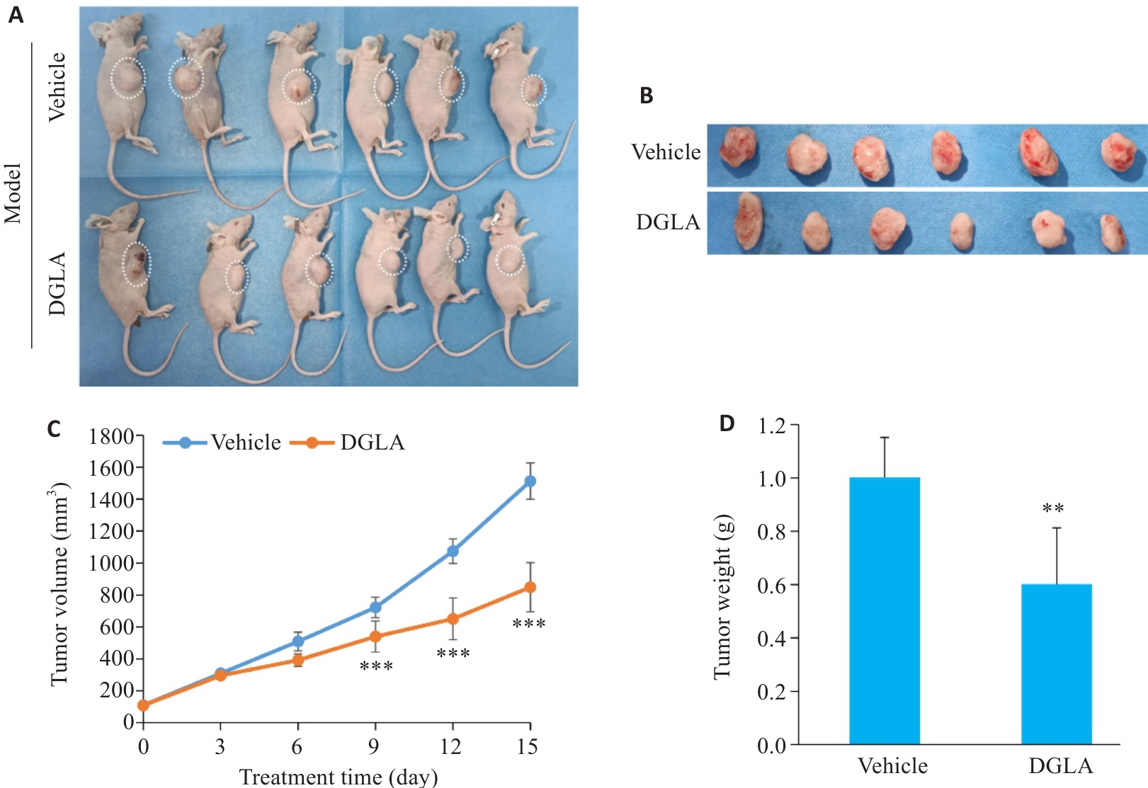
Fig.1 Establishment of nude mouse models bearing subcutaneous AML cell xenograft. A: Mouse grouping of subcutaneous xenograft model. B: Dissected tumors from DGLA and vehicle groups. C: Tumor volume in DGLA and vehicle groups. D: Tumor weight changes in DGLA and vehicle groups. **P<0.01, ***P<0.001 vs vehicle group, n=6.
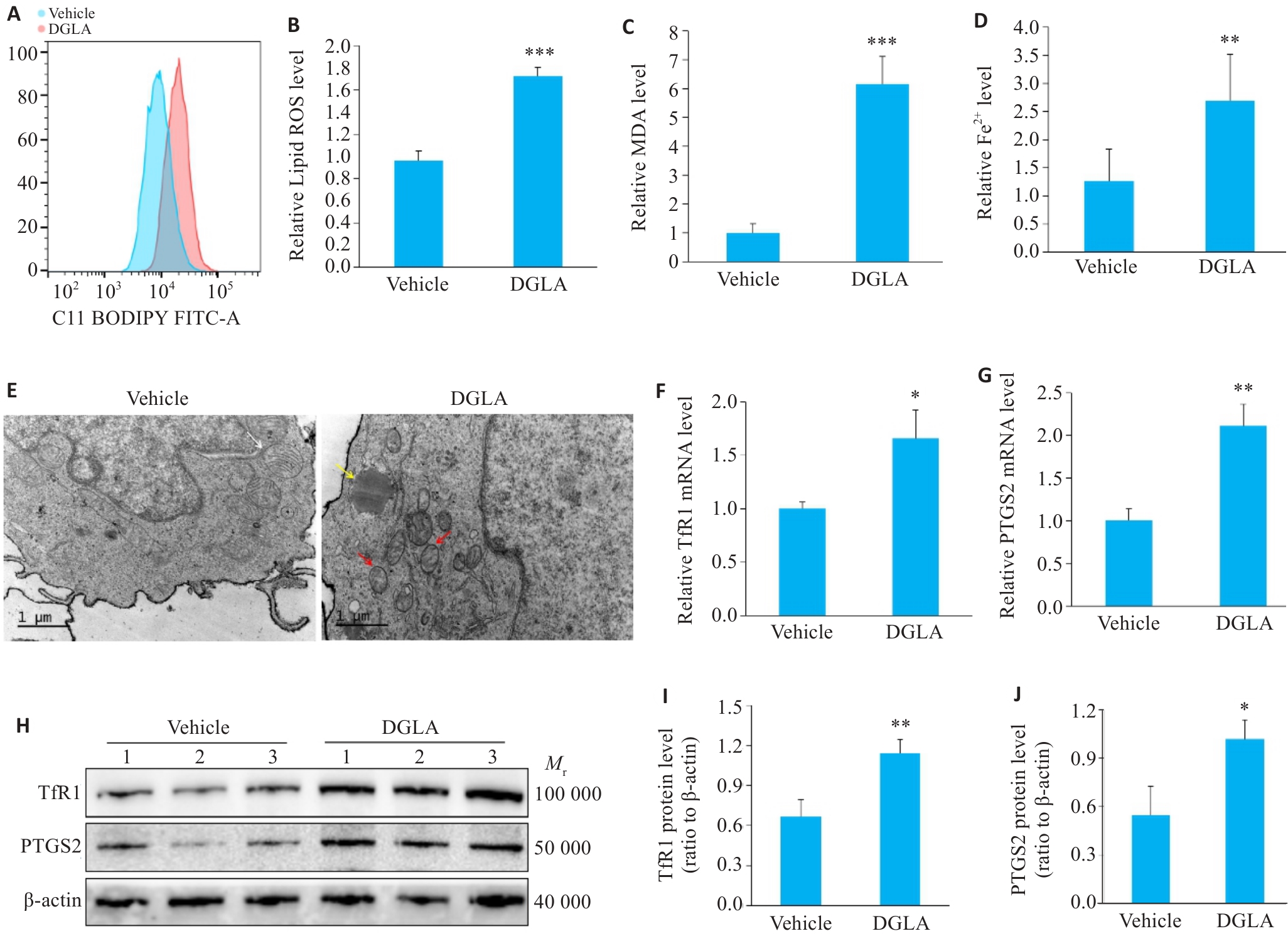
Fig.2 Detection of ferroptosis markers in subcutaneous xenografts derived from DGLA-induced AML cells. A: Flow histogram of lipid ROS levels. B: Quantitative result of ROS. C: Detection of MDA levels. D: Detection of Fe2+ levels. E: Changes of mitochondrial ultrastructural (Scale bar=1 μm). White arrows: normal mitochondria; red arrows: ferroptotic mitochondria; yellow arrows: lipid droplets. F, G: TfR1 and PTGS2 mRNA expression levels. H: TfR1 and PTGS2 protein blotting bands. I, J: TfR1 and PTGS2 protein expression levels. *P<0.05, **P<0.01, ***P<0.001 vs Vehicle group (n=6); n=3 for qRT-PCR and Western blotting analysis.
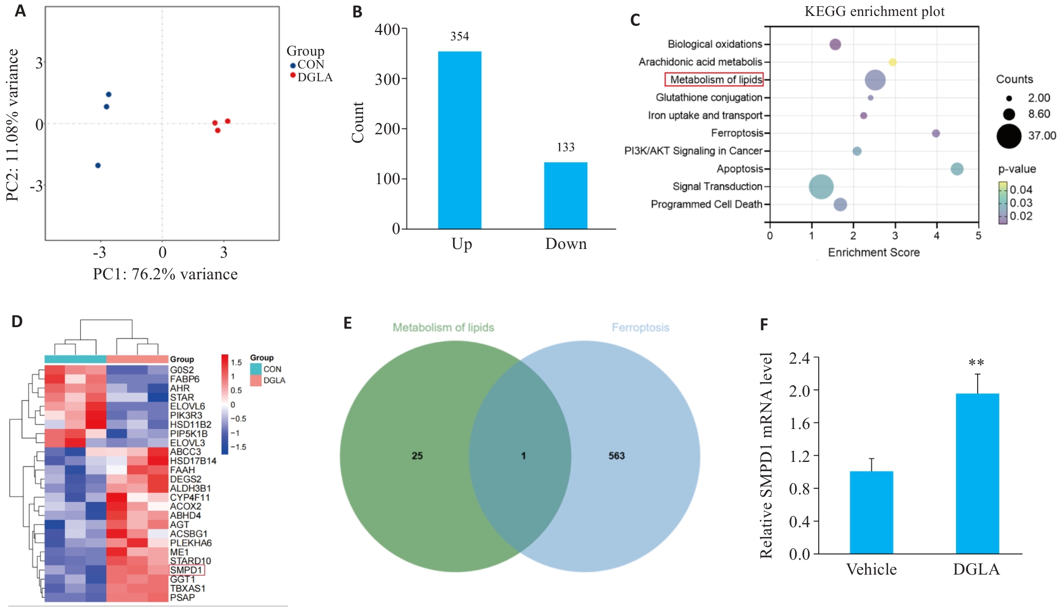
Fig.3 Significant upregulation of SMPD1 identified by RNA-seq combined with FerrDb analysis. A: PCA plot of RNA-seq. B: Bar chart of differentially expressed genes. C: Bubble chart depicting the KEGG enrichment of the differentially expressed genes. D: Heatmap of lipid metabolism. E: Venn diagram of lipid metabolism and FerrDb. F: Changes of SMPD1 mRNA level after DGLA treatment. **P<0.01 vs vehicle group (n=3).
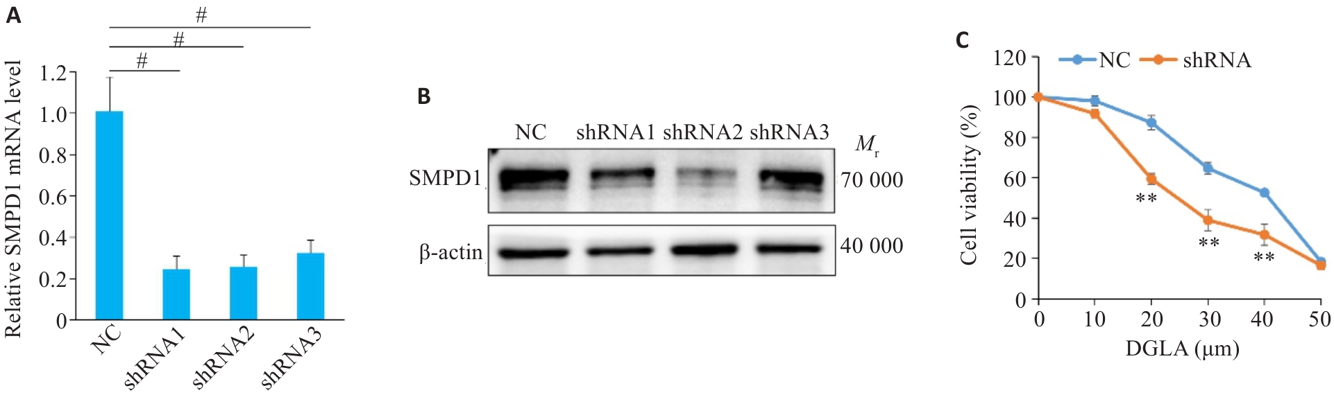
Fig.4 Downregulation of SMPD1 enhances DGLA-induced inhibition of AML cell viability in vitro. A, B: SMPD1 expression levels in THP-1 cells after lentiviral transfection. C: Effect of SMPD1 knockdown on THP-1 cell viability with DGLA treatment. **P<0.01 vs NC group, #P<0.001. n=3.
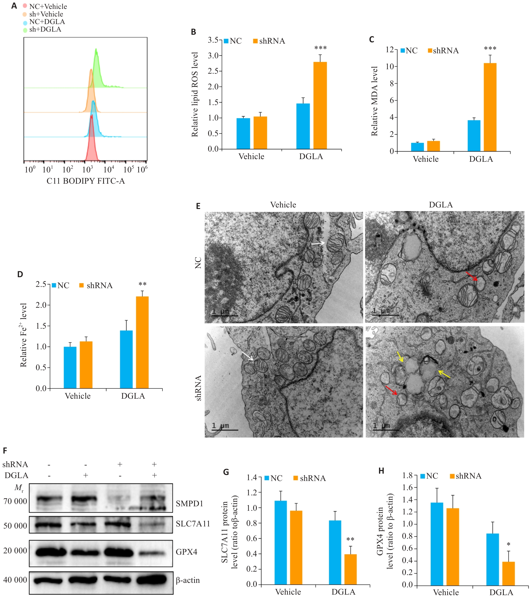
Fig.5 SMPD1 knockdown promotes DGLA-induced ferroptosis in AML cells in vitro. A-D: Effect of SMPD1 knockdown on ROS, MDA and Fe2+ in THP-1 cells with DGLA treatment. E: Effect of SMPD1 knockdown on mitochondrial ultrastructure of THP-1 with DGLA treatment (Scale bar=1 μm). White arrows: normal mitochondria; red arrows: Ferroptotic mitochondria; yellow arrows: Lipid droplets. F-H: SLC7A11 and GPX4 protein expression levels. *P<0.05, **P<0.01, ***P<0.001 vs NC+DGLA group (n=3).
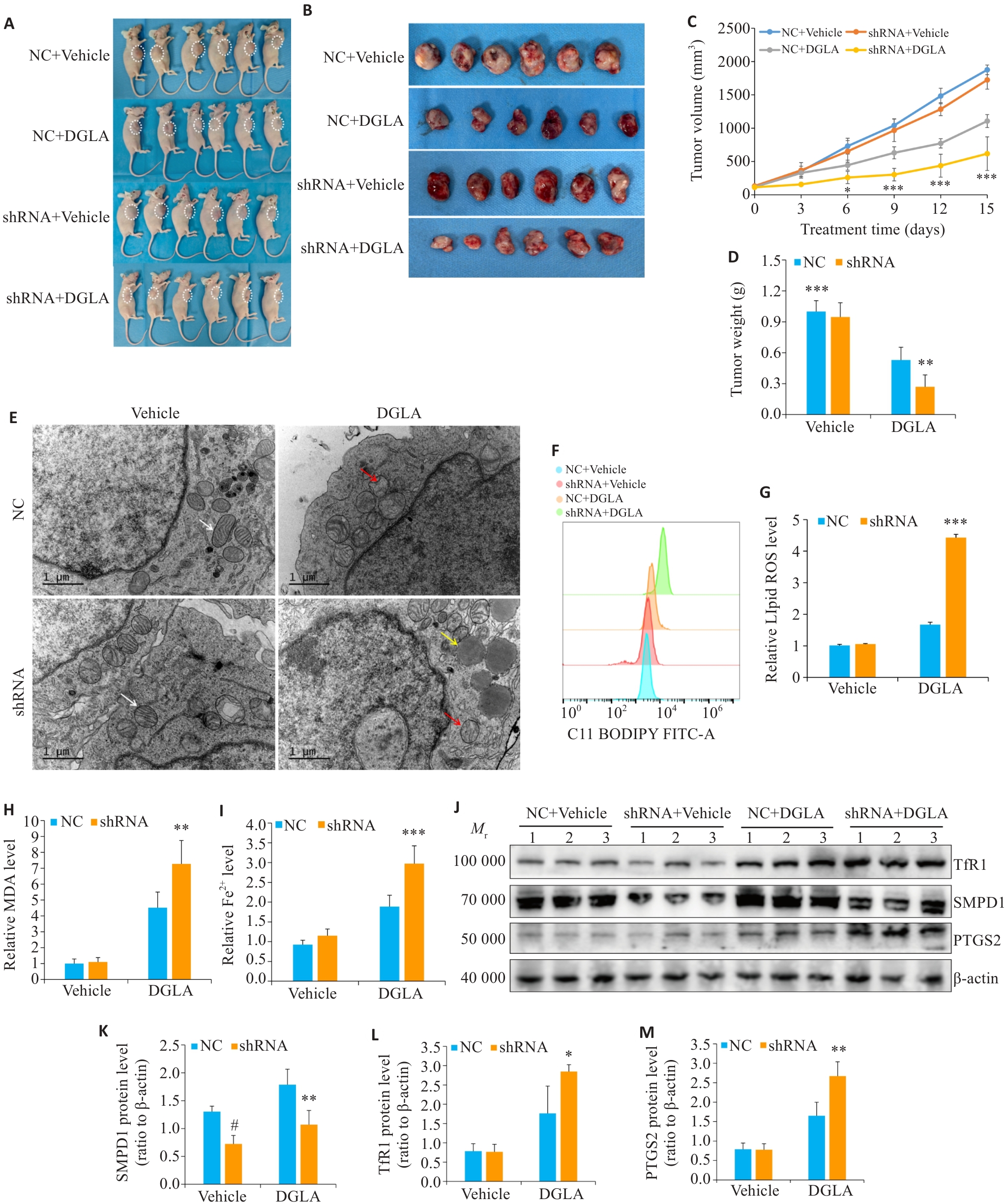
Fig.6 SMPD1 knockdown promotes DGLA-induced ferroptosis of AML cells in mice. A: Observation of subcutaneous AML tumors in the mice. B: Dissected of subcutaneous tumors from each group. C: Tumor volume. D: Tumor weight. E: Mitochondrial ultrastructure in subcutaneous tumors from each group (Scale bar=1 μm). White arrows: Normal mitochondria; red arrows: Ferroptotic mitochondria; yellow arrows: Lipid droplets. F-I: ROS, MDA and Fe2+ levels. J-M: SMPD1, TfR1 and PTGS2 protein expression levels. *P<0.05, **P<0.01, ***P<0.001 vs NC+DGLA group. #P<0.05 vs NC+Vehicle group (n=6); n=3 per group for Western blotting analysis.
| [1] | Zhang SJ, Liu MH, Yao YF, et al. Targeting LSD1 for acute myeloid leukemia (AML) treatment[J]. Pharmacol Res, 2021, 164: 105335. doi:10.1016/j.phrs.2020.105335 |
| [2] | Haubner S, Subklewe M, Sadelain M. Honing CAR T cells to tackle acute myeloid leukemia[J]. Blood, 2025, 145(11): 1113-25. doi:10.1182/blood.2024024063 |
| [3] | Feng J, Li YL, He F, et al. RBM15 silencing promotes ferroptosis by regulating the TGF‑β/Smad2 pathway in lung cancer[J]. Environ Toxicol, 2023, 38(4): 950-61. doi:10.1002/tox.23741 |
| [4] | Yang C, Lu T, Liu M, et al. Tiliroside targets TBK1 to induce ferroptosis and sensitize hepatocellular carcinoma to sorafenib[J]. Phytomedicine, 2023, 111: 154668. doi:10.1016/j.phymed.2023.154668 |
| [5] | Yang F, Xiao Y, Ding JH, et al. Ferroptosis heterogeneity in triple-negative breast cancer reveals an innovative immunotherapy combination strategy[J]. Cell Metab, 2023, 35(1): 84-100.e8. doi:10.1016/j.cmet.2022.09.021 |
| [6] | Yang WS, SriRamaratnam R, Welsch ME, et al. Regulation of ferroptotic cancer cell death by GPX4[J]. Cell, 2014, 156(1/2): 317-31. doi:10.1016/j.cell.2013.12.010 |
| [7] | Dixon SJ, Lemberg KM, Lamprecht MR, et al. Ferroptosis: an iron-dependent form of nonapoptotic cell death[J]. Cell, 2012, 149(5): 1060-72. doi:10.1016/j.cell.2012.03.042 |
| [8] | Tousignant KD, Rockstroh A, Poad BLJ, et al. Therapy-induced lipid uptake and remodeling underpin ferroptosis hypersensitivity in prostate cancer[J]. Cancer Metab, 2020, 8(1): 11. doi:10.1186/s40170-020-00217-6 |
| [9] | Han JW, Zheng DH, Liu PS, et al. Peroxisomal homeostasis in metabolic diseases and its implication in ferroptosis[J]. Cell Commun Signal, 2024, 22(1): 475. doi:10.1186/s12964-024-01862-w |
| [10] | Turchi R, Tortolici F, Benvenuto M, et al. Low sulfur amino acid, high polyunsaturated fatty acid diet inhibits breast cancer growth[J]. Int J Mol Sci, 2022, 24(1): 249. doi:10.3390/ijms24010249 |
| [11] | Habib A, Mynott RL, Best OG, et al. Novel (1S, 3R)‑RSL3-encapsulated polyunsaturated fatty acid rich liposomes sensitise multiple myeloma cells to ferroptosis-mediated cell death[J]. Int J Mol Sci, 2025, 26(14): 6579. doi:10.3390/ijms26146579 |
| [12] | Nilsen DWT, Myhre PL, Kalstad A, et al. Serum levels of dihomo-gamma (γ)-linolenic acid (DGLA) are inversely associated with linoleic acid and total death in elderly patients with a recent myocardial infarction[J]. Nutrients, 2021, 13(10): 3475. doi:10.3390/nu13103475 |
| [13] | Perez MA, Magtanong L, Dixon SJ, et al. Dietary lipids induce ferroptosis in caenorhabditiselegans and human cancer cells[J]. Dev Cell, 2020, 54(4): 447-54.e4. doi:10.1016/j.devcel.2020.06.019 |
| [14] | Sarparast M, Pourmand E, Hinman J, et al. Dihydroxy-metabolites of dihomo‑γ‑linolenic acid drive ferroptosis-mediated neuro-degeneration[J]. ACS Cent Sci, 2023, 9(5): 870-82. doi:10.1021/acscentsci.3c00052 |
| [15] | Jiang XD, Huang YY, Hong XY, et al. Exogenous dihomo‑γ-linolenic acid triggers ferroptosis via ACSL4-mediated lipid metabolic reprogramming in acute myeloid leukemia cells[J]. Transl Oncol, 2025, 52: 102227. doi:10.1016/j.tranon.2024.102227 |
| [16] | Döhner H, Wei AH, Appelbaum FR, et al. Diagnosis and management of AML in adults: 2022 recommendations from an international expert panel on behalf of the ELN[J]. Blood, 2022, 140(12): 1345-77. doi:10.1182/blood.2022016867 |
| [17] | Kantarjian H, Kadia T, DiNardo C, et al. Acute myeloid leukemia: current progress and future directions[J]. Blood Cancer J, 2021, 11(2): 41. doi:10.1038/s41408-021-00425-3 |
| [18] | Kremer DM, Nelson BS, Lin L, et al. GOT1 inhibition promotes pancreatic cancer cell death by ferroptosis[J]. Nat Commun, 2021, 12(1): 4860. doi:10.1038/s41467-021-24859-2 |
| [19] | Yang JJ, Zhou YL, Xie SD, et al. Metformin induces Ferroptosis by inhibiting UFMylation of SLC7A11 in breast cancer[J]. J Exp Clin Cancer Res, 2021, 40(1): 206. doi:10.1186/s13046-021-02012-7 |
| [20] | Peng YX, Li N, Tang FF, et al. Corosolic acid sensitizes ferroptosis by upregulating HERPUD1 in liver cancer cells[J]. Cell Death Discov, 2022, 8(1): 376. doi:10.1038/s41420-022-01169-0 |
| [21] | Mustonen AM, Nieminen P. Dihomo‑γ-linolenic acid (20: 3n-6)-metabolism, derivatives, and potential significance in chronic inflammation[J]. Int J Mol Sci, 2023, 24(3): 2116. doi:10.3390/ijms24032116 |
| [22] | Stockwell BR, Jiang XJ, Gu W. Emerging mechanisms and disease relevance of ferroptosis[J]. Trends Cell Biol, 2020, 30(6): 478-90. doi:10.1016/j.tcb.2020.02.009 |
| [23] | Zhou N, Yuan XQ, Du QS, et al. FerrDb V2: update of the manually curated database of ferroptosis regulators and ferroptosis-disease associations[J]. Nucleic Acids Res, 2023, 51(D1): D571-82. doi:10.1093/nar/gkac935 |
| [24] | Huang ZW, Li M, Gu BY, et al. Ferroptosis-related LINC02535/has-miR-30c-5p/EIF2S1 axis as a novel prognostic biomarker involved in immune infiltration and progression of PDAC[J]. Cell Signal, 2024, 123: 111338. doi:10.1016/j.cellsig.2024.111338 |
| [25] | Yang ZW, Han TX, Yang RB, et al. Dicoumarol sensitizes hepatocellular carcinoma cells to ferroptosis induced by imidazole ketone erastin[J]. Front Immunol, 2025, 16: 1531874. doi:10.3389/fimmu.2025.1531874 |
| [26] | Qin JK, Li ZD, Su LZ, et al. Expression of transferrin receptor/TFRC protein in bladder cancer cell T24 and its role in inducing iron death in bladder cancer[J]. Int J Biol Macromol, 2024, 274(Pt 1): 133323. doi:10.1016/j.ijbiomac.2024.133323 |
| [27] | Ma HD, Wang XD, Zhang WL, et al. Melatonin suppresses ferroptosis induced by high glucose via activation of the Nrf2/HO-1 signaling pathway in type 2 diabetic osteoporosis[J]. Oxid Med Cell Longev, 2020, 2020(1): 9067610. doi:10.1155/2020/9067610 |
| [28] | Chen X, Tsvetkov AS, Shen HM, et al. International consensus guidelines for the definition, detection, and interpretation of autophagy-dependent ferroptosis[J]. Autophagy, 2024, 20(6): 1213-46. doi:10.1080/15548627.2024.2319901 |
| [29] | Ye Z, Xie B, Tao YG, et al. Mechanism of ferroptosis and its role in disease development[J]. Int J Biol Sci, 2025, 21(12): 5328-60. doi:10.7150/ijbs.102859 |
| [30] | Feng HZ, Schorpp K, Jin J, et al. Transferrin receptor is a specific ferroptosis marker[J]. Cell Rep, 2020, 30(10): 3411-23.e7. doi:10.1016/j.celrep.2020.02.049 |
| [31] | Kong ZY, Liu R, Cheng YR. Artesunate alleviates liver fibrosis by regulating ferroptosis signaling pathway[J]. Biomed Pharmacother, 2019, 109: 2043-53. doi:10.1016/j.biopha.2018.11.030 |
| [32] | Luo SM, Luo Y, Wang ZM, et al. Super-enhancer mediated upregulation of MYEOV suppresses ferroptosis in lung adenocarcinoma[J]. Cancer Lett, 2024, 589: 216811. doi:10.1016/j.canlet.2024.216811 |
| [33] | Dixon SJ, Pratt DA. Ferroptosis: a flexible constellation of related biochemical mechanisms[J]. Mol Cell, 2023, 83(7): 1030-42. doi:10.1016/j.molcel.2023.03.005 |
| [34] | Aldrovandi M, Fedorova M, Conrad M. Juggling with lipids, a game of Russian roulette[J]. Trends Endocrinol Metab, 2021, 32(7): 463-73. doi:10.1016/j.tem.2021.04.012 |
| [35] | Thayyullathil F, Cheratta AR, Alakkal A, et al. Acid sphingomyelinase-dependent autophagic degradation of GPX4 is critical for the execution of ferroptosis[J]. Cell Death Dis, 2021, 12: 26. doi:10.1038/s41419-020-03297-w |
| [36] | Guo C, Liao KQ, Chen GL, et al. SMPD1 modulates malignant progress of osteosarcoma through ferroptosis pathway[J]. Tissue Cell, 2026, 102: 103588. doi:10.1016/j.tice.2026.103588 |
| [37] | Sokol KH, Lee CJ, Rogers TJ, et al. Lipid availability influences ferroptosis sensitivity in cancer cells by regulating polyunsaturated fatty acid trafficking[J]. Cell Chem Biol, 2025, 32(3): 408-22.e6. doi:10.1016/j.chembiol.2024.09.008 |
| [1] | Chunhui QI, Heqian ZHANG, Xiaoshuang WU, Yongan KANG, Jingyi YANG, Di ZHAO, Shegan GAO, Yijun QI. Dual inhibition of PSME2 and STAT3 induces ferroptosis in esophageal squamous cell carcinoma cells by regulating the SLC7A11/GPX4 axis [J]. Journal of Southern Medical University, 2026, 46(9): 2230-2241. |
| [2] | Liling HE, Dan WU, Bixuan LAI, Qixuan NIU, Junhua CHEN, Jianhong GAO, Qinghua LONG, Zhengyu WANG. Wuyou Decoction alleviates hippocampal neuronal injury and improves synaptic plasticity in rats with chronic sleep deprivation by inhibiting ferroptosis via activating the Nrf2/HO-1 pathway [J]. Journal of Southern Medical University, 2026, 46(8): 1754-1763. |
| [3] | Kun WANG, Shunfu HOU, Yong LI, Qinghua LIU, Chonggao YIN, Hongli LI. Overexpression of miR-183-5p enhances malignant phenotype and inhibits ferroptosis of triple-negative breast cancer cells by negatively regulating KLHL24 [J]. Journal of Southern Medical University, 2026, 46(8): 1790-1798. |
| [4] | Jiping YANG, Yongzeng GU, Zhenyi FU, Yuexin DONG, Yihao DENG, Zheng LI, Liangli LIU, Tao GUO. Gastrodin alleviates hypobaric hypoxia-induced brain injury in rats by reducing neuronal ferroptosis via the P53/SLC7A11/GPX4 signaling axis [J]. Journal of Southern Medical University, 2026, 46(7): 1498-1508. |
| [5] | Jinchao ZHU, Ningning SHAO, Zeyu HUANG, Yanqing LIU, Xiangyan MENG, Ziquan LIU, Yujie SUN, Jinrui DONG, Haojun FAN. Ultrafine Xiebai powder alleviates lipopolysaccharide-induced acute lung injury in mice by suppressing ferroptosis and modulating the NF-κB/STAT3 signaling axis [J]. Journal of Southern Medical University, 2026, 46(5): 1006-1017. |
| [6] | Yuanyuan CHENG, Chuanbing HUANG, Juan ZHANG, Yawen ZHU, Ai QIAN. Chonggu Granules attenuate cartilage injury in knee osteoarthritis patients with liver-kidney deficiency syndrome by regulating the Nrf2 pathway [J]. Journal of Southern Medical University, 2026, 46(5): 1028-1038. |
| [7] | Feifei ZHU, Na YAN, Jiwen YANG, Xiaolei CHEN, Yingying WANG. SENP1 reduces ferroptosis of thyroid cancer cells by regulating hypoxia-inducible factor-1α [J]. Journal of Southern Medical University, 2026, 46(5): 1093-1101. |
| [8] | Dongmin WEI, Bingbing WANG, Jianzhong GAO, Tianyu ZHAI, Xinyue BAI, Jinghui ZHU, Can ZHANG, Caizhen SHI, Qin HAO, Chen CHEN, Lin ZHAO. miR-24-3p promotes spinal cord injury repair in rats by inhibiting ferroptosis via targeting GSK-3β [J]. Journal of Southern Medical University, 2026, 46(4): 715-727. |
| [9] | Yunfei LI, Chuanbing HUANG, Lijun PANG, Ziheng ZHU, Ming LI. Jianpi Zishen Formula improves renal damage in mice with systemic lupus erythematosus by inhibiting p53-MDM2 signaling axis-mediated ferroptosis [J]. Journal of Southern Medical University, 2026, 46(4): 785-793. |
| [10] | Qile SONG, Yikai MIAO, Xiaotong FENG, Yifan WANG, Wei LIU, Qi WEI, Xinru YU, Wenwen CHEN, Xiaoyan FU. Selenocystine inhibits colon cancer cell growth by promoting reactive oxygen species generation to trigger oxidative damage [J]. Journal of Southern Medical University, 2026, 46(3): 532-540. |
| [11] | Tao GUO, Bolin CHEN, Xiao YANG, Yanli ZHAO, Xiaomin LI, Jiahao HE, Jinsha SHI, Hanjun ZUO, Juanjuan LI. Formononetin downregulates P53/SAT1/ACSL4 pathway-mediated ferroptosis to improve hypoxic-ischemic brain injury in neonatal mice [J]. Journal of Southern Medical University, 2026, 46(3): 604-614. |
| [12] | Tao GUO, Jiahao HE, Yuexin DONG, Zhao WANG, Hanjun ZUO, Yanli ZHAO, Jinsha SHI, Min ZHAO, Juanjuan LI. Schisandrin A alleviates neuronal ferroptosis in mice with hypoxic-ischemic brain injury by inhibiting the ACSL4/TFRC/PTGS2 signaling pathway [J]. Journal of Southern Medical University, 2026, 46(10): 2393-2404. |
| [13] | Rongmao HE, Zeyang FANG, Yunyun ZHANG, Youliang WU, Shixiu LIANG, Tao JI, Kequan CHEN, Siqi WANG. Diagnostic and predictive value of ferroptosis-related genes in patients with ulcerative colitis [J]. Journal of Southern Medical University, 2025, 45(9): 1927-1937. |
| [14] | Qi YUN, Ruoli DU, Yuying HE, Yixin ZHANG, Jiahui WANG, Hongwei YE, Zhenghong LI, Qin GAO. Cinnamic acid ameliorates doxorubicin-induced myocardial injury in mice by attenuating cardiomyocyte ferroptosis via inhibiting TLR4 [J]. Journal of Southern Medical University, 2025, 45(9): 1946-1958. |
| [15] | Zejin OU, Ying LI, Shi CHEN, Ziyi WANG, Meiyi HE, Zhicheng CHEN, Shihao TANG, Xiaojing MENG, Zhi WANG. Inhibition of ferroptosis alleviates acute kidney injury caused by diquat in zebrafish [J]. Journal of Southern Medical University, 2025, 45(8): 1743-1750. |
| Viewed | ||||||
|
Full text |
|
|||||
|
Abstract |
|
|||||
