
Journal of Southern Medical University ›› 2026, Vol. 46 ›› Issue (10): 2393-2404.doi: 10.12122/j.issn.1673-4254.2026.10.09
Tao GUO1,2( ), Jiahao HE2, Yuexin DONG2, Zhao WANG1, Hanjun ZUO1, Yanli ZHAO1, Jinsha SHI1, Min ZHAO1(), Juanjuan LI1()
Received:2026-03-23
Online:2026-10-20
Published:2026-10-26
Contact:
Min ZHAO, Juanjuan LI
E-mail:20190034@kust.edu.cn;zhaomin_kmmu@163.com;lijuanjuan@kmmu.edu.cn
Supported by:Tao GUO, Jiahao HE, Yuexin DONG, Zhao WANG, Hanjun ZUO, Yanli ZHAO, Jinsha SHI, Min ZHAO, Juanjuan LI. Schisandrin A alleviates neuronal ferroptosis in mice with hypoxic-ischemic brain injury by inhibiting the ACSL4/TFRC/PTGS2 signaling pathway[J]. Journal of Southern Medical University, 2026, 46(10): 2393-2404.
Add to citation manager EndNote|Ris|BibTeX
URL: https://www.j-smu.com/EN/10.12122/j.issn.1673-4254.2026.10.09
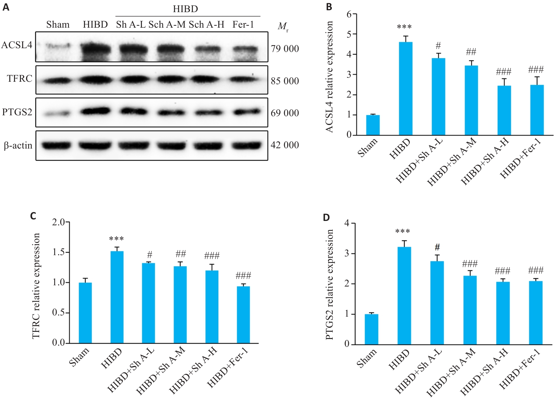
Fig.1 Effects of schisandrin A (Sch A) on expressions of ferroptosis-related proteins ACSL4, TFRC and PTGS2 in neonatal mice with hypoxic ischemic brain damage (HIBD). A: Western blotting of ACSL4, TFRC, and PTGS2 in the ischemic penumbra of the cerebral cortex of the neonatal mice in each group. B-D: Statistical analysis of the relative expression levels. ***P<0.001 vs Sham group; #P<0.05, ##P<0.01, ###P<0.001 vs HIBD group (n=3). L: Low; M: Medium; H: High; Fer-1: Ferrostatin-1.
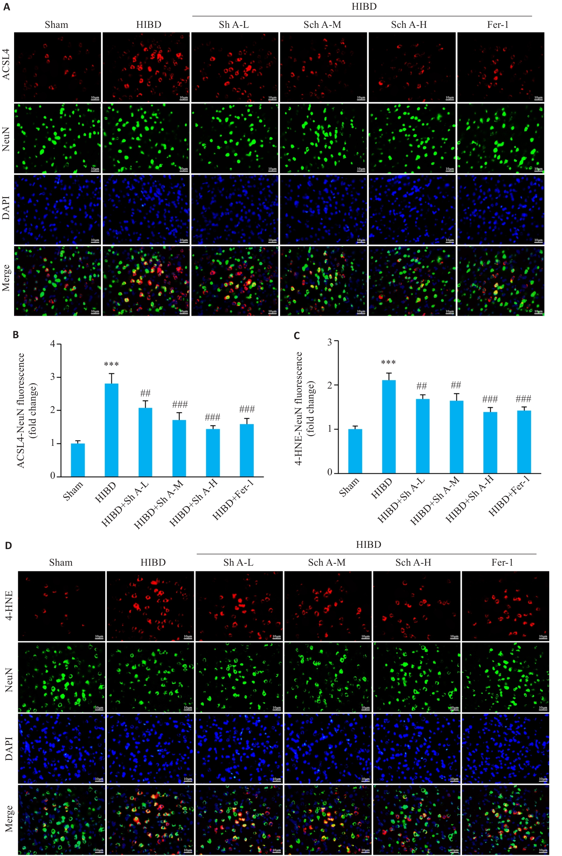
Fig.2 Effects of Sch A on expressions of ACSL4 and 4-HNE in brain neurons in mice with HIBD. A, D: Co-localization of ACSL4 or 4-HNE (red) with the neuronal marker NeuN (green) in the ischemic penumbra of the cerebral cortex detected by immunofluorescence double staining. B, C: Quantitative analysis of fluorescence intensity of ACSL4-NeuN and 4-HNE-NeuN. Scale bar=10 μm. ***P<0.001 vs Sham group; ##P<0.01, ###P<0.001 vs HIBD group (n=3).
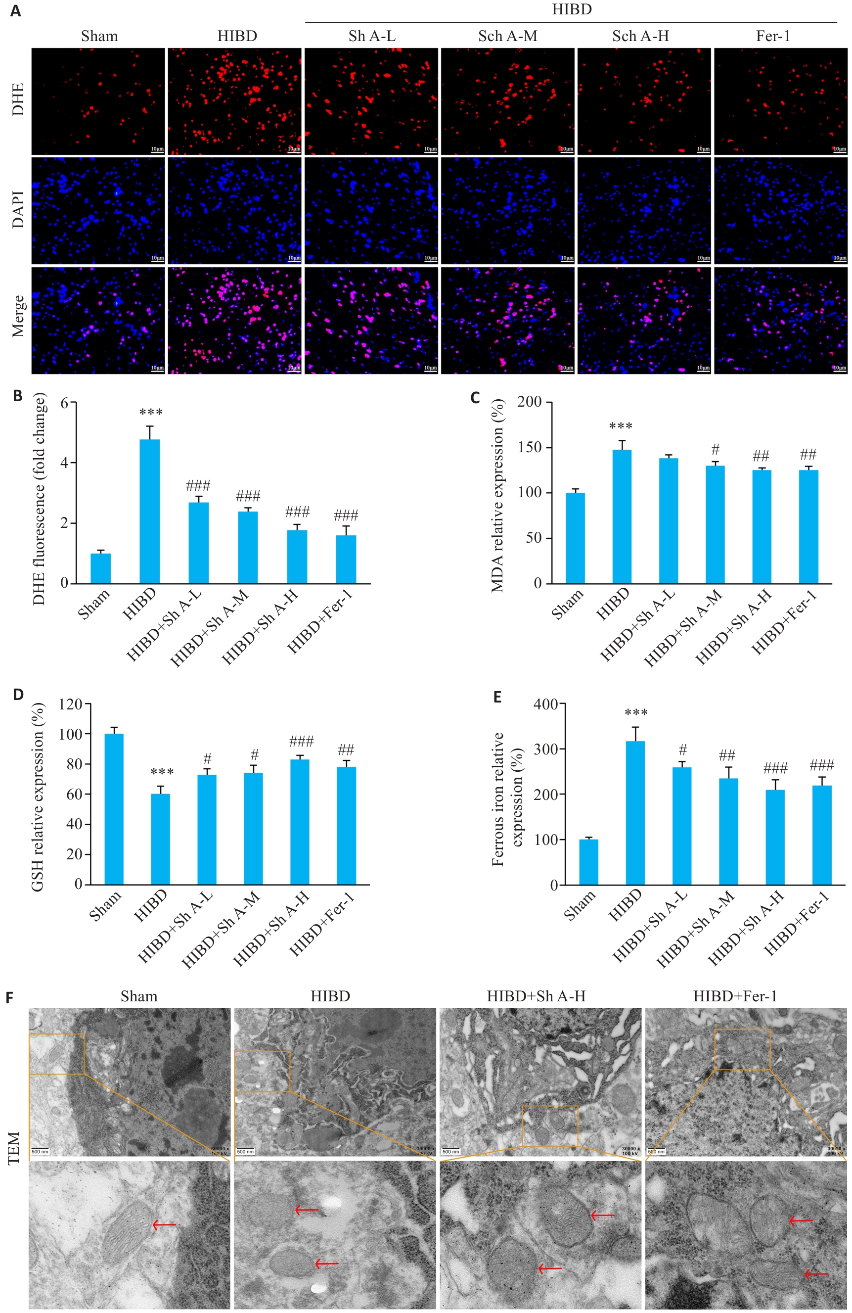
Fig.3 Effects of Sch A on oxidative stress and iron overload following HIBD. A, B: ROS levels in the ischemic penumbra of mouse cerebral cortex detected by DHE fluorescence staining and quantitative analysis (Scale bar=10 μm). C: Relative expression levels of malondialdehyde (MDA). D: Relative expression levels of glutathione (GSH). E: Levels of ferrous ions (Fe²⁺). F: Ultrastructure of the neurons in the ischemic penumbra in each group observed by transmission electron microscopy (Original magnification: ×30 000; scale bar=500 nm). Red arrows indicate mitochondria. ***P<0.001 vs Sham group; #P<0.05, ##P<0.01, ###P<0.001 vs HIBD group (n=3).
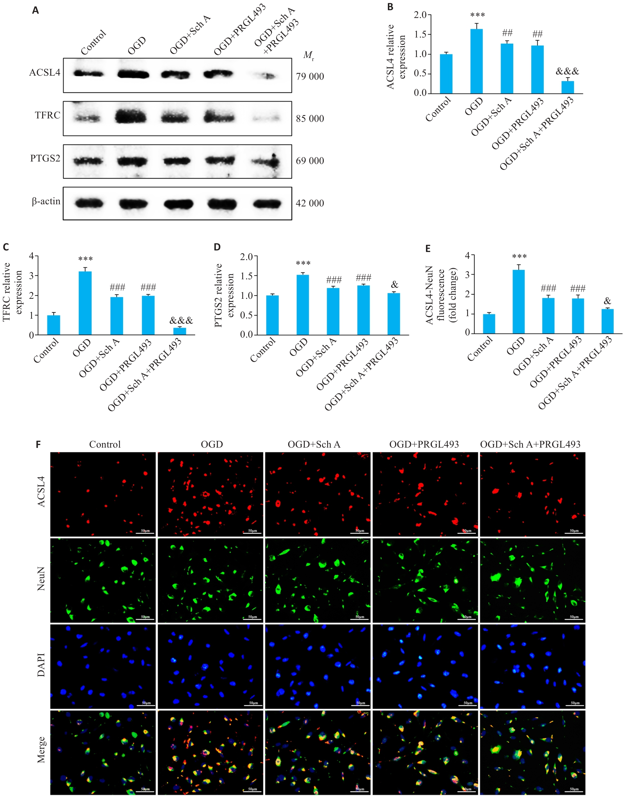
Fig.4 Effects of Sch A combined with the ACSL4 inhibitor PRGL493 on OGD‑induced neuronal ferroptosis. A: Western blotting of ACSL4, TFRC, and PTGS2 protein expression levels in the neurons of each group. B-D: Statistical analysis of the relative expression levels of ACSL4, TFRC, and PTGS2 proteins. E, F: Immunofluorescence double staining for detecting co-localization of ACSL4 (red) with the neuronal marker NeuN (green). Scale bar=50 μm. ***P<0.001 vs Control group; ##P<0.01, ###P<0.001 vs OGD group; &P<0.05, &&&P<0.001 vs OGD+Sch A group (n=3). OGD: Oxygen-Glucose Deprivation.
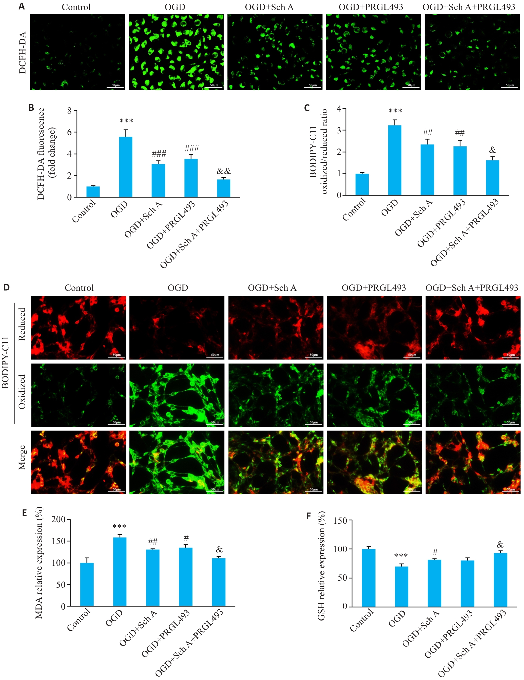
Fig.5 Effects of Sch A on OGD induced neuronal oxidative stress and lipid peroxidation. A, B: ROS levels in the neurons detected by DCFH DA fluorescence staining. C, D: Oxidized/reduced lipid peroxidation levels in the neurons detected by BODIPY C11 fluorescence staining. E: Relative expression levels of MDA. F: Relative expression levels of GSH. Scale bar=50 μm. ***P<0.001 vs Control group; #P<0.05, ##P<0.01, ###P<0.001 vs OGD group; &P<0.05, &&P<0.01 vs OGD+Sch A group (n=3). MDA: Malondialdehyde; GSH: Glutathione.
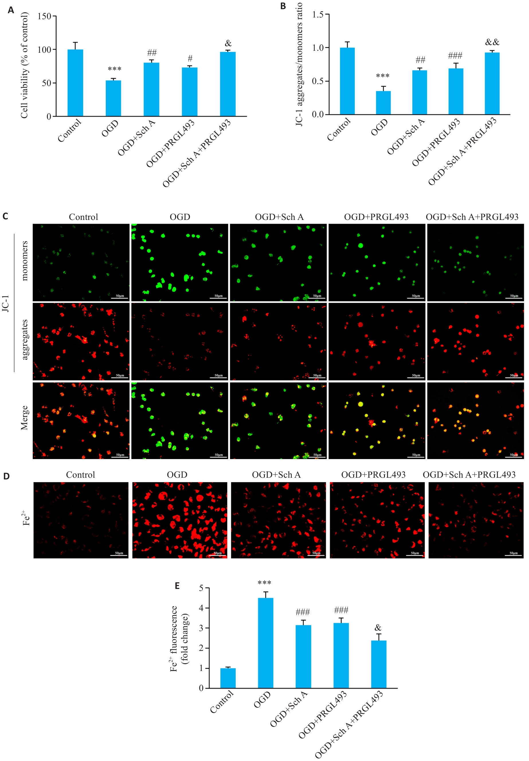
Fig.6 Effects of Sch A on OGD induced neuronal viability, mitochondrial function, and iron accumulation. A: Neuronal viability detected by CCK-8 assay. B, C: Fluorescence staining of mitochondrial membrane potential detected by JC-1 staining. D, E: Neuronal iron ion levels detected by Fe²⁺ fluorescence staining. Scale bar=50 μm. ***P<0.001 vs Control group; #P<0.05, ##P<0.01, ###P<0.001 vs OGD group; &P<0.05, &&P<0.01 vs OGD+Sch A group (n=3).
| [1] | Deng QT, Parker E, Duan R, et al. Preconditioning and posttr-eatment strategies in neonatal hypoxic-ischemic encephalopathy: recent advances and clinical challenges[J]. Mol Neurobiol, 2025, 62(8): 10020-44. doi:10.1007/s12035-025-04896-4 |
| [2] | Rondagh M, Schrama WJJ, de Vries LS, et al. Brain state of the newborn as a biomarker for brain injury in infants with hypoxic-ischemic encephalopathy[J]. J Pediatr, 2025, 285: 114702. doi:10.1016/j.jpeds.2025.114702 |
| [3] | 郭 涛, 左涵珺, 匡显锋, 等. 小胶质细胞介导的铁死亡在缺氧缺血性脑损伤中的研究进展[J]. 细胞与分子免疫学杂志, 2025, 41(6): 552-8. |
| [4] | Brunets N, Brunets V, Bokiniec R. Cardiovascular hemodynamics of hypoxic neonates during therapeutic hypothermia and the warming phase: a literature review[J]. Ther Hypothermia Temp Manag, 2025, 15(4): 161-73. doi:10.1089/ther.2025.0035 |
| [5] | Wang XX, Li M, Xu XW, et al. BNIP3-mediated mitophagy attenuates hypoxic-ischemic brain damage in neonatal rats by inhibiting ferroptosis through P62-KEAP1-NRF2 pathway activation to maintain iron and redox homeostasis[J]. Acta Pharmacol Sin, 2025, 46(1): 33-51. doi:10.1038/s41401-024-01365-x |
| [6] | Shi JS, Xiao X, Wang Z, et al. Gastrodin promotes Alkbh5 nuclear localization and Gclm m6A demethylation to alleviate ferroptosis in ischemic stroke[J]. Phytomedicine, 2025, 147: 157177. doi:10.1016/j.phymed.2025.157177 |
| [7] | Yu XX, Wang SH, Ji ZJ, et al. Ferroptosis: an important mechanism of disease mediated by the gut-liver-brain axis[J]. Life Sci, 2024, 347: 122650. doi:10.1016/j.lfs.2024.122650 |
| [8] | Peeples ES, Genaro-Mattos TC. Ferroptosis: a promising therapeutic target for neonatal hypoxic-ischemic brain injury[J]. Int J Mol Sci, 2022, 23(13): 7420. doi:10.3390/ijms23137420 |
| [9] | 张安邦, 孙秀颀, 庞 博, 等. 电针预处理通过调节肠道-大脑轴及Nrf2/HO-1信号通路抑制铁死亡减轻大鼠脑缺血再灌注损伤[J]. 南方医科大学学报, 2025, 45(5): 911-20. |
| [10] | Luo C, Fang C, Zou RJ, et al. Hyperglycemia-induced DNA damage response activates DNA-PK complex to promote endothelial ferroptosis in type 2 diabetic cardiomyopathy[J]. Theranostics, 2025, 15(10): 4507-25. doi:10.7150/thno.109514 |
| [11] | Zhou JF, Tang JP, Zhang CR, et al. ALKBH5 targets ACSL4 mRNA stability to modulate ferroptosis in hyperbilirubinemia-induced brain damage[J]. Free Radic Biol Med, 2024, 220: 271-87. doi:10.1016/j.freeradbiomed.2024.05.014 |
| [12] | Wang L, Liu HR, Liu HQ, et al. SETD1B promotes brain cell ferroptosis in ischemic stroke mice via increasing H3K4me3 enrichment on the Tfrc promoter[J]. Life Sci, 2025, 372: 123625. doi:10.1016/j.lfs.2025.123625 |
| [13] | 杨晓玲, 何宗源, 章琦鑫, 等. 小胶质细胞PTGS2/Hepcidin炎症通路在神经元铁死亡中的作用机制[J]. 西南医科大学学报, 2024, 47(1): 39-44. |
| [14] | Fu K, Zhou H, Wang C, et al. A review: Pharmacology and pharmacokinetics of Schisandrin A[J]. Phytother Res, 2022, 36(6): 2375-93. doi:10.1002/ptr.7456 |
| [15] | Zong WT, Gouda M, Cai EL, et al. The antioxidant phytochemical schisandrin a promotes neural cell proliferation and differentiation after ischemic brain injury[J]. Molecules, 2021, 26(24): 7466. doi:10.3390/molecules26247466 |
| [16] | 臧 瑞, 杨继苹, 郭 涛, 等. 五味子醇甲诱导缺血半影区神经元自噬提高脑卒中后神经保护[J]. 中国生物化学与分子生物学报, 2020, 36(5): 544-51. |
| [17] | Hassanein EHM, Althagafy HS, Baraka MA, et al. The promising antioxidant effects of lignans: Nrf2 activation comes into view[J]. Naunyn Schmiedeberg's Arch Pharmacol, 2024, 397(9): 6439-58. doi:10.1007/s00210-024-03102-x |
| [18] | Zhou F, Wang MD, Ju J, et al. Schizandrin A protects against cerebral ischemia-reperfusion injury by suppressing inflammation and oxidative stress and regulating the AMPK/Nrf2 pathway regulation[J]. Am J Transl Res, 2019, 11(1): 199-209. |
| [19] | Zhang TL, Zhang ZW, Geng JY, et al. A new approach for exploring reperfusion brain damage in hypoxic ischemic encephalopathy[J]. Mol Neurobiol, 2024, 61(3): 1417-32. doi:10.1007/s12035-023-03645-9 |
| [20] | Zhu X, Shi MR, Ma CY, et al. Activation of the serotonin-1A receptor promotes neural regeneration in the rat model of neonatal hypoxic-ischemic brain damage[J]. Brain Res Bull, 2026, 235: 111739. doi:10.1016/j.brainresbull.2026.111739 |
| [21] | 郭 涛, 陈柏霖, 石金沙, 等. 天麻素通过激活GPX4/SLC7A11/FTH1信号抑制铁死亡减轻新生小鼠缺氧缺血性脑损伤[J]. 南方医科大学学报, 2025, 45(10): 2071-81. |
| [22] | Wu XQ, He SQ, Lu JZ, et al. Untargeted metabolomics and transcriptomics study reveals an activated ferroptosis metabolic spectrum in recurrent spontaneous abortion[J]. Free Radic Biol Med, 2025, 239: 145-54. doi:10.1016/j.freeradbiomed.2025.07.029 |
| [23] | Jin JX, Chen MS, Wang HC, et al. Schizandrin A attenuates early brain injury following subarachnoid hemorrhage through suppressing neuroinflammation[J]. Mol Biol Rep, 2024, 51(1): 236. doi:10.1007/s11033-023-08956-7 |
| [24] | 马 强, 欧阳心怡, 颜思阳, 等. 芒柄花素通过影响ACSL4/LPCAT3/GPX4表达缓解脑缺血再灌注损伤中神经元铁死亡[J]. 湖南中医药大学学报, 2025, 45(6): 989-99. doi:10.3969/j.issn.1674-070X.2025.06.001 |
| [25] | Lin W, Zhang TL, Zheng JY, et al. Ferroptosis is involved in hypoxic-ischemic brain damage in neonatal rats[J]. Neuroscience, 2022, 487: 131-42. doi:10.1016/j.neuroscience.2022.02.013 |
| [26] | Hou D, Hu YJ, Yun T, et al. The deubiquitinase OTUD3 stabilizes IRP2 expression to reduce hippocampal neuron ferroptosis via the p53/PTGS2 pathway to ameliorate cerebral ischemia-reperfusion injury[J]. Eur J Med Res, 2024, 29(1): 498. doi:10.1186/s40001-024-02095-w |
| [27] | Chen Y, Guo XY, Zeng YC, et al. Oxidative stress induces mitochondrial iron overload and ferroptotic cell death[J]. Sci Rep, 2023, 13: 15515. doi:10.1038/s41598-023-42760-4 |
| [28] | Tan XY, Zhang T, Ding XJ, et al. Iron overload facilitates neonatal hypoxic-ischemic brain damage via SLC7A11‐mediated ferroptosis[J]. J Neurosci Res, 2023, 101: 1107-24. doi:10.1002/jnr.25184/v4/decision1 |
| [29] | Zheng JY, Fang Y, Zhang M, et al. Mechanisms of ferroptosis in hypoxic-ischemic brain damage in neonatal rats[J]. Exp Neurol, 2024, 372: 114641. doi:10.1016/j.expneurol.2023.114641 |
| [30] | Jiang DY, Zhao J, Zheng J, et al. LOX-mediated ECM mechanical stress induces Piezo1 activation in hypoxic-ischemic brain damage and identification of novel inhibitor of LOX[J]. Redox Biol, 2024, 76: 103346. doi:10.1016/j.redox.2024.103346 |
| [31] | Gan BY. Mitochondrial regulation of ferroptosis[J]. J Cell Biol, 2021, 220(9): e202105043. doi:10.1083/jcb.202105043 |
| [32] | Ahmed Selim N, Wojtovich AP. Mitochondrial membrane potential and compartmentalized signaling: Calcium, ROS, and beyond[J]. Redox Biol, 2025, 86: 103859. doi:10.1016/j.redox.2025.103859 |
| [1] | Quan YUAN, Dongdong ZHANG, Jingtao WANG, Hanjun SONG. Social isolation induces social avoidance in mice: the mediating role of the medial prefrontal cortex glutamatergic neurons [J]. Journal of Southern Medical University, 2026, 46(9): 1993-2005. |
| [2] | Lixuan YE, Yuxin LIAO, Qiong LIU, Jia ZHANG, Lei GAO, Zhaoyang DONG. Xiaoyao San alleviates abnormal α‑syn aggregation and neuronal injury in a Caenorhabditis elegans model of Parkinson disease via the SKN-1 antioxidative pathway [J]. Journal of Southern Medical University, 2026, 46(9): 2023-2034. |
| [3] | Chunhui QI, Heqian ZHANG, Xiaoshuang WU, Yongan KANG, Jingyi YANG, Di ZHAO, Shegan GAO, Yijun QI. Dual inhibition of PSME2 and STAT3 induces ferroptosis in esophageal squamous cell carcinoma cells by regulating the SLC7A11/GPX4 axis [J]. Journal of Southern Medical University, 2026, 46(9): 2230-2241. |
| [4] | Liling HE, Dan WU, Bixuan LAI, Qixuan NIU, Junhua CHEN, Jianhong GAO, Qinghua LONG, Zhengyu WANG. Wuyou Decoction alleviates hippocampal neuronal injury and improves synaptic plasticity in rats with chronic sleep deprivation by inhibiting ferroptosis via activating the Nrf2/HO-1 pathway [J]. Journal of Southern Medical University, 2026, 46(8): 1754-1763. |
| [5] | Kun WANG, Shunfu HOU, Yong LI, Qinghua LIU, Chonggao YIN, Hongli LI. Overexpression of miR-183-5p enhances malignant phenotype and inhibits ferroptosis of triple-negative breast cancer cells by negatively regulating KLHL24 [J]. Journal of Southern Medical University, 2026, 46(8): 1790-1798. |
| [6] | Dongkun YU, Shijie ZHI, Yanbo LIU, Zhilun YANG, Juan LIU, Yu SHAO. MiR-124 ameliorates MK-801-induced anxiety-like behaviors in mice by promoting hippocampal neural stem cell proliferation and differentiation [J]. Journal of Southern Medical University, 2026, 46(8): 1878-1885. |
| [7] | Jiping YANG, Yongzeng GU, Zhenyi FU, Yuexin DONG, Yihao DENG, Zheng LI, Liangli LIU, Tao GUO. Gastrodin alleviates hypobaric hypoxia-induced brain injury in rats by reducing neuronal ferroptosis via the P53/SLC7A11/GPX4 signaling axis [J]. Journal of Southern Medical University, 2026, 46(7): 1498-1508. |
| [8] | Weishuai YIN, Fei LI, Yi CAO, Qiqi YANG, Zetian DENG, Shijie CAO, Yongqi SUN, Xinyang BI, Zhengpeng LIANG, Sijia WU, Baoguo WANG. Effect of Huayu Tongluo moxibustion combined with neural stem cell transplantation on hippocampal Drp1/Mfn proteins and mitochondrial dynamics in rats with vascular dementia [J]. Journal of Southern Medical University, 2026, 46(7): 1520-1532. |
| [9] | Xueke SHI, Tingyu CHEN, Jing GAO, Mingli WU, Ruiqing LI, Kaiqi SU, Zhuan LÜ, Xiaolei SONG, Xiaodong FENG. Serum from rats with electroacupuncture activates the PI3K/Akt signaling pathway to improve synaptic plasticity in HT22 neurons with oxygen and glucose deprivation [J]. Journal of Southern Medical University, 2026, 46(6): 1407-1415. |
| [10] | Jinchao ZHU, Ningning SHAO, Zeyu HUANG, Yanqing LIU, Xiangyan MENG, Ziquan LIU, Yujie SUN, Jinrui DONG, Haojun FAN. Ultrafine Xiebai powder alleviates lipopolysaccharide-induced acute lung injury in mice by suppressing ferroptosis and modulating the NF-κB/STAT3 signaling axis [J]. Journal of Southern Medical University, 2026, 46(5): 1006-1017. |
| [11] | Yuanyuan CHENG, Chuanbing HUANG, Juan ZHANG, Yawen ZHU, Ai QIAN. Chonggu Granules attenuate cartilage injury in knee osteoarthritis patients with liver-kidney deficiency syndrome by regulating the Nrf2 pathway [J]. Journal of Southern Medical University, 2026, 46(5): 1028-1038. |
| [12] | Feifei ZHU, Na YAN, Jiwen YANG, Xiaolei CHEN, Yingying WANG. SENP1 reduces ferroptosis of thyroid cancer cells by regulating hypoxia-inducible factor-1α [J]. Journal of Southern Medical University, 2026, 46(5): 1093-1101. |
| [13] | Dongmin WEI, Bingbing WANG, Jianzhong GAO, Tianyu ZHAI, Xinyue BAI, Jinghui ZHU, Can ZHANG, Caizhen SHI, Qin HAO, Chen CHEN, Lin ZHAO. miR-24-3p promotes spinal cord injury repair in rats by inhibiting ferroptosis via targeting GSK-3β [J]. Journal of Southern Medical University, 2026, 46(4): 715-727. |
| [14] | Yunfei LI, Chuanbing HUANG, Lijun PANG, Ziheng ZHU, Ming LI. Jianpi Zishen Formula improves renal damage in mice with systemic lupus erythematosus by inhibiting p53-MDM2 signaling axis-mediated ferroptosis [J]. Journal of Southern Medical University, 2026, 46(4): 785-793. |
| [15] | Zhuoning ZHANG, Xinyu HAO, Fuyang CAO, Yongxin GUO, Shuting GUO, Chen CAI, Weidong MI, Li TONG. Esketamine alleviates depression-like behaviors in mice with chronic restraint stress by activating glutamatergic neurons in the medial prefrontal cortex [J]. Journal of Southern Medical University, 2026, 46(3): 489-496. |
| Viewed | ||||||
|
Full text |
|
|||||
|
Abstract |
|
|||||
