
Journal of Southern Medical University ›› 2026, Vol. 46 ›› Issue (9): 1993-2005.doi: 10.12122/j.issn.1673-4254.2026.09.01
Quan YUAN( ), Dongdong ZHANG, Jingtao WANG, Hanjun SONG()
Received:2026-04-16
Online:2026-09-20
Published:2026-09-30
Contact:
Hanjun SONG
E-mail:18245506700@163.com;songhanjun1973@163.com
Supported by:Quan YUAN, Dongdong ZHANG, Jingtao WANG, Hanjun SONG. Social isolation induces social avoidance in mice: the mediating role of the medial prefrontal cortex glutamatergic neurons[J]. Journal of Southern Medical University, 2026, 46(9): 1993-2005.
Add to citation manager EndNote|Ris|BibTeX
URL: https://www.j-smu.com/EN/10.12122/j.issn.1673-4254.2026.09.01
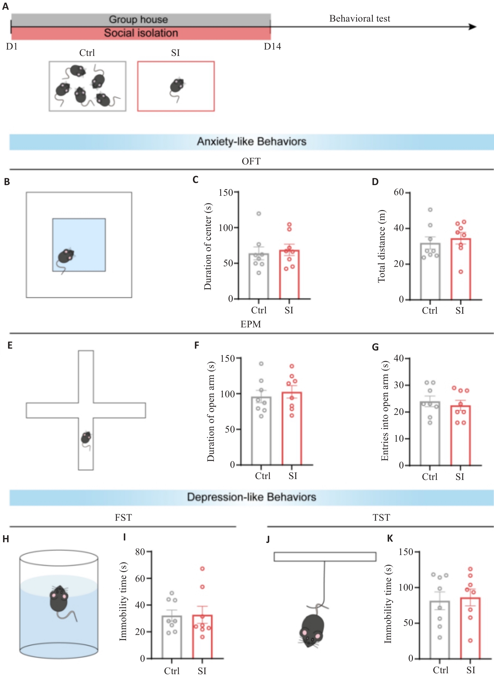
Fig.1 Social isolation (SI) does not cause anxiety- or depressive-like behaviors in mice. A: Schematic diagram of SI modeling in mice (n=8). B: Diagram of open field test (OFT) of the mice. C: Statistical plot of exploration time in the central areas of the mice during OFT. D: Statistical plot of total distance traveled of the mice in OFT. E: Diagram of elevated plus-maze (EPM) test of the mice. F: Statistical plot of exploration time in the open arms of the mice in EPM test. G: Statistical plot of open arm entry frequency of the mice in EPM test. H: Diagram of forced swimming test (FST) of the mice. I: Statistical plot of inactivity duration of the mice during the final 4 min in FST. J: Behavioral patterns of the mice in tail suspension test (TST). K: Statistical plot of inactivity duration of the mice during the final 4 min in TST.
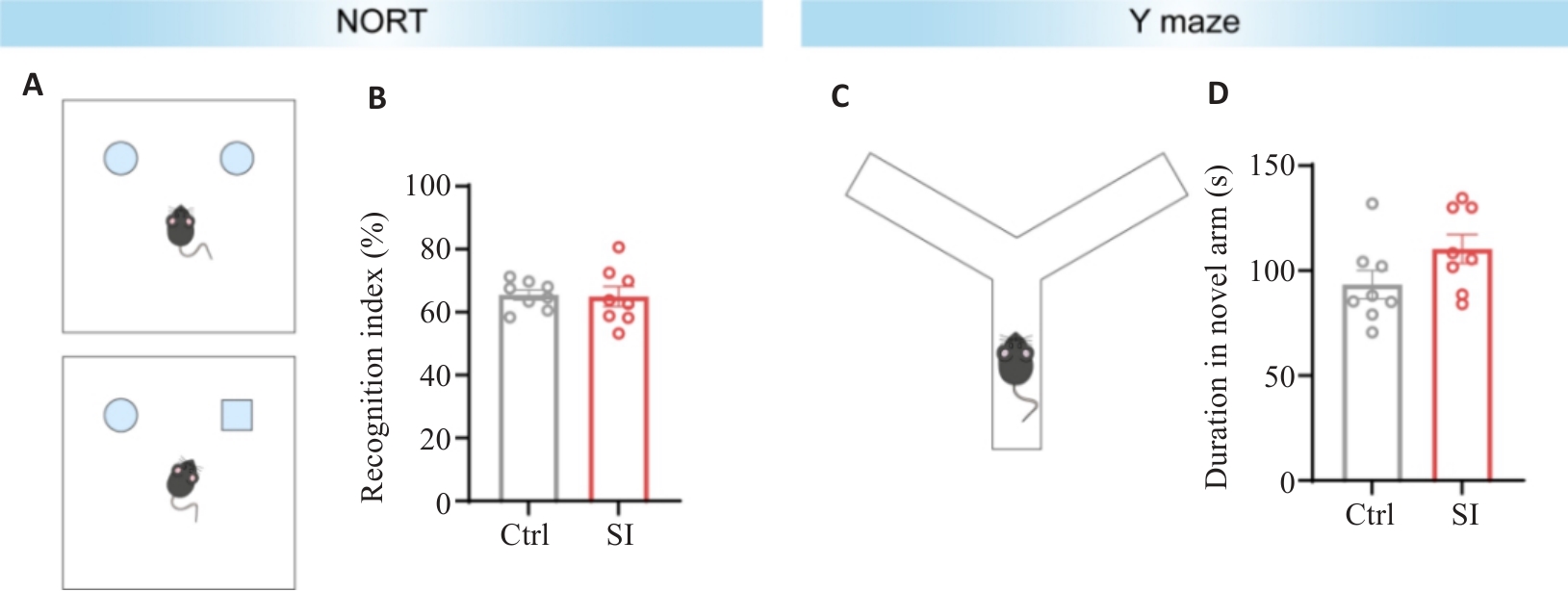
Fig.2 SI does not affect object recognition and working memory of mice. A: Diagram of novel object recognition test (NORT) of the mice. B: Statistical chart of cognitive indices in the two groups of mice during the second phase of NORT. C: Diagram of Y-maze test of the mice. D: Statistical chart of exploration time of the mice in the novel arm in Y-maze test (n=8).
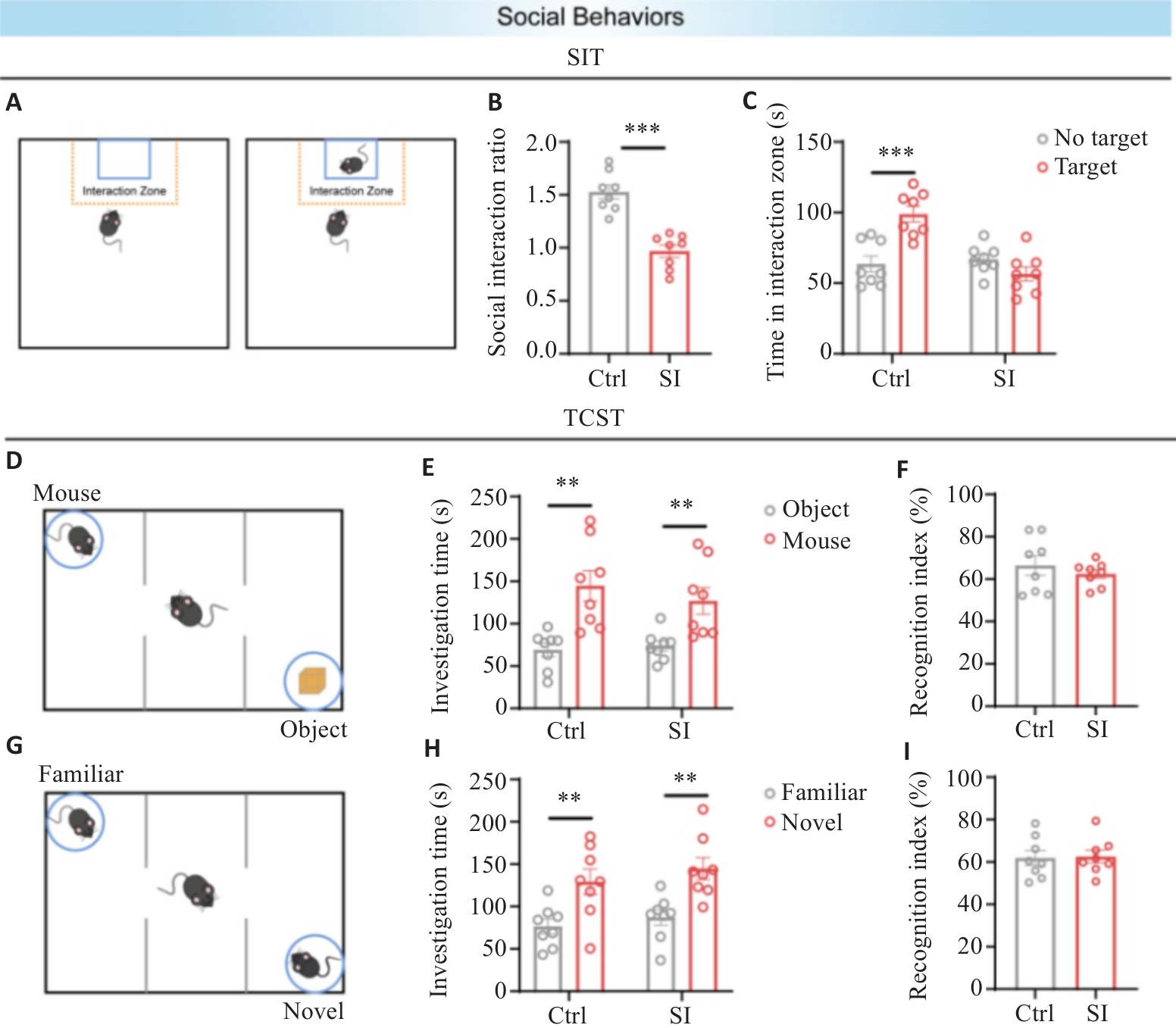
Fig.3 SI does not affect social competence or social memory, but may cause social avoidance in mice. A: Diagram of social interaction test (SIT) of the mice. B: Statistical chart of social interaction ratios of the mice in SIT. C: Statistical chart of time spent by target mice in the social interaction zone in the two groups. D: Diagram of TCST social competence stage. E: Statistical chart of exploration time of the mice toward the social mice and objects. F: Statistical chart of social preference index of the social mice in the two groups. G: Diagram of TCST social memory stage. H: Statistical chart of exploration time toward familiar mice and new mice in the two groups. I: Statistical chart of social preference index of the new mice in the two groups. **P<0.01, ***P<0.001 (n=8).
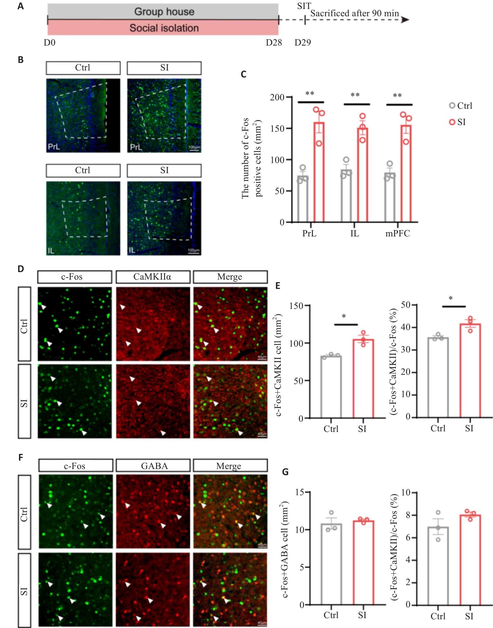
Fig.4 SI leads to increased c-Fos expression in the glutamatergic neurons in the medial prefrontal cortex (mPFC) of mice. A: Flowchart of c-Fos staining experiments in mice. B: Representative immunofluorescence images of c-Fos staining in PrL and IL subregions of the mPFC (Scale bar=100 μm). C: Statistical plot of average density of c-Fos-positive neurons in the PrL, IL subregions and mPFC brain region of the mice. D: Representative immunofluorescence images of co-staining for c-Fos and CaMKIIα in the mPFC brain region of the mice (Scale bar=40 μm). White arrows indicate co-localized cells expressing c-Fos and CaMKIIα. E: Statistical plot of co-localization of c-Fos- and CaMKIIα-positive neurons in the two groups. F: Representative immunofluorescence images of co-staining for c-Fos and GABA in the mPFC brain region of the mice (Scale bar=40 μm). White arrows indicate co-localized cells expressing c-Fos and GABA. G: Statistical plot of co-localization of c-Fos- and GABA-positive neurons in the two groups. *P<0.05, **P<0.01 (n=3).
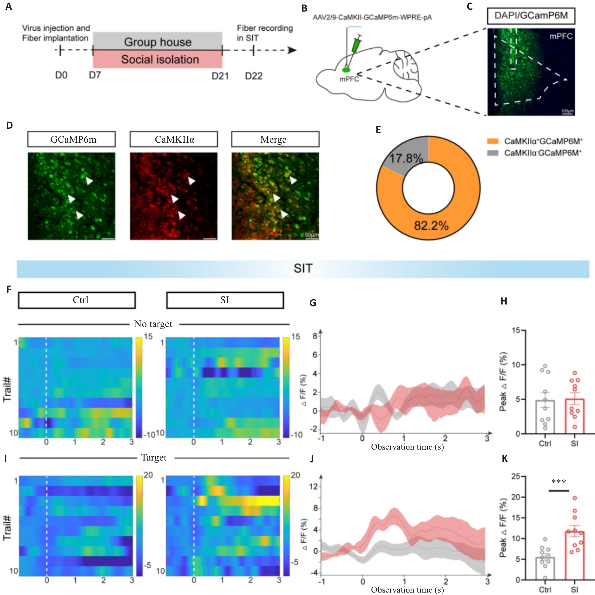
Fig.5 Increased calcium activity in mPFC glutamatergic neurons in SI mice during SIT. A: Flowchart of optical fiber recording in the two groups of mice. B: Schematic diagram of calcium indicator GCaMP6m injection and fiber embedding in mPFC neurons expressing CaMKIIα. C: Schematic diagram of viral injection sites (Scale bar=100 μm). D: Co-staining of GCaMP6m (green) and CaMKIIα (red). White arrows indicate co-localized cells expressing GCaMP6m and CaMKIIα (Scale bar=50 μm). E: Pie chart showing co-labeling rates of GCaMP6m and CaMKIIα. F: Heat map illustrating Ca2+ signal changes during optical fiber recording in the first stage of SIT in the two groups of mice. G: Curve plots of average Ca2+ fluorescence signals in the first stage in the two groups. H: Statistical plot of ΔF/F peak signals in the first stage in the two groups. I: Heat map illustrating Ca2+ signal changes during optical fiber recording in the second stage of SIT in the two groups. J: Curve plots of average Ca2+ fluorescence signals in the second stage in the two groups. K: Statistical plot of ΔF/F peak signals in the second stage in the two groups. ***P<0.001 (n=5).
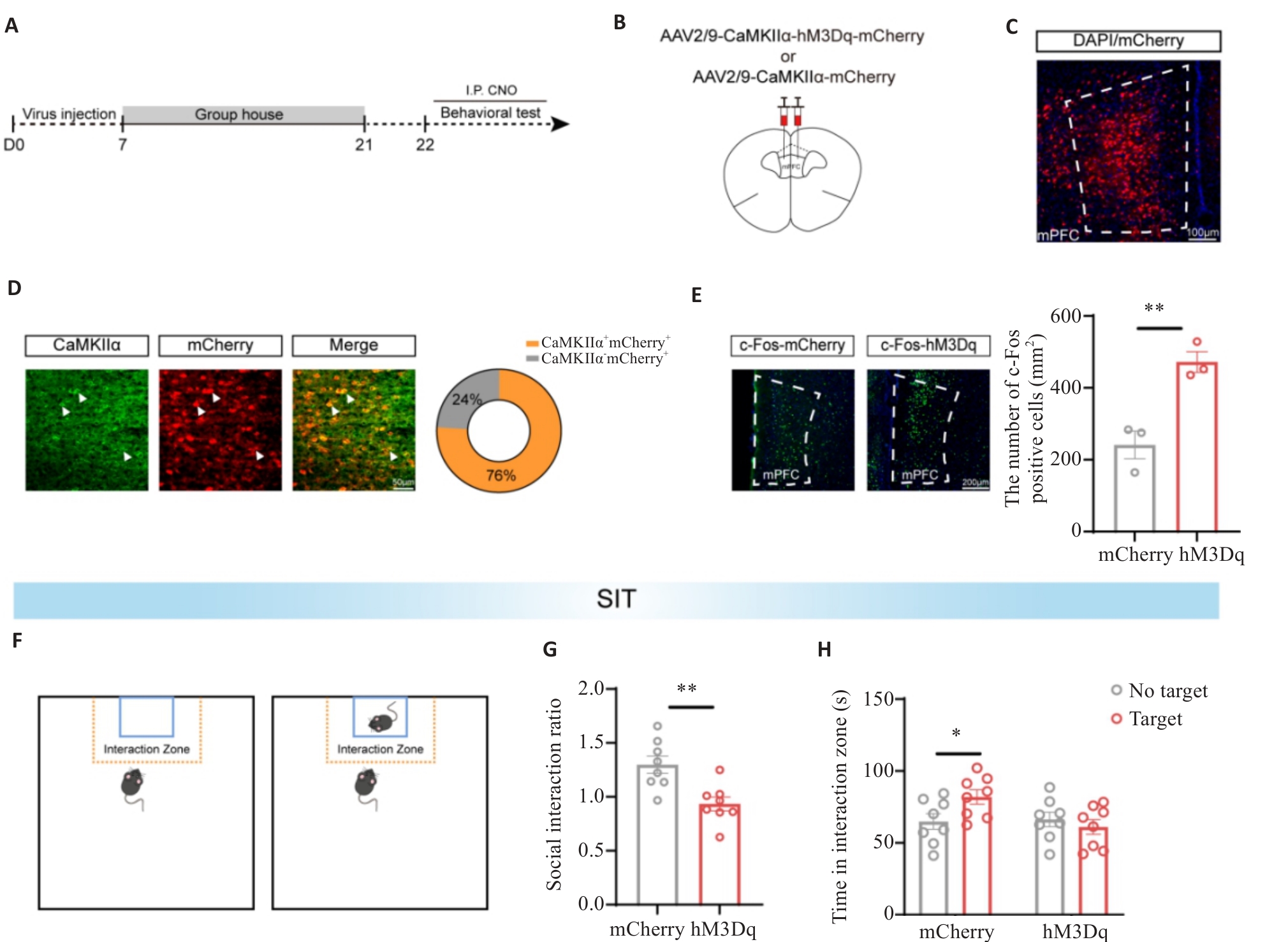
Fig.6 Chemical genetics activation of mPFC glutamatergic neurons induces social avoidance in mice. A: Experimental flowchart of chemogenetic activation of mPFC glutamatergic neurons. B: Schematic diagram of viral injection protocol and injection sites. C: Fluorescence image showing viral expression of hM3Dq in mPFC (Scale bar=100 μm). D: Fluorescence image of co-staining with hM3Dq-mCherry (red) and CaMKIIα (green) in mPFC sections. White arrows indicate co-localized cells, and the pie chart displays co-localization rates of hM3Dq and CaMKIIα (Scale bar= 50 μm). E: Representative immunofluorescence images of c-Fos staining in mPFC regions and statistical plot of average c-Fos density in mPFC of the mice (Scale bar=200 μm; n=3). F: SIT diagram. G: Statistical plot of social interaction rates during social interaction tests in SIT in the two groups. H: Statistical plot of time spent in social interaction areas by target mice in SIT in the two groups. *P<0.05, **P<0.01 (n=8).
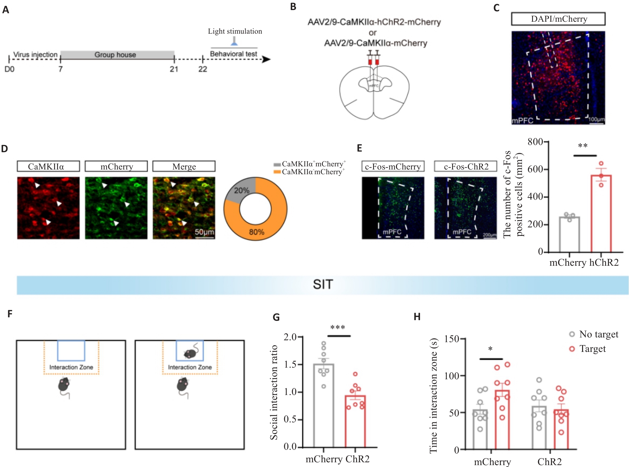
Fig.7 Optogenetic activation of mPFC glutamatergic neurons induces social avoidance in mice. A: Experimental flowchart for optogenetic activation of mPFC glutamatergic neurons. B: Schematic diagram of viral injection protocol and injection sites. C: Fluorescence image showing viral expression of hChR2 in mPFC (Scale bar=100 μm). D: Fluorescence image of co-staining hChR2-mCherry (red) and CaMKIIα (green) in mPFC sections (Scale bar=50 μm). White arrows indicate co-localized cells. The pie chart displays co-localization rates of hChR2 and CaMKIIα. E: Representative immunofluorescence images of c-Fos staining in mPFC regions and statistical plot of average c-Fos density in the mPFC of the mice (Scale bar=200 μm; n=3). F: SIT pattern diagram. G: Statistical plot of social interaction rates during social interaction tests in SIT in the two groups. H: Statistical plot of time spent in social interaction areas by target mice in SIT in the two groups. *P<0.05, **P<0.01, ***P<0.001 (n=8).
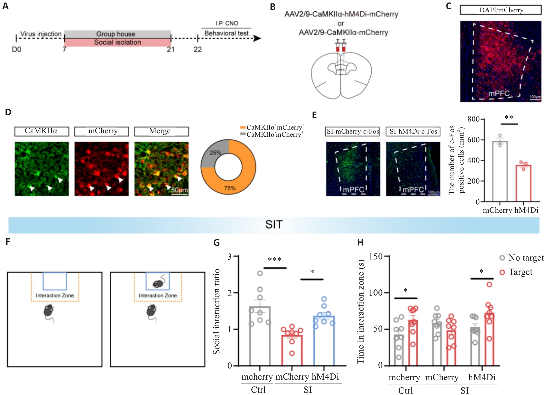
Fig.8 Chemical genetics inhibition of mPFC glutamatergic neurons alleviates social avoidance in SI mice. A: Flowchart of the experimental protocol for chemogenetic inhibition of mPFC glutamatergic neurons. B: Schematic diagram of the viral injection protocol and injection sites. C: Fluorescence image showing the localization of hM4Di virus expression in the mPFC (Scale bar= 100 μm). D: Fluorescence image of co-staining with hM4Di-mCherry (red) and CaMKIIα (green) in mPFC sections (Scale bar= 50 μm). White arrows indicate co-localized cells. The pie chart shows the co-localization rate of hM4Di and CaMKIIα. E: Representative immunofluorescence images of c-Fos staining in the mPFC regions (Scale bar=100 μm) and statistical plot of the average c-Fos density in the mPFC in the two groups (n=3). F: SIT protocol diagram. G: Statistical plot of social interaction rates among the 3 groups during the social interaction test in SIT. H: Statistical plot of time spent by target mice in the social interaction regions in SIT. *P<0.05, **P<0.01, ***P<0.001 (n=8).
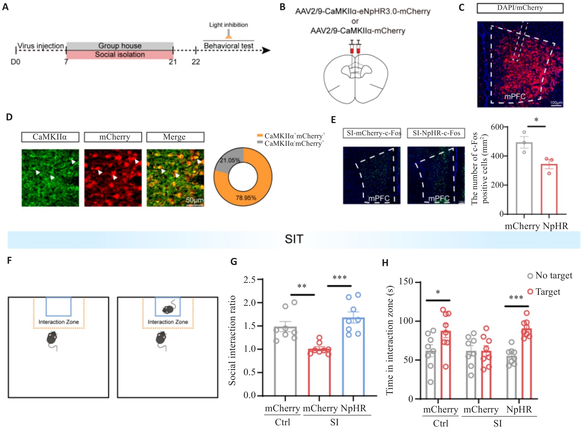
Fig.9 Optogenetic inhibition of mPFC glutamatergic neurons alleviates social avoidance in SI mice. A: Flowchart of the optogenetic inhibition of mPFC glutamatergic neurons. B: Schematic diagram of viral injection protocol and injection sites. C: Fluorescence image showing the injection site of NpHR virus in the mPFC (Scale bar=100 μm). D: Fluorescence image demonstrating co-staining of NpHR-mCherry (red) and CaMKIIα (green) in mPFC sections (Scale bar=50 μm). White arrows indicate co-localized cells. The pie chart shows the co-localization rate of NpHR and CaMKIIα. E: Representative immunofluorescence images of c-Fos staining in the mPFC regions (Scale bar=100 μm) and average c-Fos density in the mPFC in the two groups (n=3). F: SIT protocol diagram. G: Statistical plot of social interaction rates among the 3 groups during the social interaction test in the SIT. H: Statistical plot of time spent by target mice in the social interaction regions in SIT. *P<0.05, **P<0.01, ***P<0.001 (n=8).
| [1] | Albasheer O, Abdelwahab SI, Zaino MR, et al. The impact of social isolation and loneliness on cardiovascular disease risk factors: a systematic review, meta-analysis, and bibliometric investigation[J]. Sci Rep, 2024, 14: 12871. doi:10.1038/s41598-024-63528-4 |
| [2] | Holt-Lunstad J. Social connection as a critical factor for mental and physical health: evidence, trends, challenges, and future implications[J]. World Psychiatry, 2024, 23(3): 312-32. doi:10.1002/wps.21224 |
| [3] | Ran ZR, Wei JJ, Yang GJ, et al. Prevalence of social isolation in the elderly: a systematic review and meta-analysis[J]. Geriatr Nurs, 2024, 58: 87-97. doi:10.1016/j.gerinurse.2024.05.008 |
| [4] | 赵嘉盈, 纪小小, 潘玉峰, 等. 社交隔离调控本能行为的神经机制研究进展[J]. 生理学报, 2024, 76(2): 309-18. |
| [5] | Boyraz G, Legros DN, Tigershtrom A. COVID-19 and traumatic stress: The role of perceived vulnerability, COVID-19-related worries, and social isolation[J]. J Anxiety Disord, 2020, 76: 102307. doi:10.1016/j.janxdis.2020.102307 |
| [6] | Adepoju OE, Ojinnaka CO, Pieratt J, et al. Racial and ethnic differences in predictors of participation in an intergenerational social connectedness intervention for older adults[J]. BMC Geriatr, 2024, 24(1): 70. doi:10.1186/s12877-024-04679-x |
| [7] | Li WH, Wang ZK, Syed S, et al. Chronic social isolation signals starvation and reduces sleep in Drosophila [J]. Nature, 2021, 597(7875): 239-44. doi:10.1038/s41586-021-03837-0 |
| [8] | Xiong Y, Hong HL, Liu CR, et al. Social isolation and the brain: effects and mechanisms[J]. Mol Psychiatry, 2023, 28(1): 191-201. doi:10.1038/s41380-022-01835-w |
| [9] | 马 雪, 董莹莹, 刘 鹏, 等. 成年期社会隔离对雄性BALB/C小鼠神经行为学的影响[J]. 中国医药导报, 2016, 13(18): 13-6, 41. |
| [10] | Clemenza K, Weiss SH, Cheslack K, et al. Social isolation is closely linked to a marked reduction in physical activity in male mice[J]. J Neurosci Res, 2021, 99(4): 1099-107. doi:10.1002/jnr.24777 |
| [11] | Zelikowsky M, Hui M, Karigo T, et al. The neuropeptide Tac2 controls a distributed brain state induced by chronic social isolation stress[J]. Cell, 2018, 173(5): 1265-79.e19. doi:10.1016/j.cell.2018.03.037 |
| [12] | Carlén M. What constitutes the prefrontal cortex?[J]. Science, 2017, 358(6362): 478-82. doi:10.1126/science.aan8868 |
| [13] | Liu H, Jia X, Huang ZQ, et al. A top-down neural circuit for affective-motivational responses of pain relief induced by electroacupuncture[J]. Chin Med, 2026, 21(1): 73. doi:10.1186/s13020-026-01349-5 |
| [14] | Yang SS, Mack NR, Shu YS, et al. Prefrontal GABAergic interneurons gate long-range afferents to regulate prefrontal cortex-associated complex behaviors[J]. Front Neural Circuits, 2021, 15: 716408. doi:10.3389/fncir.2021.716408 |
| [15] | 克丽比努尔·艾尔肯. 生命早期社交孤立破坏社会功能及内侧前额叶皮层调控机制[J]. 社会科学前沿, 2024(4): 398-405. doi:10.12677/ass.2024.134314 |
| [16] | Fruehwirth JC, Biswas S, Perreira KM. The Covid-19 pandemic and mental health of first-year college students: Examining the effect of Covid-19 stressors using longitudinal data[J]. PLoS One, 2021, 16(3): e0247999. doi:10.1371/journal.pone.0247999 |
| [17] | Cauberghe V, Van Wesenbeeck I, De Jans S, et al. How adolescents use social media to cope with feelings of loneliness and anxiety during COVID-19 lockdown[J]. Cyberpsychol Behav Soc Netw, 2021, 24(4): 250-7. doi:10.1089/cyber.2020.0478 |
| [18] | Smith L, Jacob L, Yakkundi A, et al. Correlates of symptoms of anxiety and depression and mental wellbeing associated with COVID-19: a cross-sectional study of UK-based respondents[J]. Psychiatry Res, 2020, 291: 113138. doi:10.1016/j.psychres.2020.113138 |
| [19] | Sepúlveda-Loyola W, Rodríguez-Sánchez I, Pérez-Rodríguez P, et al. Impact of social isolation due to COVID-19 on health in older people: mental and physical effects and recommendations[J]. J Nutr Health Aging, 2020, 24(9): 938-47. doi:10.1007/s12603-020-1500-7 |
| [20] | Liu CH, Zhang E, Wong GTF, et al. Factors associated with depression, anxiety, and PTSD symptomatology during the COVID-19 pandemic: Clinical implications for U.S. young adult mental health[J]. Psychiatry Res, 2020, 290: 113172. doi:10.1016/j.psychres.2020.113172 |
| [21] | Morrissette M. School closures and social anxiety during the COVID-19 pandemic[J]. J Am Acad Child Adolesc Psychiatry, 2021, 60(1): 6-7. doi:10.1016/j.jaac.2020.08.436 |
| [22] | Wu X, Ding ZB, Fan TT, et al. Childhood social isolation causes anxiety-like behaviors via the damage of blood-brain barrier in amygdala in female mice[J]. Front Cell Dev Biol, 2022, 10: 943067. doi:10.3389/fcell.2022.943067 |
| [23] | Fosnocht AQ, Lucerne KE, Ellis AS, et al. Adolescent social isolation increases cocaine seeking in male and female mice[J]. Behav Brain Res, 2019, 359: 589-96. doi:10.1016/j.bbr.2018.10.007 |
| [24] | Lin S, Li X, Chen YH, et al. Social isolation during adolescence induces anxiety behaviors and enhances firing activity in BLA pyramidal neurons via mGluR5 upregulation[J]. Mol Neurobiol, 2018, 55(6): 5310-20. doi:10.1007/s12035-017-0766-1 |
| [25] | Yin T, Yuan JY, Liu L, et al. Confronting the anxiety of Generation Z: electroacupuncture therapy regulates oxidative stress and microglia activity in amygdala-basolateral of socially isolated mice[J]. Front Psychiatry, 2025, 15: 1496201. doi:10.3389/fpsyt.2024.1496201 |
| [26] | Locci A, Geoffroy P, Miesch M, et al. Social isolation in early versus late adolescent mice is associated with persistent behavioral deficits that can be improved by neurosteroid-based treatment[J]. Front Cell Neurosci, 2017, 11: 208. doi:10.3389/fncel.2017.00208 |
| [27] | Wang XN, Zhan Y. Regulation of social recognition memory in the hippocampal circuits[J]. Front Neural Circuits, 2022, 16: 839931. doi:10.3389/fncir.2022.839931 |
| [28] | Zhao JL, Ye LH, Liu ZY, et al. Protective effects of resveratrol on adolescent social isolation-induced anxiety-like behaviors via modulating nucleus accumbens spine plasticity and mitochondrial function in female rats[J]. Nutrients, 2022, 14(21): 4542. doi:10.3390/nu14214542 |
| [29] | Hu YY, Ding XS, Yang G, et al. Analysis of the influences of social isolation on cognition and the therapeutic potential of deep brain stimulation in a mouse model[J]. Front Psychiatry, 2023, 14: 1186073. doi:10.3389/fpsyt.2023.1186073 |
| [30] | Halldorsdottir T. Youth mental health crisis: a translational view of the COVID-19 pandemic[J]. Neurobiol Stress, 2025, 37: 100738. doi:10.1016/j.ynstr.2025.100738 |
| [31] | Khodr CE, Chen LH, Dave S, et al. Combined chronic blockade of hyper-active L-type calcium channels and NMDA receptors ameliorates HIV-1 associated hyper-excitability of mPFC pyramidal neurons[J]. Neurobiol Dis, 2016, 94: 85-94. doi:10.1016/j.nbd.2016.06.008 |
| [32] | Reeves KC, Shah N, Muñoz B, et al. Opioid receptor-mediated regulation of neurotransmission in the brain[J]. Front Mol Neurosci, 2022, 15: 919773. doi:10.3389/fnmol.2022.919773 |
| [33] | Li H, Zhao Z, Jiang SF, et al. Brain circuits that regulate social behavior[J]. Mol Psychiatry, 2025, 30(7): 3240-56. doi:10.1038/s41380-025-03037-6 |
| [34] | Shirenova SD, Khlebnikova NN, Krupina NA. Long-term social isolation reduces expression of the BDNF precursor and prolyl endopeptidase in the rat brain[J]. Biochemistry, 2021, 86(6): 704-15. doi:10.1134/s0006297921060080 |
| [35] | Yang XK, Shi YL, Li M, et al. Divergent BLA engram circuits orchestrate social preference dynamics in bystander male mice with self-experienced stress[J]. Proc Natl Acad Sci USA, 2026, 123(11): e2516904123. doi:10.1073/pnas.2516904123 |
| [36] | Park G, Ryu C, Kim S, et al. Social isolation impairs the prefrontal-nucleus accumbens circuit subserving social recognition in mice[J]. Cell Rep, 2021, 35(6): 109104. doi:10.1016/j.celrep.2021.109104 |
| [37] | Lai CH, Park G, Xu P, et al. Functional specialization of mPFC-BLA and mPFC-NAc pathways in affective state representation[J/OL]. eLife, 2026. DOI:10.7554/eLife.105528 . |
| [38] | Cui WP, Shen C, Xiong WC, et al. Prefrontal ErbB4-positive interneurons for avoidance[J]. Cell Rep, 2025, 44(5): 115628. doi:10.1016/j.celrep.2025.115628 |
| [39] | Liu L, Xu HF, Wang J, et al. Cell type-differential modulation of prefrontal cortical GABAergic interneurons on low gamma rhythm and social interaction[J]. Sci Adv, 2020, 6(30): eaay4073. doi:10.1126/sciadv.aay4073 |
| [40] | Xu HF, Liu L, Tian YY, et al. A disinhibitory microcircuit mediates conditioned social fear in the prefrontal cortex[J]. Neuron, 2019, 102(3): 668-82.e5. doi:10.1016/j.neuron.2019.02.026 |
| Viewed | ||||||
|
Full text |
|
|||||
|
Abstract |
|
|||||
