Journal of Southern Medical University ›› 2025, Vol. 45 ›› Issue (7): 1363-1371.doi: 10.12122/j.issn.1673-4254.2025.07.02
Previous Articles Next Articles
Xinyuan CHEN1( ), Chengting WU1, Ruidi LI2, Xueqin PAN3, Yaodan ZHANG3, Junyu TAO1,4, Caizhi LIN1,2(
 )
)
Received:2025-01-25
Online:2025-07-20
Published:2025-07-17
Contact:
Caizhi LIN
E-mail:839029828@qq.com;lincaizhi710103@163.com
Supported by:Xinyuan CHEN, Chengting WU, Ruidi LI, Xueqin PAN, Yaodan ZHANG, Junyu TAO, Caizhi LIN. Shuangshu Decoction inhibits growth of gastric cancer cell xenografts by promoting cell ferroptosis via the P53/SLC7A11/GPX4 axis[J]. Journal of Southern Medical University, 2025, 45(7): 1363-1371.
Add to citation manager EndNote|Ris|BibTeX
URL: https://www.j-smu.com/EN/10.12122/j.issn.1673-4254.2025.07.02
| Primer | The sequence (5'→3') |
|---|---|
| P53-F | TCAGATAGCGATGGTCTGGC |
| P53-R | CTCATAGGGACCCACCACAC |
| SLC7A11-F | GGCAGTTGCTGGGCTGATTT |
| SLC7A11-R | GGGCAACCATGAAGAGGCAT |
| GPX4-F | GCGGGCTACAACGTCAAATTC |
| GPX4-R | TCCACTTGATGGCATTTCCCAG |
| β-actin-F | CCTGGCACCCAGCACAAT |
| β-actin-R | GGGCCGGACTCGTCATAC |
Tab.1 Primers sequences and the product length
| Primer | The sequence (5'→3') |
|---|---|
| P53-F | TCAGATAGCGATGGTCTGGC |
| P53-R | CTCATAGGGACCCACCACAC |
| SLC7A11-F | GGCAGTTGCTGGGCTGATTT |
| SLC7A11-R | GGGCAACCATGAAGAGGCAT |
| GPX4-F | GCGGGCTACAACGTCAAATTC |
| GPX4-R | TCCACTTGATGGCATTTCCCAG |
| β-actin-F | CCTGGCACCCAGCACAAT |
| β-actin-R | GGGCCGGACTCGTCATAC |
| Molecule name | Degree | Molecular weight | Atom-based log P | Oral bioavailability (%) | Drug-likeness | Main source |
|---|---|---|---|---|---|---|
| Quercetin | 199 | 302 | 1.5 | 46.43 | 0.28 | Yanhusuo, Chuanlianzi |
| Wogonin | 146 | 284.28 | 2.59 | 30.68 | 0.23 | Cangshu |
| Naringenin | 127 | 314.31 | 2.57 | 41.17 | 0.30 | Chenpi |
| Cavidine | 125 | 353.45 | 3.72 | 35.64 | 0.8 | Banxia, Yanhusuo |
| Hyndarin | 107 | 355.47 | 3.6 | 73.93 | 0.64 | Yanhusuo |
| Mandenol | 108 | 308.56 | 6.99 | 41.99 | 0.19 | Yanhusuo, Chuanlianzi |
| Cryptopin | 109 | 369.45 | 3.15 | 78.74 | 0.72 | Yanhusuo |
| (r)-canadine | 106 | 339.42 | 3.40 | 55.37 | 0.77 | Yanhusuo |
| Bicuculline | 105 | 367.38 | 2.83 | 69.67 | 0.88 | Yanhusuo |
| Berberine | 105 | 336.39 | 3.39 | 36.86 | 0.77 | Yanhusuo |
Tab.2 Top 10 active ingredients of Shuangshu Decoction
| Molecule name | Degree | Molecular weight | Atom-based log P | Oral bioavailability (%) | Drug-likeness | Main source |
|---|---|---|---|---|---|---|
| Quercetin | 199 | 302 | 1.5 | 46.43 | 0.28 | Yanhusuo, Chuanlianzi |
| Wogonin | 146 | 284.28 | 2.59 | 30.68 | 0.23 | Cangshu |
| Naringenin | 127 | 314.31 | 2.57 | 41.17 | 0.30 | Chenpi |
| Cavidine | 125 | 353.45 | 3.72 | 35.64 | 0.8 | Banxia, Yanhusuo |
| Hyndarin | 107 | 355.47 | 3.6 | 73.93 | 0.64 | Yanhusuo |
| Mandenol | 108 | 308.56 | 6.99 | 41.99 | 0.19 | Yanhusuo, Chuanlianzi |
| Cryptopin | 109 | 369.45 | 3.15 | 78.74 | 0.72 | Yanhusuo |
| (r)-canadine | 106 | 339.42 | 3.40 | 55.37 | 0.77 | Yanhusuo |
| Bicuculline | 105 | 367.38 | 2.83 | 69.67 | 0.88 | Yanhusuo |
| Berberine | 105 | 336.39 | 3.39 | 36.86 | 0.77 | Yanhusuo |

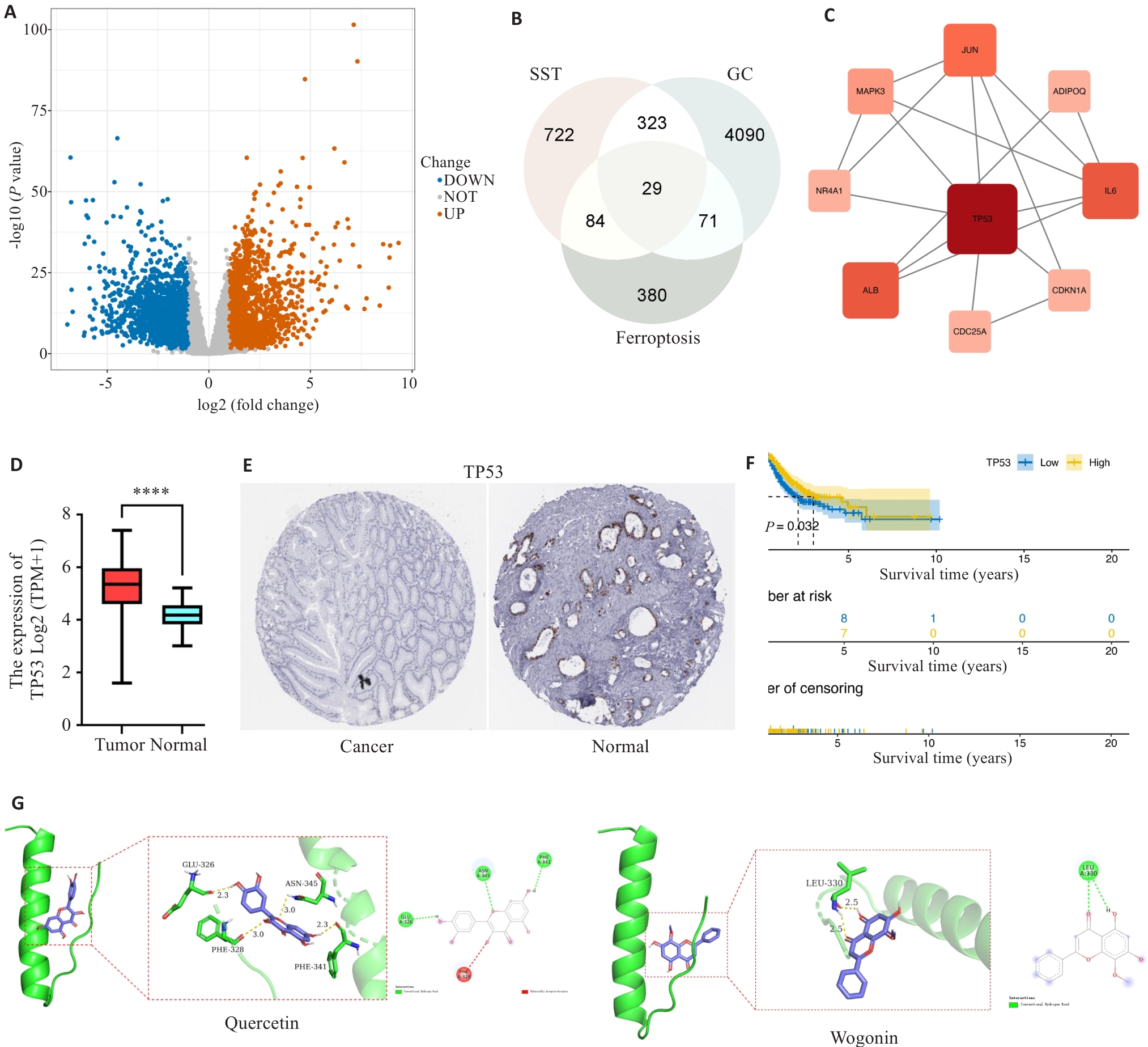
Fig.1 Network pharmacology analysis of gastric cancer-related genes and the mechanism of Shuangshu Decoction. A: Differential gene screening for gastric cancer. B: Wayne diagram of Shuangshu Decoction, gastric cancer (GC) and ferroptopsis. C: PPI diagram of the core targets. D: Differential analysis of P53. E: Representative P53 protein expression pattern in human gastric cancer and normal tissues (Original magnification: ×400). F: Survival analysis of gastric cancer patients with different P53 expression levels. G: Molecular docking analysis of the interaction between the compounds and the core proteins. ****P<0.0001.

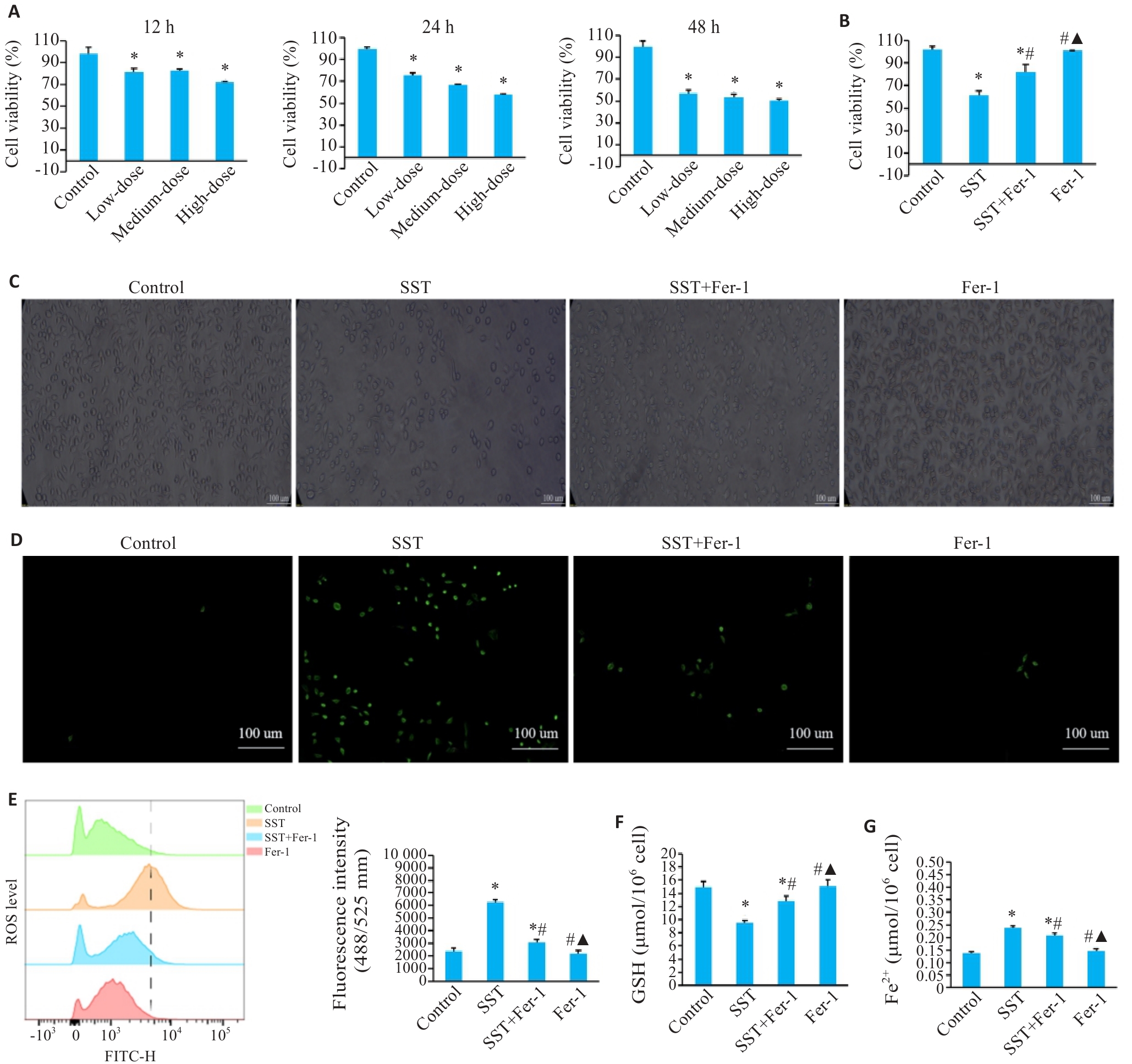
Fig.2 Shuangshu Decoction-medicated serum promotes ferroptosis of AGS cells. A: Determination of the optimal concentration and time of the medicated serum for cell treatment. B: Effect of Shuangshu Decoction on ferroptosis of AGS cells. C: Effect of Shuangshu Decoction-medicated serum on AGS cell morphology (×100). D: Observation of ROS production in the cells under fluorescence microscope (×200). E: Influence of serum of Shuangshu Decoction on ROS production analyzed by flow cytometry and fluorescence intensity. F: Expression of GSH in AGS cells in each group. G: Expression of Fe2+ in AGS cells in each group. *P<0.05 vs control group; #P<0.05 vs SST group; ▲P<0.05 vs SST+Fer-1 group.

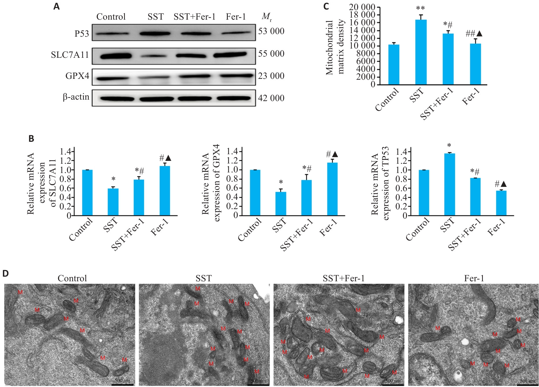
Fig.3 Shuangshu Decoction promotes ferroptosis in AGS cells via the P53/SLC7A11/GPX4 pathway. A: Protein expression levels of P53, SLC7A11 and GPX4 in the treated cells. B: P53, SLC7A11 and GPX4 mRNA expressions in the treated cells. C: Mitochondrial matrix density in each group. D: Mitochondrial damage in each group (TEM, ×20000). *P<0.05, **P<0.01 vs control group; #P<0.05, ##P<0.01 vs SST group; ▲P<0.05 vs SST+Fer-1 group; M: Mitochondria.

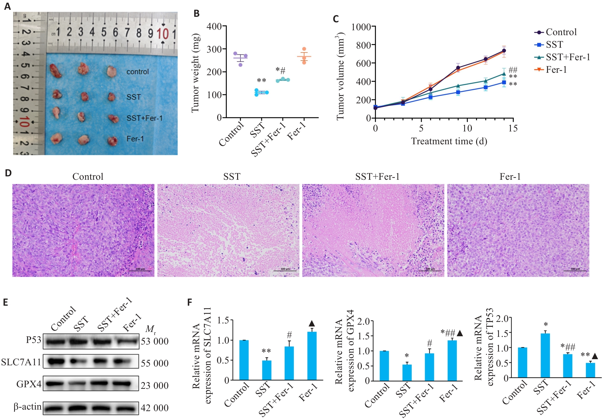
Fig.4 Inhibitory effect of Shuangshu Decoction on xenograft tumor growth in nude mice and its regulation of the P53/SLC7A11/GPX4 pathway. A: Tumor volume in each group. B: Tumor weight in each group. C: Tumor growth curve in each group. D: Pathological examination of the tumors in each group (HE staining, ×200). E: Protein expression levels of P53, SLC7A11, and GPX4 in the tumor tissues from each group detected by Western blotting. F: SLC7A11, GPX4, and P53 mRNA expression levels in the tumor tissues. *P<0.05, **P<0.01 vs control group; #P<0.05, ##P<0.05 vs SST group; ▲P<0.05 vs SST+Fer-1 group.
| [1] | Morgan E, Arnold M, Camargo MC, et al. The current and future incidence and mortality of gastric cancer in 185 countries, 2020-40: a population-based modelling study[J]. EClinicalMedicine, 2022, 47: 101404. doi:10.1016/j.eclinm.2022.101404 |
| [2] | Zheng RS, Chen R, Han BF, et al. Cancer incidence and mortality in China, 2022[J]. Zhonghua Zhong Liu Za Zhi Chin J Oncol, 2024, 46(3): 221-31. |
| [3] | Wang FH, Zhang XT, Tang L, et al. The Chinese society of clinical oncology (CSCO): clinical guidelines for the diagnosis and treatment of gastric cancer, 2023[J]. Cancer Commun (Lond), 2024, 44(1): 127-72. doi:10.1002/cac2.12516 |
| [4] | Zhang X, Li M, Chen S, et al. Effectiveness of endoscopic screening for gastric cancer in China: A population-based study[J]. Gastrointest Endosc, 2023, 97(2): 231-240. |
| [5] | Qiu HB, Cao SM, Xu RH. Cancer incidence, mortality, and burden in China: a time-trend analysis and comparison with the United States and United Kingdom based on the global epidemiological data released in 2020[J]. Cancer Commun (Lond), 2021, 41(10): 1037-48. doi:10.1002/cac2.12197 |
| [6] | Camargo MC, Figueiredo C, Machado JC. Review: gastric malignancies: basic aspects[J]. Helicobacter, 2019, 24(): e12642. doi:10.1111/hel.12642 |
| [7] | Dixon SJ, Lemberg KM, Lamprecht MR, et al. Ferroptosis: an iron-dependent form of nonapoptotic cell death[J]. Cell, 2012, 149(5): 1060-72. doi:10.1016/j.cell.2012.03.042 |
| [8] | Xie Y, Hou W, Song X, et al. Ferroptosis: process and function[J]. Cell Death Differ, 2016, 23(3): 369-79. doi:10.1038/cdd.2015.158 |
| [9] | Yang WS, Stockwell BR. Ferroptosis: death by lipid peroxidation[J]. Trends Cell Biol, 2016, 26(3): 165-76. doi:10.1016/j.tcb.2015.10.014 |
| [10] | Liang C, Zhang XL, Yang MS, et al. Recent progress in ferroptosis inducers for cancer therapy[J]. Adv Mater, 2019, 31(51): e1904197. doi:10.1002/adma.201904197 |
| [11] | Liang YY, Qiu SJ, Zou YW, et al. Ferroptosis-modulating natural products for targeting inflammation-related diseases: challenges and opportunities in manipulating redox signaling[J]. Antioxid Redox Signal, 2024, 41(13/14/15): 976-91. doi:10.1089/ars.2024.0556 |
| [12] | Li S, Zhang B. Traditional Chinese medicine network pharm-acology: theory, methodology and application[J]. Chin J Nat Med, 2013, 11(2): 110-20. doi:10.3724/sp.j.1009.2013.00110 |
| [13] | Li X, Yang G, Li X, et al. Traditional Chinese medicine in cancer care: a review of controlled clinical studies published in Chinese[J]. PLoS One, 2013, 8(4): e60338. doi:10.1371/journal.pone.0060338 |
| [14] | Zhang LL, Zhang DJ, Shi JX, et al. Immunogenic cell death inducers for cancer therapy: an emerging focus on natural products[J]. Phytomedicine, 2024, 132: 155828. doi:10.1016/j.phymed.2024.155828 |
| [15] | Yuan JW, Khan SU, Yan JF, et al. Baicalin enhances the efficacy of 5-Fluorouracil in gastric cancer by promoting ROS-mediated ferroptosis[J]. Biomed Pharmacother, 2023, 164: 114986. doi:10.1016/j.biopha.2023.114986 |
| [16] | Hu LY, Zhang ZY, Zhu F, et al. Schizandrin A enhances the sensitivity of gastric cancer cells to 5-FU by promoting ferroptosis[J]. Cytotechnology, 2024, 76(3): 329-40. doi:10.1007/s10616-024-00623-4 |
| [17] | 杨建宇, 林才志, 冯 利. 抗癌秘验方[M]. 3版. 北京: 化学工业出版社, 2019: 376. |
| [18] | 王春鹏, 程伟玲, 戴明明, 等. 林才志治疗慢性萎缩性胃炎验案举隅[J]. 中国民间疗法, 2021, 29(5): 107-8. doi:10.19621/j.cnki.11-3555/r.2021.0543 |
| [19] | 陈浩彬, 曹育启, 林才志. 林才志治疗大肠癌临床经验采撷[J]. 广西中医药, 2024, 47(1): 35-8. |
| [20] | Huang J, Chen J, Li JN. Quercetin promotes ATG5-mediating autophagy-dependent ferroptosis in gastric cancer[J]. J Mol Histol, 2024, 55(2): 211-25. doi:10.1007/s10735-024-10186-5 |
| [21] | Ding LX, Dang SW, Sun MJ, et al. Quercetin induces ferroptosis in gastric cancer cells by targeting SLC1A5 and regulating the p-Camk2/p-DRP1 and NRF2/GPX4 Axes[J]. Free Radic Biol Med, 2024, 213: 150-63. doi:10.1016/j.freeradbiomed.2024.01.002 |
| [22] | Liu X, Peng XH, Cen S, et al. Wogonin induces ferroptosis in pancreatic cancer cells by inhibiting the Nrf2/GPX4 axis[J]. Front Pharmacol, 2023, 14: 1129662. doi:10.3389/fphar.2023.1129662 |
| [23] | Warias P, Plewa P, Poniewierska-Baran A. Resveratrol, piceatannol, curcumin, and quercetin as therapeutic targets in gastric cancer-mechanisms and clinical implications for natural products[J]. Molecules, 2024, 30(1): 3. doi:10.3390/molecules30010003 |
| [24] | Qian JY, Lou CY, Chen YL, et al. A prospective therapeutic strategy: GPX4-targeted ferroptosis mediators[J]. Eur J Med Chem, 2025, 281: 117015. doi:10.1016/j.ejmech.2024.117015 |
| [25] | Huang WJ, Wen F, Yang PP, et al. Yi-qi-Hua-yu-Jie-du decoction induces ferroptosis in cisplatin-resistant gastric cancer via the AKT/GSK3β/NRF2/GPX4 axis[J]. Phytomedicine, 2024, 123: 155220. doi:10.1016/j.phymed.2023.155220 |
| [26] | Zheng GT, Liu XY, Abuduwufuer A, et al. Poria cocos inhibits the invasion, migration, and epithelial-mesenchymal transition of gastric cancer cells by inducing ferroptosis in cells[J]. Eur J Med Res, 2024, 29(1): 531. doi:10.1186/s40001-024-02110-0 |
| [27] | Liu YQ, Gu W. p53 in ferroptosis regulation: the new weapon for the old guardian[J]. Cell Death Differ, 2022, 29(5): 895-910. doi:10.1038/s41418-022-00943-y |
| [28] | Jiang L, Kon N, Li TY, et al. Ferroptosis as a p53-mediated activity during tumour suppression[J]. Nature, 2015, 520(7545): 57-62. doi:10.1038/nature14344 |
| [29] | Zhou BS, Wang HL, Zhang B, et al. Licochalcone B attenuates neuronal injury through anti-oxidant effect and enhancement of Nrf2 pathway in MCAO rat model of stroke[J]. Int Immunopharmacol, 2021, 100: 108073. doi:10.1016/j.intimp.2021.108073 |
| [30] | Koppula P, Zhang YL, Zhuang L, et al. Amino acid transporter SLC7A11/xCT at the crossroads of regulating redox homeostasis and nutrient dependency of cancer[J]. Cancer Commun (Lond), 2018, 38(1): 12. doi:10.1186/s40880-018-0288-x |
| [31] | Hirschhorn T, Stockwell BR. The development of the concept of ferroptosis[J]. Free Radic Biol Med, 2019, 133: 130-43. doi:10.1016/j.freeradbiomed.2018.09.043 |
| [1] | Liming WANG, Hongrui CHEN, Yan DU, Peng ZHAO, Yujie WANG, Yange TIAN, Xinguang LIU, Jiansheng LI. Yiqi Zishen Formula ameliorates inflammation in mice with chronic obstructive pulmonary disease by inhibiting the PI3K/Akt/NF-κB signaling pathway [J]. Journal of Southern Medical University, 2025, 45(7): 1409-1422. |
| [2] | Yinfu ZHU, Yiran LI, Yi WANG, Yinger HUANG, Kunxiang GONG, Wenbo HAO, Lingling SUN. Therapeutic mechanism of hederagenin, an active component in Guizhi Fuling Pellets, against cervical cancer in nude mice [J]. Journal of Southern Medical University, 2025, 45(7): 1423-1433. |
| [3] | Mengying ZHANG, Chenling ZHAO, Liwei TIAN, Guofang YU, Wenming YANG, Ting DONG. Gandou Fumu Decoction improves liver steatosis by inhibiting hepatocyte ferroptosis in mice with Wilson's disease through the GPX4/ACSL4/ALOX15 signaling pathway [J]. Journal of Southern Medical University, 2025, 45(7): 1471-1478. |
| [4] | Xuan WU, Jiamin FANG, Weiwei HAN, Lin CHEN, Jing SUN, Qili JIN. High PRELID1 expression promotes epithelial-mesenchymal transition in gastric cancer cells and is associated with poor prognosis [J]. Journal of Southern Medical University, 2025, 45(7): 1535-1542. |
| [5] | Kang WANG, Haibin LI, Jing YU, Yuan MENG, Hongli ZHANG. High expression of ELFN1 is a prognostic biomarker and promotes proliferation and metastasis of colorectal cancer cells [J]. Journal of Southern Medical University, 2025, 45(7): 1543-1553. |
| [6] | Lijun HE, Xiaofei CHEN, Chenxin YAN, Lin SHI. Inhibitory effect of Fuzheng Huaji Decoction against non-small cell lung cancer cells in vitro and the possible molecular mechanism [J]. Journal of Southern Medical University, 2025, 45(6): 1143-1152. |
| [7] | Guoyong LI, Renling LI, Yiting LIU, Hongxia KE, Jing LI, Xinhua WANG. Therapeutic mechanism of Arctium lappa extract for post-viral pneumonia pulmonary fibrosis: a metabolomics, network pharmacology analysis and experimental verification [J]. Journal of Southern Medical University, 2025, 45(6): 1185-1199. |
| [8] | Xinrui HOU, Zhendong ZHANG, Mingyuan CAO, Yuxin DU, Xiaoping WANG. Salidroside inhibits proliferation of gastric cancer cells by regulating the miR-1343-3p-OGDHL/PDHB glucose metabolic axis [J]. Journal of Southern Medical University, 2025, 45(6): 1226-1239. |
| [9] | Liping GUAN, Yan YAN, Xinyi LU, Zhifeng LI, Hui GAO, Dong CAO, Chenxi HOU, Jingyu ZENG, Xinyi LI, Yang ZHAO, Junjie WANG, Huilong FANG. Compound Centella asiatica formula alleviates Schistosoma japonicum-induced liver fibrosis in mice by inhibiting the inflammation-fibrosis cascade via regulating the TLR4/MyD88 pathway [J]. Journal of Southern Medical University, 2025, 45(6): 1307-1316. |
| [10] | Anbang ZHANG, Xiuqi SUN, Bo PANG, Yuanhua WU, Jingyu SHI, Ning ZHANG, Tao YE. Electroacupuncture pretreatment alleviates cerebral ischemia-reperfusion injury in rats by inhibiting ferroptosis through the gut-brain axis and the Nrf2/HO-1 signaling pathway [J]. Journal of Southern Medical University, 2025, 45(5): 911-920. |
| [11] | Peipei TANG, Yong TAN, Yanyun YIN, Xiaowei NIE, Jingyu HUANG, Wenting ZUO, Yuling LI. Tiaozhou Ziyin recipe for treatment of premature ovarian insufficiency: efficacy, safety and mechanism [J]. Journal of Southern Medical University, 2025, 45(5): 929-941. |
| [12] | Xiaotao LIANG, Yifan XIONG, Xueqi LIU, Xiaoshan LIANG, Xiaoyu ZHU, Wei XIE. Huoxue Shufeng Granule alleviates central sensitization in chronic migraine mice via TLR4/NF-κB inflammatory pathway [J]. Journal of Southern Medical University, 2025, 45(5): 986-994. |
| [13] | Niandong RAN, Jie LIU, Jian XU, Yongping ZHANG, Jiangtao GUO. n-butanol fraction of ethanol extract of Periploca forrestii Schltr.: its active components, targets and pathways for treating Alcheimer's disease in rats [J]. Journal of Southern Medical University, 2025, 45(4): 785-798. |
| [14] | Linluo ZHANG, Changqing LI, Lingling HUANG, Xueping ZHOU, Yuanyuan LOU. Catalpol reduces liver toxicity of triptolide in mice by inhibiting hepatocyte ferroptosis through the SLC7A11/GPX4 pathway: testing the Fuzheng Zhidu theory for detoxification [J]. Journal of Southern Medical University, 2025, 45(4): 810-818. |
| [15] | Yi ZHANG, Yu SHEN, Zhiqiang WAN, Song TAO, Yakui LIU, Shuanhu WANG. High expression of CDKN3 promotes migration and invasion of gastric cancer cells by regulating the p53/NF-κB signaling pathway and inhibiting cell apoptosis [J]. Journal of Southern Medical University, 2025, 45(4): 853-861. |
| Viewed | ||||||
|
Full text |
|
|||||
|
Abstract |
|
|||||