
南方医科大学学报 ›› 2026, Vol. 46 ›› Issue (8): 1956-1966.doi: 10.12122/j.issn.1673-4254.2026.08.23
• • 上一篇
黄凌霄1,4,5( ), 徐海喆1,4,5(), 黄凌燕2,3, 姚新波1,4,5, 周开元1,4,5, 高勇占1,4,5
收稿日期:2025-12-27
出版日期:2026-08-20
发布日期:2026-08-01
通讯作者:
徐海喆
E-mail:huanglx@nxu.edu.cn;xhz246824@163.com
作者简介:黄凌霄,博士,副教授,E-mail: huanglx@nxu.edu.cn
基金资助:
Lingxiao HUANG1,4,5(), Haizhe XU1,4,5(), Lingyan HUANG2,3, Xinbo YAO1,4,5, Kaiyuan ZHOU1,4,5, Yongzhan GAO1,4,5
Received:2025-12-27
Online:2026-08-20
Published:2026-08-01
Contact:
Haizhe XU
E-mail:huanglx@nxu.edu.cn;xhz246824@163.com
Supported by:摘要:
目的 构建在极低标注率条件下的高价值区域引导双网络半监督分割方法(HVASS),针对MRI肿瘤亚区异质性及边界模糊难题,提升心脏和脑胶质瘤复杂边界分割的准确性和可靠性。 方法 提出一种双网络协同学习框架,通过预测一致性差异自动识别两类临床高风险区域:双网络预测不一致但至少一方置信度较高的“高置信歧义区”;双网络预测一致但整体置信度较低的“低置信稳定区”。针对上述区域构建自适应双教师互导机制以优化伪标签质量。同时,引入高价值区域感知卷积模块加强对肿瘤边界模糊和异质性区域的结构捕获,并结合小波频域特征精炼模块强化边缘细节。模型在BraTS2019脑肿瘤MRI数据集及ACDC心脏MRI数据集上进行验证。 结果 在ACDC数据集HVASS平均相似系数(Dice)达90.34%、95HD2.46 mm,较当前最佳方法提升Dice3.24%、95HD下降2.68 mm;在BraTS2019Dice达85.07%、95HD7.68 mm,增强肿瘤区Dice提升约3.8%,边界误差显著减小。 结论 HVASS在极少标注下显著提升心脏和脑胶质瘤MRI分割精度,能够有效识别MRI肿瘤中的边界模糊与异质性区域,尤其对临床最关注的模糊边界和小病灶效果突出,可有效减轻医师标注负担,具有较高临床应用价值。
黄凌霄, 徐海喆, 黄凌燕, 姚新波, 周开元, 高勇占. 面向边界模糊与异质性区域的半监督MRI图像分割双网络模型[J]. 南方医科大学学报, 2026, 46(8): 1956-1966.
Lingxiao HUANG, Haizhe XU, Lingyan HUANG, Xinbo YAO, Kaiyuan ZHOU, Yongzhan GAO. A semi-supervised MRI image segmentation dual-network model for regions with ambiguous boundaries and heterogeneous regions[J]. Journal of Southern Medical University, 2026, 46(8): 1956-1966.
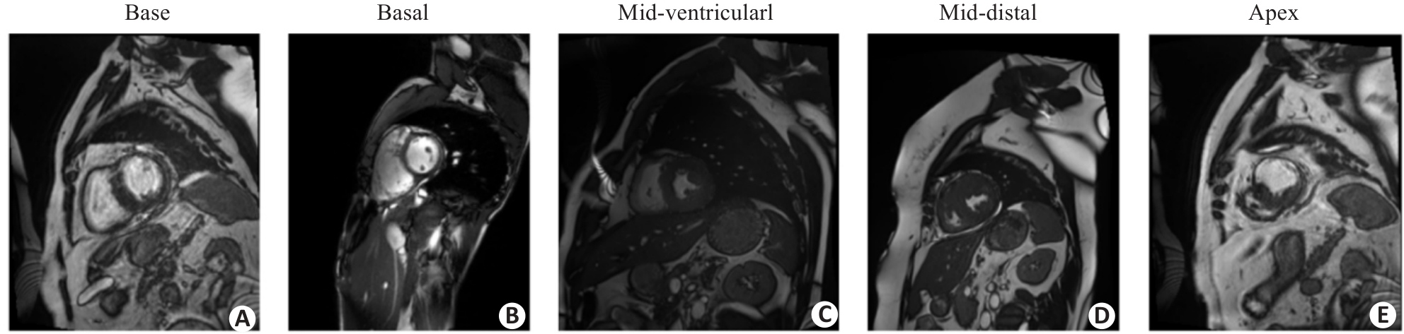
图1 ACDC数据集2D切片图像样例
Fig.1 Sample two-dimensional slice images of the ACDC dataset. A: Transverse section slice of the base of the heart. B: Transverse section tissue slice of the middle part of the heart. C: Transverse section tissue slice of the middle part of the ventricular wall of the heart. D: Transverse section tissue slice of the distal part of the heart. E: Transverse section tissue slice of the apex of the heart.
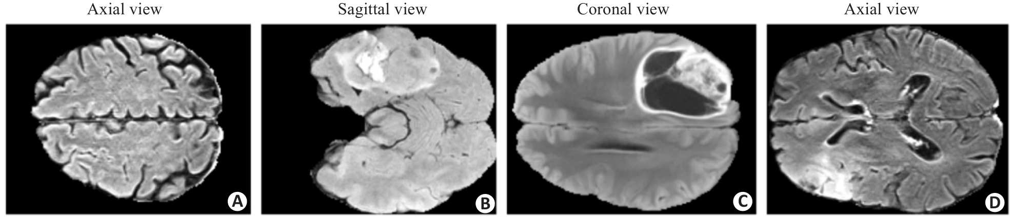
图2 BraTS2019数据集2D切片图像样例
Fig.2 Sample 2D slice images of the BraTS2019 dataset. A: Axial horizontal cross-section of the brain. B: Parasagittal sagittal section of the brain. C: Coronal section of the brain. D: Axial horizontal cross-section of the brain.
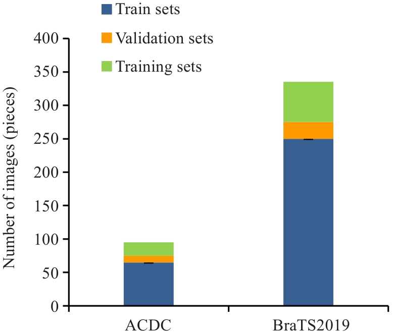
图3 ACDC、BraTS2019数据集图像样本分布图
Fig.3 Distribution of image samples in ACDC and BraTS2019 datasets.
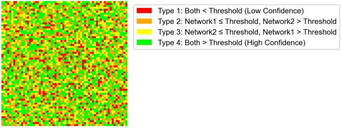
图4 双网络像素级置信度类型空间分布热力图
Fig.4 Heatmap of double-network pixel-level confidence type spatial distribution.
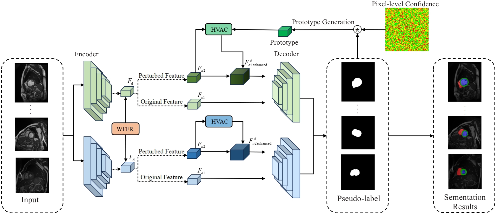
图5 HVASS模型网络结构
Fig.5 HVASS model network architecture.
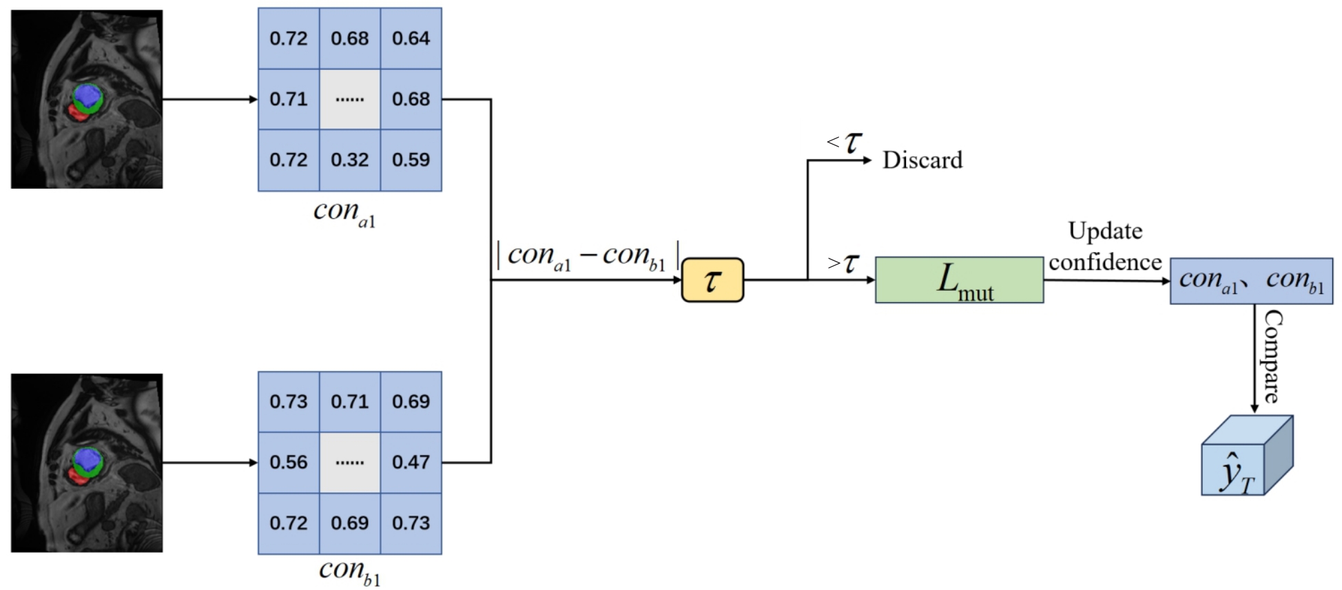
图6 ADTMG模块结构图
Fig.6 ADTMG module structure diagram.
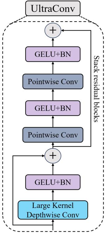
图7 HVAC模块结构图
Fig.7 HVAC module structure diagram.
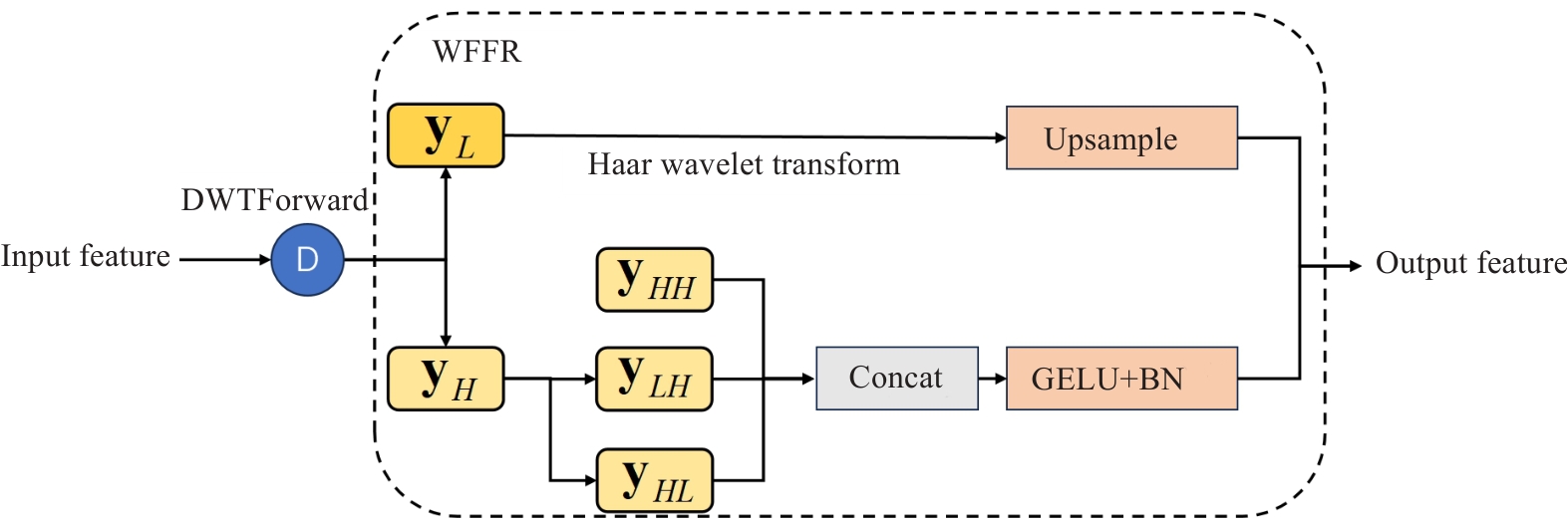
图8 WFFR模块结构图
Fig.8 WFFR module structure diagram.
| Configuration name | Model version |
|---|---|
| System environment | Ubuntu20.04 |
| Central processing unit | Intel(R) Xeon(R) Gold 5418Y |
| Graphics processing unit | NVIDIA GeForce RTX 3090 24GB |
| Graphics processing unit acceleration library | CUDA 12.2 |
| Random access memory | 32 GB |
| Deep learning framework | Pytorch 2.0.0 |
表1 实验环境配置
Tab.1 Experimental environment configuration
| Configuration name | Model version |
|---|---|
| System environment | Ubuntu20.04 |
| Central processing unit | Intel(R) Xeon(R) Gold 5418Y |
| Graphics processing unit | NVIDIA GeForce RTX 3090 24GB |
| Graphics processing unit acceleration library | CUDA 12.2 |
| Random access memory | 32 GB |
| Deep learning framework | Pytorch 2.0.0 |
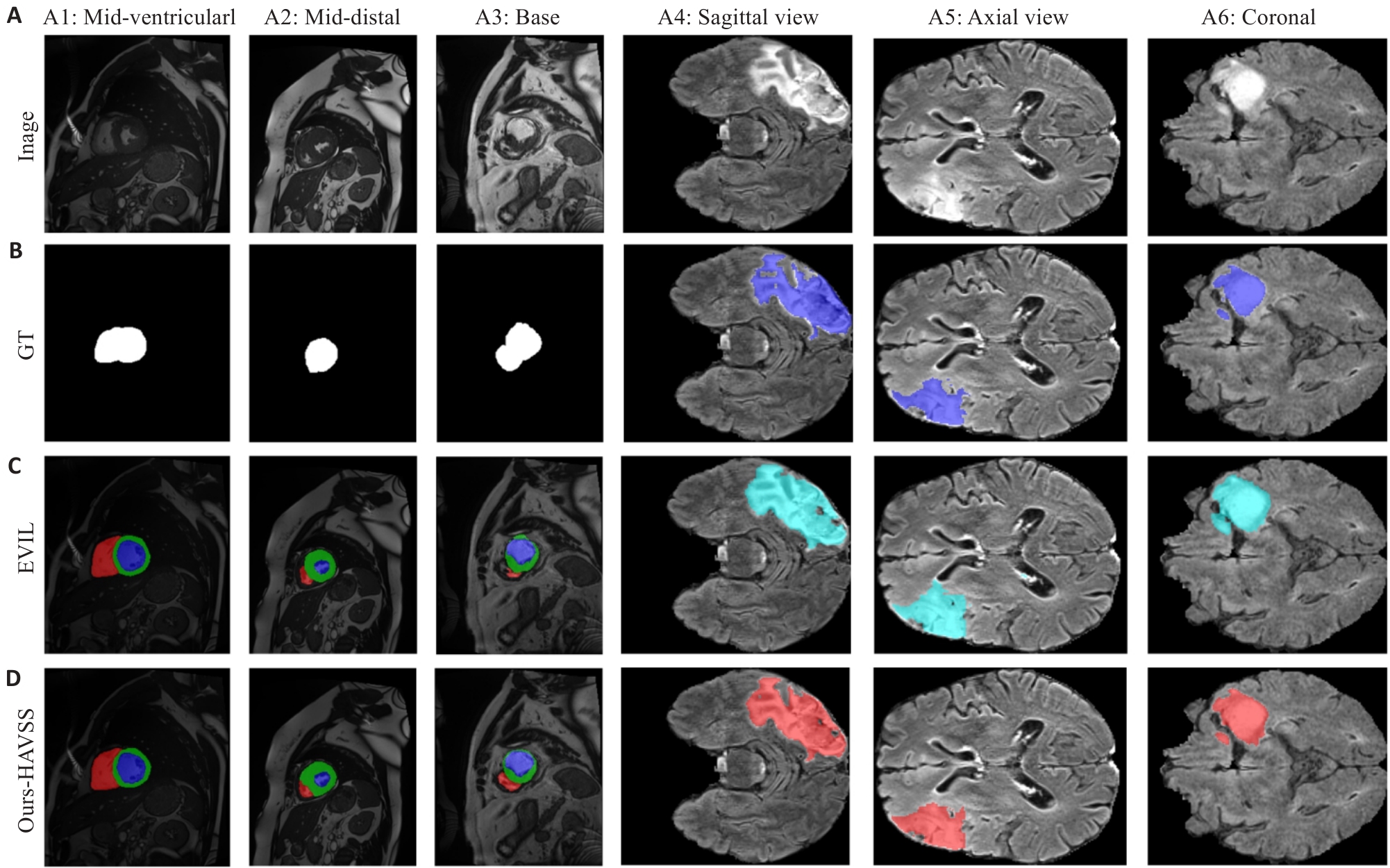
图9 不同算法的分割可视化结果
Fig.9 Visualization results of segmentation by different algorithms. A: Original 2D axial MRI image (A1: Two-dimensional transverse section tissue slice of the middle part of the ventricular wall of the heart. A2: Two-dimensional transverse section tissue slice of the distal part of the heart. A3: Two-dimensional transverse section slice of the base of the heart. A4: Parasagittal sagittal section of the brain. A5: Axial horizontal cross-section of the brain. A6: Coronal section of the brain.). B: Ground truth annotation map. C: Lesion localization heatmap generated by the EVIL method. D: Lesion localization heatmap generated by the HAVSS method proposed in this study.
| Method | Labeled | Unlabeled | Dice | IoU | 95HD | ASD |
|---|---|---|---|---|---|---|
| MTNIPS'2017 | 7 (10%) | 63 (90%) | 83.65 | 73.15 | 13.45 | 3.61 |
| MCFCVPR'2023 | 85.18 | 75.22 | 10.73 | 2.78 | ||
| CPSCVPR'2021 | 85.34 | 75.50 | 8.78 | 2.37 | ||
| MC-NetMICCAI'2021 | 86.07 | 76.58 | 11.48 | 3.37 | ||
| AC-MTMIA'2023 | 86.36 | 76.86 | 9.64 | 2.59 | ||
| AAUMIA'2024 | 86.46 | 76.99 | 8.00 | 2.06 | ||
| UG-MCLAIIM'2023 | 86.84 | 77.60 | 7.77 | 1.94 | ||
| MLRPMIA'2024 | 87.10 | 78.16 | 4.91 | 1.30 | ||
| EVILCBM'2024 | 87.25 | 78.18 | 5.31 | 1.35 | ||
| Ours-HVASS | 90.34 | 83.13 | 2.46 | 0.79 | ||
| MTNIPS'2017 | 14 (20%) | 56 (80%) | 86.55 | 77.49 | 6.86 | 2.15 |
| MC-NetMICCAI'2021 | 86.58 | 77.68 | 15.99 | 4.93 | ||
| CPSCVPR'2021 | 87.03 | 78.13 | 6.66 | 2.12 | ||
| MCFCVPR'2023 | 87.32 | 78.43 | 6.53 | 2.00 | ||
| AAUMIA'2024 | 87.71 | 78.92 | 8.51 | 2.14 | ||
| AC-MTMIA'2023 | 87.99 | 79.34 | 7.85 | 2.14 | ||
| MLRPMIA'2024 | 88.00 | 79.54 | 5.41 | 1.58 | ||
| EVILCBM'2024 | 88.34 | 79.90 | 6.46 | 1.68 | ||
| UG-MCLAIIM'2023 | 88.40 | 79.86 | 9.12 | 2.44 | ||
| Ours-HVASS | 91.02 | 83.65 | 2.74 | 0.78 |
表2 ACDC数据集上的HVASS对比实验结果
Tab.2 Comparison results of HVASS on the ACDC dataset
| Method | Labeled | Unlabeled | Dice | IoU | 95HD | ASD |
|---|---|---|---|---|---|---|
| MTNIPS'2017 | 7 (10%) | 63 (90%) | 83.65 | 73.15 | 13.45 | 3.61 |
| MCFCVPR'2023 | 85.18 | 75.22 | 10.73 | 2.78 | ||
| CPSCVPR'2021 | 85.34 | 75.50 | 8.78 | 2.37 | ||
| MC-NetMICCAI'2021 | 86.07 | 76.58 | 11.48 | 3.37 | ||
| AC-MTMIA'2023 | 86.36 | 76.86 | 9.64 | 2.59 | ||
| AAUMIA'2024 | 86.46 | 76.99 | 8.00 | 2.06 | ||
| UG-MCLAIIM'2023 | 86.84 | 77.60 | 7.77 | 1.94 | ||
| MLRPMIA'2024 | 87.10 | 78.16 | 4.91 | 1.30 | ||
| EVILCBM'2024 | 87.25 | 78.18 | 5.31 | 1.35 | ||
| Ours-HVASS | 90.34 | 83.13 | 2.46 | 0.79 | ||
| MTNIPS'2017 | 14 (20%) | 56 (80%) | 86.55 | 77.49 | 6.86 | 2.15 |
| MC-NetMICCAI'2021 | 86.58 | 77.68 | 15.99 | 4.93 | ||
| CPSCVPR'2021 | 87.03 | 78.13 | 6.66 | 2.12 | ||
| MCFCVPR'2023 | 87.32 | 78.43 | 6.53 | 2.00 | ||
| AAUMIA'2024 | 87.71 | 78.92 | 8.51 | 2.14 | ||
| AC-MTMIA'2023 | 87.99 | 79.34 | 7.85 | 2.14 | ||
| MLRPMIA'2024 | 88.00 | 79.54 | 5.41 | 1.58 | ||
| EVILCBM'2024 | 88.34 | 79.90 | 6.46 | 1.68 | ||
| UG-MCLAIIM'2023 | 88.40 | 79.86 | 9.12 | 2.44 | ||
| Ours-HVASS | 91.02 | 83.65 | 2.74 | 0.78 |
| Method | Labeled | Unlabeled | Dice | IoU | 95HD | ASD |
|---|---|---|---|---|---|---|
| MTNIPS'2017 | 50 (20%) | 200 (80%) | 80.49 | 69.99 | 11.71 | 3.46 |
| MCFCVPR'2023 | 81.96 | 71.76 | 13.96 | 3.99 | ||
| AC-MTMIA'2023 | 82.09 | 71.65 | 15.91 | 4.81 | ||
| AAUMIA'2024 | 82.43 | 72.33 | 13.83 | 4.18 | ||
| MC-NetMICCAI'2021 | 82.51 | 72.98 | 14.57 | 3.78 | ||
| EVILCBM'2024 | 82.60 | 72.46 | 11.44 | 2.76 | ||
| CPSCVPR'2021 | 82.70 | 72.30 | 12.99 | 3.46 | ||
| UG-MCLAIIM'2023 | 82.85 | 72.69 | 11.12 | 2.28 | ||
| MLRPMIA'2024 | 83.96 | 74.00 | 15.16 | 4.21 | ||
| Ours-HVASS | 85.07 | 75.80 | 7.68 | 2.03 |
表3 BraTS2019数据集上的HVASS对比实验结果
Tab.3 Comparison results of HVASS on the BraTS2019 dataset
| Method | Labeled | Unlabeled | Dice | IoU | 95HD | ASD |
|---|---|---|---|---|---|---|
| MTNIPS'2017 | 50 (20%) | 200 (80%) | 80.49 | 69.99 | 11.71 | 3.46 |
| MCFCVPR'2023 | 81.96 | 71.76 | 13.96 | 3.99 | ||
| AC-MTMIA'2023 | 82.09 | 71.65 | 15.91 | 4.81 | ||
| AAUMIA'2024 | 82.43 | 72.33 | 13.83 | 4.18 | ||
| MC-NetMICCAI'2021 | 82.51 | 72.98 | 14.57 | 3.78 | ||
| EVILCBM'2024 | 82.60 | 72.46 | 11.44 | 2.76 | ||
| CPSCVPR'2021 | 82.70 | 72.30 | 12.99 | 3.46 | ||
| UG-MCLAIIM'2023 | 82.85 | 72.69 | 11.12 | 2.28 | ||
| MLRPMIA'2024 | 83.96 | 74.00 | 15.16 | 4.21 | ||
| Ours-HVASS | 85.07 | 75.80 | 7.68 | 2.03 |
| Datasets | ADTMG | HVAC | WFFR | Dice | IoU | 95HD | ASD |
|---|---|---|---|---|---|---|---|
| ACDC | 84.38 | 71.45 | 18.23 | 4.61 | |||
| √ | 87.23 | 80.30 | 8.78 | 1.87 | |||
| √ | 86.97 | 79.28 | 11.48 | 3.37 | |||
| √ | 85.56 | 79.42 | 10.73 | 3.97 | |||
| √ | √ | 88.78 | 80.22 | 3.86 | 0.98 | ||
| √ | √ | 89.08 | 82.59 | 3.79 | 1.23 | ||
| √ | √ | 88.89 | 81.43 | 5.31 | 1.58 | ||
| √ | √ | √ | 90.34 | 83.13 | 2.46 | 0.79 |
表4 HVASS在10%的标注下ACDC和BraTS2019 数据集上的消融实验结果
Tab.4 Ablation experiment results of HVASS on the ACDC and BraTS2019 datasets under 10% annotation
| Datasets | ADTMG | HVAC | WFFR | Dice | IoU | 95HD | ASD |
|---|---|---|---|---|---|---|---|
| ACDC | 84.38 | 71.45 | 18.23 | 4.61 | |||
| √ | 87.23 | 80.30 | 8.78 | 1.87 | |||
| √ | 86.97 | 79.28 | 11.48 | 3.37 | |||
| √ | 85.56 | 79.42 | 10.73 | 3.97 | |||
| √ | √ | 88.78 | 80.22 | 3.86 | 0.98 | ||
| √ | √ | 89.08 | 82.59 | 3.79 | 1.23 | ||
| √ | √ | 88.89 | 81.43 | 5.31 | 1.58 | ||
| √ | √ | √ | 90.34 | 83.13 | 2.46 | 0.79 |
| [1] | Litjens G, Ciompi F, Wolterink JM, et al. State-of-the-art deep learning in cardiovascular image analysis[J]. JACC Cardiovasc Imaging, 2019, 12(8 Pt 1): 1549-65. doi:10.1016/j.jcmg.2019.06.009 |
| [2] | Bai WJ, Sinclair M, Tarroni G, et al. Automated cardiovascular magnetic resonance image analysis with fully convolutional networks[J]. J Cardiovasc Magn Reson, 2018, 20(1): 65. doi:10.1186/s12968-018-0471-x |
| [3] | 周 昊, 曾 栋, 边兆英, 等. 基于半监督网络的组织感知CT图像对比度的增强方法[J]. 南方医科大学学报, 2023, 43(6): 985-93. |
| [4] | 丁家满, 刘 楠, 周蜀杰, 等. 基于正则化的半监督弱标签分类方法[J]. 计算机学报, 2022, 45(1): 69-81 |
| [5] | 翟德明, 沈斯娴, 周 雄, 等. 基于最优传输理论的深度半监督学习伪标签生成算法[J]. 软件学报, 2024, 35(11): 5196-209. |
| [6] | Li CC, Zhang JS, Niu DM, et al. Boundary-aware uncertainty suppression for semi-supervised medical image segmentation[J]. IEEE Trans Artif Intell, 2024, 5(8): 4074-86. doi:10.1109/tai.2024.3359576 |
| [7] | 姚宗亮, 黄 荣, 董爱华, 等. 基于多模态融合和自适应剪枝Transformer的脑肿瘤图像分割算法[J]. 宁夏大学学报: 自然科学版, 2024, 45(1): 16-24. |
| [8] | 徐晗晗, 张印辉, 何自芬, 等. 伪标签置信度调控结直肠癌病理图像半监督语义分割[J]. 光学精密工程, 2025, 33(4): 591-609. |
| [9] | Chen XK, Yuan YH, Zeng G, et al. Semi-supervised semantic segmentation with cross pseudo supervision[C]//2021 IEEE/CVF Conference on Computer Vision and Pattern Recognition (CVPR). June 20-25, 2021. Nashville, TN, USA. IEEE, 2021: 2613-22. doi:10.1109/cvpr46437.2021.00264 |
| [10] | Wu YC, Xu MF, Ge ZY, et al. Semi-supervised left atrium segmentation with mutual ConsistencyTraining[C]//Medical Image Computing and Computer Assisted Intervention-MICCAI 2021. Cham: Springer, 2021: 297-306. doi:10.1007/978-3-030-87196-3_28 |
| [11] | Adiga V S, Dolz J, Lombaert H. Anatomically-aware uncertainty for semi-supervised image segmentation[J]. Med Image Anal, 2024, 91: 103011. doi:10.1016/j.media.2023.103011 |
| [12] | Bernard O, Lalande A, Zotti C, et al. Deep learning techniques for automatic MRI cardiac multi-structures segmentation and diagnosis: is the problem solved?[J]. IEEE Trans Med Imaging, 2018, 37(11): 2514-25. doi:10.1109/tmi.2018.2837502 |
| [13] | Bakas S, Akbari H, Sotiras A, et al. Advancing The Cancer Genome Atlas glioma MRI collections with expert segmentation labels and radiomic features[J]. Sci Data, 2017, 4: 170117. doi:10.1038/sdata.2017.117 |
| [14] | 权 笑, 戴鹏程, 辛 潮, 等. LTE网络高价值区域识别与分析方法研究[J].电信工程技术与标准化, 2018, 31(5):18-21. |
| [15] | Zhou ZW, Rahman Siddiquee MM, Tajbakhsh N, et al. UNet++: a nested U-Net architecture for medical image segmentation[C]//Deep Learning in Medical Image Analysis and Multimodal Learning for Clinical Decision Support. Cham: Springer, 2018: 3-11. doi:10.1007/978-3-030-00889-5_1 |
| [16] | Zhao CJ, Xiang S, Wang YQ, et al. Context-aware network fusing transformer and V-Net for semi-supervised segmentation of 3D left atrium[J]. Expert Syst Appl, 2023, 214: 119105. doi:10.1016/j.eswa.2022.119105 |
| [17] | Topol EJ. High-performance medicine: the convergence of human and artificial intelligence[J]. Nat Med, 2019, 25(1): 44-56. doi:10.1038/s41591-018-0300-7 |
| [18] | Tarvainen A, Valpola H. Mean teachers are better role models: Weight-averaged consistency targets improve semi-supervised deep learning results[C]//Neural Information Processing Systems., 2017 |
| [19] | Xu Z, Wang YX, Lu DH, et al. Ambiguity-selective consistency regularization for mean-teacher semi-supervised medical image segmentation[J]. Med Image Anal, 2023, 88: 102880. doi:10.1016/j.media.2023.102880 |
| [20] | Zhang YC, Jiao RS, Liao QC, et al. Uncertainty-guided mutual consistency learning for semi-supervised medical image segmentation[J]. Artif Intell Med, 2023, 138: 102476. doi:10.1016/j.artmed.2022.102476 |
| [21] | Wang YC, Xiao B, Bi XL, et al. MCF: mutual correction framework for semi-supervised medical image segmentation[C]//2023 IEEE/CVF Conference on Computer Vision and Pattern Recognition (CVPR). June 17-24, 2023, Vancouver, BC, Canada. IEEE, 2023: 15651-60. doi:10.1109/cvpr52729.2023.01502 |
| [22] | Chen YY, Yang ZY, Shen CY, et al. Evidence-based uncertainty-aware semi-supervised medical image segmentation[J]. Comput Biol Med, 2024, 170: 108004. doi:10.1016/j.compbiomed.2024.108004 |
| [23] | 郑孙易, 刘佳鑫, 崔效楠, 等. 人工智能在肿瘤影像学中的应用进展[J]. 放射学实践, 2025, 40(9): 1093-7. |
| [24] | Menze BH, Jakab A, Bauer S, et al. The multimodal brain tumor image segmentation benchmark (BRATS)[J]. IEEE Trans Med Imaging, 2015, 34(10): 1993-2024. |
| [25] | Bauer S, Wiest R, Nolte LP, et al. A survey of MRI-based medical image analysis for brain tumor studies[J]. Phys Med Biol, 2013, 58(13): R97-R129. doi:10.1088/0031-9155/58/13/r97 |
| [26] | Salvador A, Bellver M, Campos V, et al. Recurrent neural networks for semantic instance segmentation[J]. arXiv preprint arXiv:, 2017. |
| [27] | Taha AA, Hanbury A. Metrics for evaluating 3D medical image segmentation: analysis, selection, and tool[J]. BMC Med Imaging, 2015, 15: 29. doi:10.1186/s12880-015-0068-x |
| [28] | Petitjean C, Dacher JN. A review of segmentation methods in short axis cardiac MR images[J]. Med Image Anal, 2011, 15(2): 169-84. doi:10.1016/j.media.2010.12.004 |
| [29] | Zhuang XH, Shen J. Multi-scale patch and multi-modality atlases for whole heart segmentation of MRI[J]. Med Image Anal, 2016, 31: 77-87. doi:10.1016/j.media.2016.02.006 |
| [30] | Isensee F, Jaeger PF, Kohl SAA, et al. nnU-Net: a self-configuring method for deep learning-based biomedical image segmentation[J]. Nat Methods, 2021, 18(2): 203-11. doi:10.1038/s41592-020-01008-z |
| [31] | Esteva A, Robicquet A, Ramsundar B, et al. A guide to deep learning in healthcare[J]. Nat Med, 2019, 25(1): 24-9. doi:10.1038/s41591-018-0316-z |
| [1] | 朱君, 谭顺谦, 黄方俊, 蔡光瑶, 甄鑫. 医疗先验引导的TabMap深度学习模型用于卵巢癌预测及可解释性研究[J]. 南方医科大学学报, 2026, 46(7): 1714-1722. |
| [2] | 苏柏尔, 张颖, 闫智娴, 邹颖, 成禹帆, 李天舒, 李晋铭, 陈晓彤, 梁春祺, 徐镕, 杨晟明, 彭渤, 陈豪, 余江, 胡彦锋. 人工智能在微创外科手术技能评估与质量控制中的应用研究进展[J]. 南方医科大学学报, 2026, 46(7): 1723-1730. |
| [3] | 张明旭, 吴迪恒, 林嘉泽, 吕祎君, 周凌宏, 甄鑫, 周浩, 秦耿耿. 基于Transformer的影像与检验数据多模态预测模型在医院获得性感染的应用研究[J]. 南方医科大学学报, 2026, 46(6): 1423-1433. |
| [4] | 许家宁, 毛钇骁, 张煜. 基于分层注意力驱动多实例学习的肾透明细胞癌数字病理分级分期[J]. 南方医科大学学报, 2026, 46(5): 1175-1184. |
| [5] | 王童, 段曹辉, 娄昕, 冯前进. 基于血管增强一致性保持扩散模型的3T到7T SWI生成方法[J]. 南方医科大学学报, 2026, 46(5): 1185-1192. |
| [6] | 陈博湧, 汪新怡, 赵新新, 宋婷, 李永宝. 基于Swin-ResViT网络的低质量动态cine-MR至高质量定位MR图像实时生成研究[J]. 南方医科大学学报, 2026, 46(4): 929-938. |
| [7] | 谭顺谦, 卓俐, 曾敏, 黄方俊, 朱君, 蔡光瑶, 甄鑫. 基于实验室指标的Transformer模型可高效鉴别卵巢癌[J]. 南方医科大学学报, 2026, 46(4): 939-945. |
| [8] | 卢学麒, 陈华元, 吴秋岑, 温耀棋, 刘国光, 陈超敏. 一种基于深度特征融合的可解释性12导联心电图自动诊断模型研究[J]. 南方医科大学学报, 2026, 46(1): 208-218. |
| [9] | 卓俐, 曾敏, 谭顺谦, 梁涛, 肖巍魏, 甄鑫. 基于扩散循环一致性生成对抗网络的骨盆活跃骨髓区域分割方法[J]. 南方医科大学学报, 2026, 46(1): 219-230. |
| [10] | 吴秋岑, 卢学麒, 温耀棋, 洪永, 吴煜良, 陈超敏. II导联心电图中心肌梗死检测与定位:基于多尺度残差模块融合改进通道注意力模型[J]. 南方医科大学学报, 2025, 45(8): 1777-1790. |
| [11] | 郑子瑜, 杨夏颖, 吴圣杰, 张诗婕, 吕国荣, 柳培忠, 王珺, 何韶铮. 多特征融合的产时超声胎方位识别模型[J]. 南方医科大学学报, 2025, 45(7): 1563-1570. |
| [12] | 计寰宇, 王蕊, 高盛祥, 车文刚. SG-UNet:基于全局注意力和自校准卷积增强的黑色素瘤分割模型[J]. 南方医科大学学报, 2025, 45(6): 1317-1326. |
| [13] | 渠梦, 傅蓉. ResLSTM-TemporalSE:多导联心电信号的自动分类[J]. 南方医科大学学报, 2025, 45(12): 2708-2717. |
| [14] | 谢辉荣, 胡潮滨, 梁国华, 韩红喆, 黄牧, 冯前进. 大学生心理压力智能评估:基于融合文本与影像的多模态模型的设计及验证[J]. 南方医科大学学报, 2025, 45(11): 2504-2510. |
| [15] | 贺亚迪, 周炫汝, 金锦辉, 宋婷. 基于PE-CycleGAN网络的鼻咽癌自适应放疗CBCT-sCT生成研究[J]. 南方医科大学学报, 2025, 45(1): 179-186. |
| 阅读次数 | ||||||
|
全文 |
|
|||||
|
摘要 |
|
|||||
