
南方医科大学学报 ›› 2026, Vol. 46 ›› Issue (10): 2482-2493.doi: 10.12122/j.issn.1673-4254.2026.10.17
• • 上一篇
张冠男1( ), 李想2, 米源2, 田丹阳2, 孟晋启3()
收稿日期:2026-03-06
出版日期:2026-10-20
发布日期:2026-10-26
通讯作者:
孟晋启
E-mail:13483111223@163.com;MengJinqi@hebmu.edu.cn
作者简介:张冠男,硕士,副研究员,E-mail: 13483111223@163.com
基金资助:
Guannan ZHANG1(), Xiang LI2, Yuan MI2, Danyang TIAN2, Jinqi MENG3()
Received:2026-03-06
Online:2026-10-20
Published:2026-10-26
Contact:
Jinqi MENG
E-mail:13483111223@163.com;MengJinqi@hebmu.edu.cn
Supported by:摘要:
目的 探究有氧运动与无氧力竭运动对糖尿病大鼠多器官糖代谢及相关功能的影响及其潜在机制。 方法 将40只雄性SD大鼠随机分为空白对照组(10只)与糖尿病造模组(30只)。采用高脂高糖饮食联合一次性小剂量链脲佐菌素(STZ)注射建立2型糖尿病大鼠模型。造模成功后,随机分为糖尿病对照组(10只)、糖尿病有氧运动组(10只)和糖尿病无氧力竭运动组(10只),并进行8周运动干预。干预结束后,对大鼠一般情况进行测试分析;取肝脏、股四头肌、心脏、脑组织前额叶皮质进行HE染色、Masson染色、PAS染色及免疫组化分析;采用Western blotting和qPCR技术检测肝脏关键代谢酶及基因表达水平。 结果 与空白对照组相比,糖尿病对照组大鼠体质量、肝脏指数、空腹血糖及血清乳酸、丙酮酸、尿素氮、肌酐、肌酸激酶水平均显著升高(P<0.01);肝脏出现脂肪变性,显著纤维化及糖原减少(P<0.01);骨骼肌出现结构损伤,显著纤维化及糖原减少(P<0.01);心肌结构损伤、炎症浸润;前额叶皮质多巴胺受体DRD1/DRD5、5-HT、TH表达显著下降(P<0.01)。与糖尿病对照组相比,糖尿病有氧组大鼠体质量、肝脏指数、空腹血糖及血清乳酸、丙酮酸、肌酐、尿素氮、肌酸激酶均显著降低(P<0.01);肝脏纤维化减轻(P<0.01),糖原沉积增加(P<0.01),糖异生关键酶G6PC及糖酵解酶表达下调(P<0.01),Sirt1 mRNA表达上调(P<0.01);骨骼肌糖原含量增加(P<0.01);心肌结构改善,炎症减轻;前额叶皮质DRD1、DRD5、5-HT、TH表达显著回升(P<0.01)。与糖尿病对照组相比,糖尿病力竭组体质量、空腹血糖下降(P<0.01),但肝脏指数、血清乳酸、丙酮酸、尿素氮、肌酐及肌酸激酶水平进一步升高(P<0.05或P<0.01);肝脏纤维化加剧(P<0.01),糖原沉积减少(P<0.01),糖酵解酶表达上调(P<0.01);骨骼肌纤维化加剧(P<0.01);心肌损伤加重;前额叶皮质DRD1、TH表达进一步降低(P<0.05)。 结论 有氧运动通过调节糖脂代谢、减轻炎症纤维化及改善神经递质功能,全面保护糖尿病大鼠多器官功能;而无氧力竭运动加剧了代谢紊乱与组织损伤。
张冠男, 李想, 米源, 田丹阳, 孟晋启. 有氧运动通过调控Sirt1/Creb1/Ppargc1a表达改善糖尿病大鼠代谢紊乱及多器官损伤[J]. 南方医科大学学报, 2026, 46(10): 2482-2493.
Guannan ZHANG, Xiang LI, Yuan MI, Danyang TIAN, Jinqi MENG. Aerobic exercise improves metabolic disorders and multi-organ damage in diabetic rats by activating the Sirt1/Creb1/Ppargc1a axis[J]. Journal of Southern Medical University, 2026, 46(10): 2482-2493.
| Primer | Sequence |
|---|---|
| Rat-Btubulin-F1 | TAGCAGCGTTGCTGGGTATC |
| Rat-Btubulin-R1 | GACTGGAATGCCTCGCTCTT |
| Rat-Sirt1-F1 | TCAGAACCACCAAAGCGGAA |
| Rat-Sirt1-R1 | GCAAGGCGAGCATAAATACCA |
| Rat-Creb1-F1 | TCCTCCCCAGCACTTCCTAC |
| Rat-Creb1-R1 | CCAATCCAGTCCATTTTCCA |
| Rat-Ppargc1a-F2 | AACAGAAACAGCAGCAGAGACAA |
| Rat-Ppargc1a-R2 | GCAGTTCCAGAGAGTTCCACAC |
表1 目的基因引物序列
Tab.1 Primer sequences for qPCR of the target genes
| Primer | Sequence |
|---|---|
| Rat-Btubulin-F1 | TAGCAGCGTTGCTGGGTATC |
| Rat-Btubulin-R1 | GACTGGAATGCCTCGCTCTT |
| Rat-Sirt1-F1 | TCAGAACCACCAAAGCGGAA |
| Rat-Sirt1-R1 | GCAAGGCGAGCATAAATACCA |
| Rat-Creb1-F1 | TCCTCCCCAGCACTTCCTAC |
| Rat-Creb1-R1 | CCAATCCAGTCCATTTTCCA |
| Rat-Ppargc1a-F2 | AACAGAAACAGCAGCAGAGACAA |
| Rat-Ppargc1a-R2 | GCAGTTCCAGAGAGTTCCACAC |
| Variables | Blank control group | Diabetes control group | Diabetes aerobic group | Diabetes exhaustion group |
|---|---|---|---|---|
| Weight (g) | 328.90±10.73 | 379.50±11.88** | 350.10±12.68## | 296.60±14.16## |
| Liver wet weight (g) | 14.22±1.52 | 26.49±2.64** | 19.26±2.49## | 29.60±3.12# |
| Liver index (%) | 4.33±0.51 | 6.99±0.75** | 5.50±0.64## | 9.97±0.78## |
表2 各组大鼠体质量、肝脏湿质量、肝脏指数变化
Tab.2 Changes in body weight, liver wet weight, and liver index of the rats in each group (Mean±SD, n=10)
| Variables | Blank control group | Diabetes control group | Diabetes aerobic group | Diabetes exhaustion group |
|---|---|---|---|---|
| Weight (g) | 328.90±10.73 | 379.50±11.88** | 350.10±12.68## | 296.60±14.16## |
| Liver wet weight (g) | 14.22±1.52 | 26.49±2.64** | 19.26±2.49## | 29.60±3.12# |
| Liver index (%) | 4.33±0.51 | 6.99±0.75** | 5.50±0.64## | 9.97±0.78## |
| Variables | Blank control group | Diabetes control group | Diabetes aerobic group | Diabetes exhaustion group |
|---|---|---|---|---|
| FBG (mmol/L) | 6.24±0.72 | 13.09±1.06** | 8.11±0.85## | 10.29±0.72## |
| Lactic acid (mmol/L) | 2.55±0.39 | 3.97±0.50** | 3.05±0.46## | 6.46±0.65## |
| Pyruvic acid (mmol/L) | 0.42±0.05 | 0.82±0.11** | 0.61±0.10## | 1.11±0.17## |
| Creatinine (mg/dL) | 0.61±0.08 | 1.55±0.16** | 0.82±0.13## | 2.35±0.27## |
| Urea nitrogen (mmol/L) | 3.70±0.28 | 6.19±0.98** | 4.73±0.65## | 7.46±0.97# |
| Creatine kinase (U/L) | 115.70±24.00 | 305.30±30.42** | 191.30±24.41## | 508.90±58.60## |
表3 各组大鼠空腹血糖、血乳酸、丙酮酸、肌酐、尿素氮、肌酸激酶水平变化
Tab. 3 Changes in fasting blood glucose, blood lactate, pyruvate, creatinine, blood urea nitrogen, and creatine kinase levels in each group of the rats (Mean±SD, n=10)
| Variables | Blank control group | Diabetes control group | Diabetes aerobic group | Diabetes exhaustion group |
|---|---|---|---|---|
| FBG (mmol/L) | 6.24±0.72 | 13.09±1.06** | 8.11±0.85## | 10.29±0.72## |
| Lactic acid (mmol/L) | 2.55±0.39 | 3.97±0.50** | 3.05±0.46## | 6.46±0.65## |
| Pyruvic acid (mmol/L) | 0.42±0.05 | 0.82±0.11** | 0.61±0.10## | 1.11±0.17## |
| Creatinine (mg/dL) | 0.61±0.08 | 1.55±0.16** | 0.82±0.13## | 2.35±0.27## |
| Urea nitrogen (mmol/L) | 3.70±0.28 | 6.19±0.98** | 4.73±0.65## | 7.46±0.97# |
| Creatine kinase (U/L) | 115.70±24.00 | 305.30±30.42** | 191.30±24.41## | 508.90±58.60## |
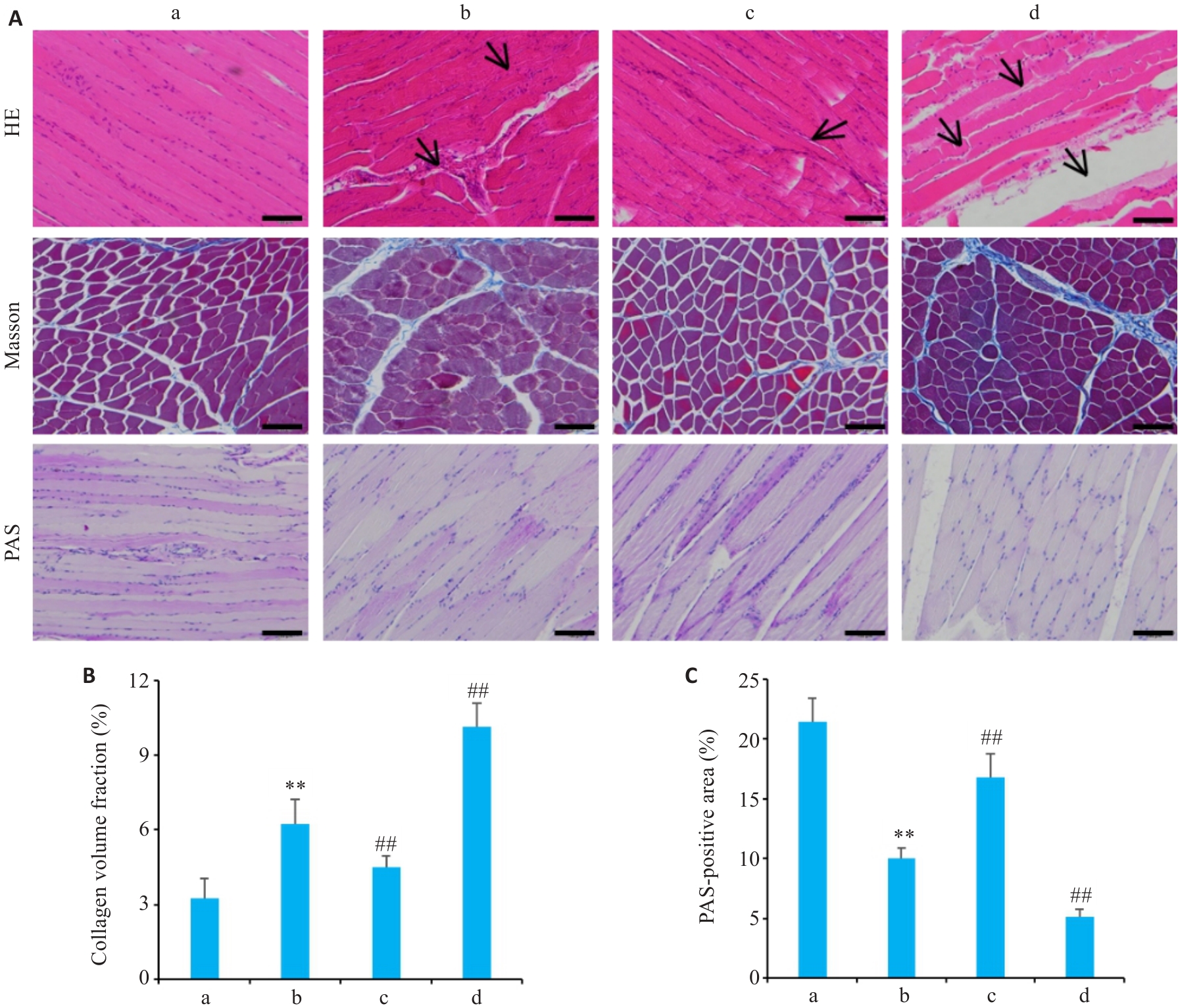
图1 各组大鼠骨骼肌组织HE染色、Masson染色、PAS染色
Fig.1 Histopathological changes in skeletal muscles of the rats in each group. A: HE staining, Masson staining, and PAS staining of mouse skeletal muscle tissues (Scale bar=100 μm). B: Collagen volume fraction of the rats in each group. C: Percentage of PAS-positive area in each group of rats. Data are presented as Mean±SD (n=10). a: Blank control group; b: Diabetes control group; c: Diabetes aerobic group; d: Diabetes exhaustive group. Arrows indicate damaged muscle fibers, inflammatory cell infiltration, and vacuolar degeneration. **P<0.01 vs blank control group; ##P<0.01 vs the diabetes control group.
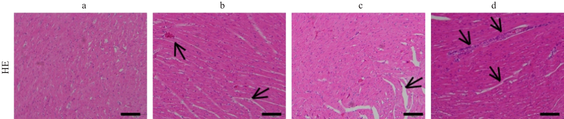
图2 各组大鼠心肌组织HE染色
Fig.2 HE staining of myocardial tissue from rats in each group (Scale bar=100 μm). a: Blank control group; b: Diabetes control group; c: Diabetes aerobic group; d: Diabetes exhaustive group. Arrows indicate disordered and ruptured myocardial fibers, widened intercellular spaces, and necrotic areas.
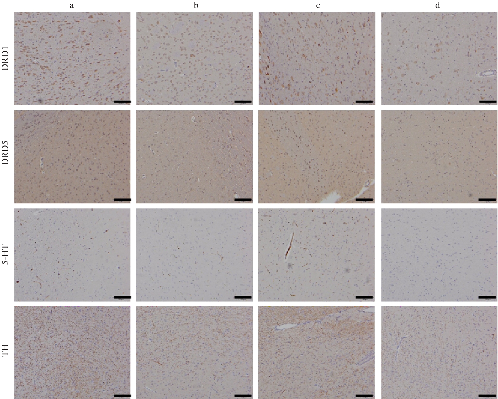
图3 各组大鼠前额叶皮质DRD1、DRD5、5-HT、TH的表达分布
Fig.3 Expression distribution of DRD1, DRD5, 5-HT, and TH in the prefrontal cortex of rats in each group (Scale bar=100 μm. a: Blank control group; b: Diabetes control group; c: Diabetes aerobic group; d: Diabetes exhaustive group.
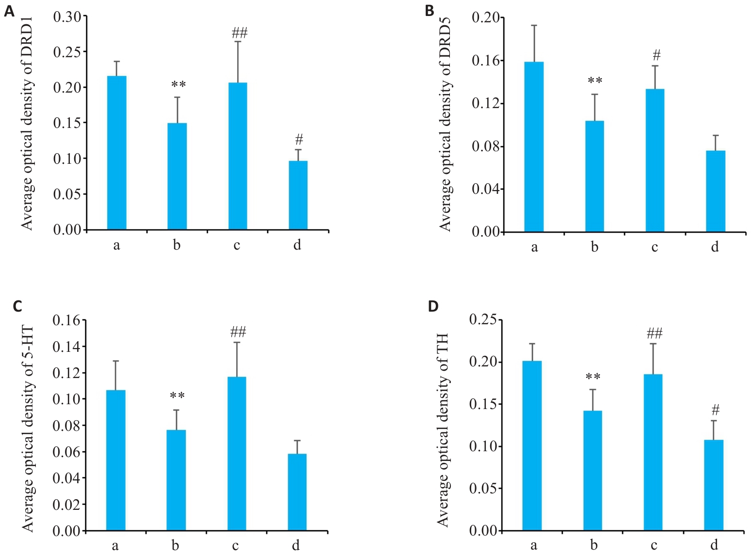
图4 各组大鼠前额叶皮质DRD1、DRD5、5-HT、TH的平均光密度
Fig.4 Average optical density of DRD1 (A), DRD5 (B), 5-HT (C) and TH (D) in the prefrontal cortex of the rats in each group (Mean±SD, n=10). **P<0.01 vs the blank control group; #P<0.05, ##P<0.01 vs the diabetes control group. a. Blank control group; b. Diabetes control group; c. Diabetes aerobic group; d. Diabetes exhaustive group.
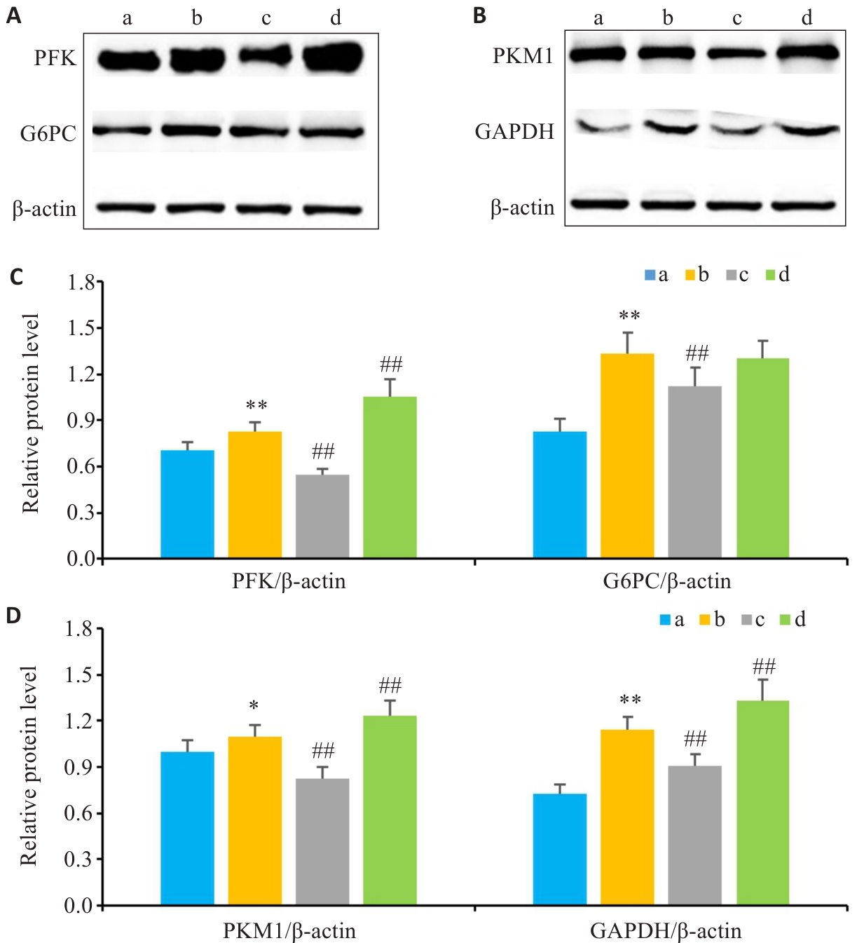
图5 各组大鼠肝脏组织G6PC、PFK、GAPDH、PKM1蛋白表达
Fig.5 Protein expressions of G6PC, PFK, GAPDH and PKM1 in liver tissues of the rats in each group. A: Protein expression levels of G6PC and PFK detected by Western blotting. B: Protein expression levels of GAPDH and PKM1 detected by Western blotting. C: Expression levels of G6PC and PFK in the liver tissues of different groups. D: Expression levels of GAPDH and PKM1 in liver tissues of different groups. Data are presented as Mean±SD (n=10). *P<0.05, **P<0.01 vs the blank control group; ##P<0.01 vs the diabetes control group. a: Blank control group; b: Diabetes control group; c: Diabetes aerobic group; d: Diabetes exhaustive group.
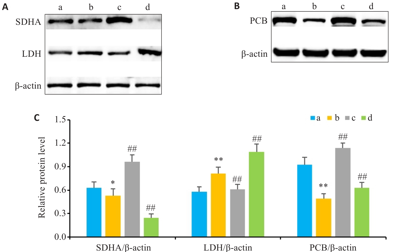
图6 各组大鼠肝脏组织LDH、 SDHA、PCB蛋白表达
Fig.6 Protein expression of LDH, SDHA and PCB in liver tissues of rats in each group. A: Detection of protein expression levels of LDH and SDHA by Western blotting. B: Detection of protein expression levels of PCB by Western blotting. C: Expression levels of SDHA, LDH and PCB in liver tissues of different groups (Mean±SD, n=10).*P<0.05, **P<0.01 vs blank control group; ##P<0.01 vs the diabetes control group. a: Blank control group; b: Diabetes control group; c: Diabetes aerobic group; d: Diabetes exhaustive group.
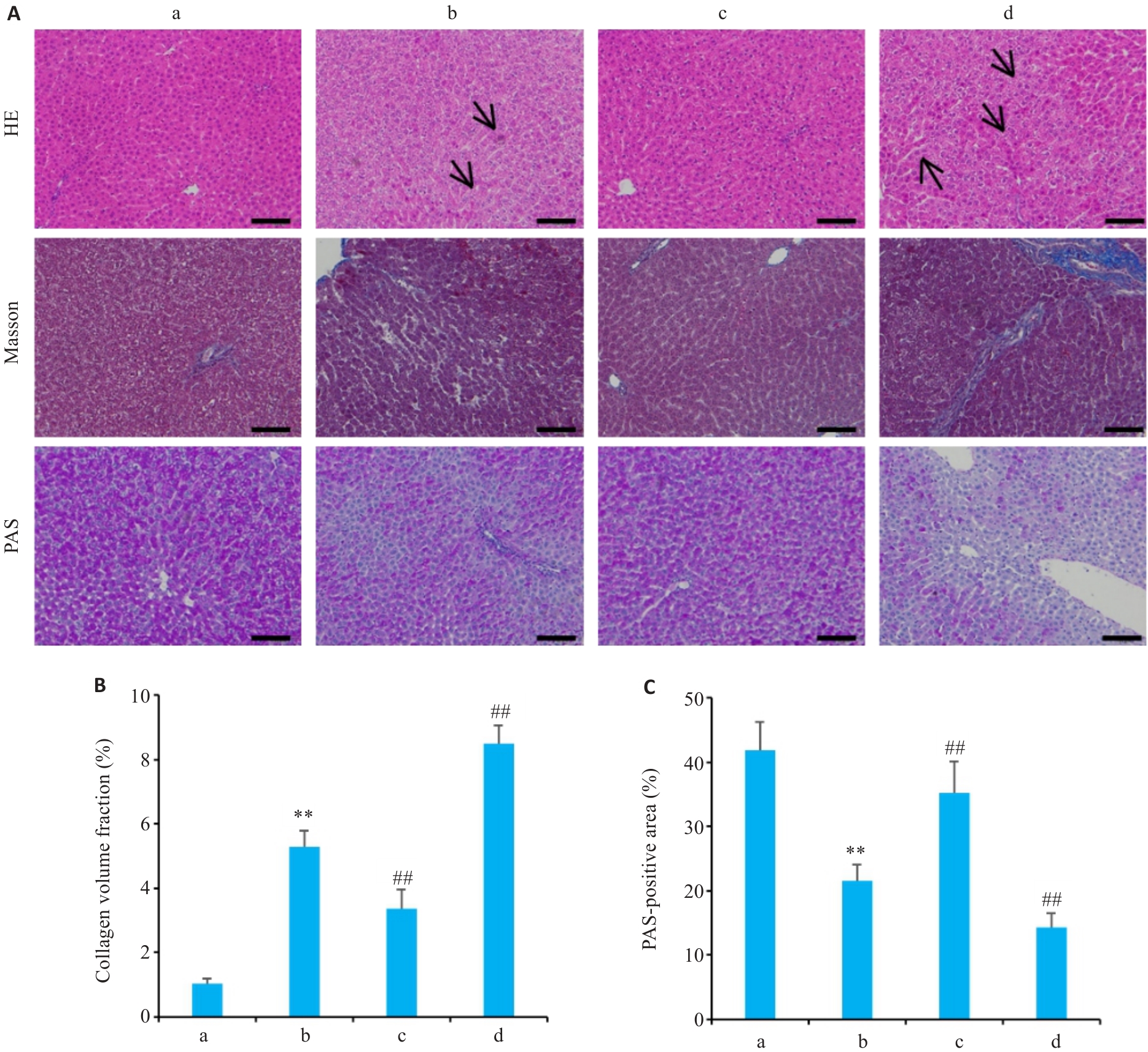
图7 各组大鼠肝脏组织HE染色、Masson染色、PAS染色
Fig.7 HE, Masson, and PAS staining (A) of liver tissues, collagen volume fraction (B) and percentage of PAS-positive area in each group (Scale bar=100 μm). Data are presented as Mean±SD (n=10). a: Blank control group; b: Diabetes control group; c: Diabetes aerobic group; d: Diabetes exhaustive group. Arrows indicate lipid vacuoles of varying sizes and inflammatory cell infiltration in hepatocytes. **P<0.01 vs blank control group; ##P<0.01 vs the diabetes control group.
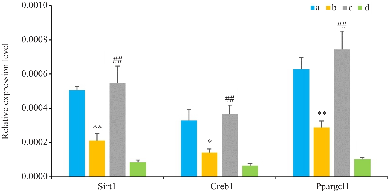
图8 各组大鼠肝脏Sirt1、Creb1、Ppargc1a的基因表达水平变化
Fig.8 Changes in mRNA expression of Sirt1, Creb1, and Ppargc1a in the livers of rats in each group (Mean±SD, n=10).*P<0.05, **P<0.01 vs the blank control group; ##P<0.01 vs the diabetes control group. a: Blank control group; b: Diabetes control group; c: Diabetes aerobic group; d: Diabetes exhaustive group.
| [1] | Risk Factor CollaborationNCD. Worldwide trends in diabetes prevalence and treatment from 1990 to 2022: a pooled analysis of 1108 population-representative studies with 141 million participants[J]. Lancet, 2024, 404(10467): 2077-93. |
| [2] | Cao R, Tian HM, Zhang Y, et al. Signaling pathways and intervention for therapy of type 2 diabetes mellitus[J]. MedComm, 2023, 4(3): e283. doi:10.1002/mco2.283 |
| [3] | Zhou Z, Sun B, Yu DS, et al. Gut microbiota: an important player in type 2 diabetes mellitus[J]. Front Cell Infect Microbiol, 2022, 12: 834485. doi:10.3389/fcimb.2022.834485 |
| [4] | Dutta BJ, Singh S, Seksaria S, et al. Inside the diabetic brain: Insulin resistance and molecular mechanism associated with cognitive impairment and its possible therapeutic strategies[J]. Pharmacol Res, 2022, 182: 106358. doi:10.1016/j.phrs.2022.106358 |
| [5] | Liu RJ, Shi XF, Zhang X. The effects of resistance training on glycemic control, cardiometabolic health, and body composition in middle-aged and older women with type 2 diabetes and overweight or obesity: a systematic review and meta-analysis[J]. Maturitas, 2025, 201: 108691. doi:10.1016/j.maturitas.2025.108691 |
| [6] | Mohd S, Kumar LL, Harish V, et al. Diabetes mellitus: Complications, emerging therapeutic targets, and evolving treatment approaches[J]. Obes Med, 2025, 58: 100652. doi:10.1016/j.obmed.2025.100652 |
| [7] | Xing HM, Lu JH, Yoong SQ, et al. Effect of aerobic and resistant exercise intervention on inflammaging of type 2 diabetes mellitus in middle-aged and older adults: a systematic review and meta-analysis[J]. J Am Med Dir Assoc, 2022, 23(5): 823-30.e13. doi:10.1016/j.jamda.2022.01.055 |
| [8] | Zhao RX, Li JY, Du FB, et al. Effects of long-term moderate-intensity exercise on autonomic nervous system dysfunction induced by cardiac neurovascular interface deterioration[J]. Rev Cardiovasc Med, 2025, 26(10): 40714. doi:10.31083/rcm40714 |
| [9] | Gao HY, Zhao YL, Zhao LL, et al. The role of oxidative stress in multiple exercise-regulated bone homeostasis[J]. Aging Dis, 2023, 14(5): 1555-82. doi:10.14336/ad.2023.0223 |
| [10] | Amar D, Gay NR, Jimenez-Morales D, et al. The mitochondrial multi-omic response to exercise training across rat tissues[J]. Cell Metab, 2024, 36(6): 1411-29.e10. |
| [11] | Araki E, Goto A, Kondo T, et al. Japanese clinical practice guideline for diabetes 2019[J]. Diabetol Int, 2020, 11(3): 165-223. doi:10.1007/s13340-020-00439-5 |
| [12] | Sheikh R, Shakerian S, Fatemi Tabatabaei SR, et al. Moderate and high-intensity interval training protect against diabetes-induced modulation of hepatic CD86 and CD206 expression associated with the amelioration of insulin resistance and inflammation in rats[J]. Immunobiology, 2023, 228(6): 152745. doi:10.1016/j.imbio.2023.152745 |
| [13] | Yap KH, Yee GS, Candasamy M, et al. Catalpol ameliorates insulin sensitivity and mitochondrial respiration in skeletal muscle of type-2 diabetic mice through insulin signaling pathway and AMPK/SIRT1/PGC-1α/PPAR-γ activation[J]. Biomolecules, 2020, 10(10): 1360. doi:10.3390/biom10101360 |
| [14] | Ma XT, Gao HY, Wang Z, et al. Beneficial effects of different types of exercise on diabetic cardiomyopathy[J]. Biomolecules, 2025, 15(9): 1223. doi:10.3390/biom15091223 |
| [15] | Wang GD. Aerobic exercise ameliorates myocardial ischemia/reperfusion injury and thrombosis of diabetic rats via activation of AMPK/Sirt1/PGC-1α pathway[J]. Gen Physiol Biophys, 2022, 41(4): 319-28. doi:10.4149/gpb_2022010 |
| [16] | Zheng LF, Qin RT, Rao ZJ, et al. High-intensity interval training induces renal injury and fibrosis in type 2 diabetic mice[J]. Life Sci, 2023, 324: 121740. doi:10.1016/j.lfs.2023.121740 |
| [17] | Sahin AM, Sonmez OF, et al. Effects of acute exhaustive exercise on oxidant and antioxidant system parameters in rats with streptozotocin induced diabetes mellitus[J]. TurkJSportsMed, 2020, 55(2): 131-7. doi:10.5152/tjsm.2020.169 |
| [18] | Tan LS, Lau HH, Abdelalim EM, et al. The role of glucose-6-phosphatase activity in glucose homeostasis and its potential for diabetes therapy[J]. Trends Mol Med, 2025, 31(2): 152-64. doi:10.1016/j.molmed.2024.09.005 |
| [19] | Luu L, Dai FF, Prentice KJ, et al. The loss of Sirt1 in mouse pancreatic beta cells impairs insulin secretion by disrupting glucose sensing[J]. Diabetologia, 2013, 56(9): 2010-20. doi:10.1007/s00125-013-2946-5 |
| [20] | Yu Y, Zhao Y, Teng F, et al. Berberine improves cognitive deficiency and muscular dysfunction via activation of the AMPK/SIRT1/PGC-1a pathway in skeletal muscle from naturally aging rats[J]. J Nutr Health Aging, 2018, 22(6): 710-7. doi:10.1007/s12603-018-1015-7 |
| [21] | Chen C, Zhou M, Ge YC, et al. SIRT1 and aging related signaling pathways[J]. Mech Ageing Dev, 2020, 187: 111215. doi:10.1016/j.mad.2020.111215 |
| [22] | Shen YH, Mao ZJ, Chen HM, et al. Exercise-specific post-translational modification signatures: unveiling precise regulatory mechanisms of molecular exercise language and cellular adaptation[J]. Front Sports Act Living, 2026, 8: 1765170. doi:10.3389/fspor.2026.1765170 |
| [23] | Rodrigues B, Figueroa DM, Mostarda CT, et al. Maximal exercise test is a useful method for physical capacity and oxygen consumption determination in streptozotocin-diabetic rats[J]. Cardiovasc Diabetol, 2007, 6: 38. doi:10.1186/1475-2840-6-38 |
| [24] | Qin F, Dong YN, Wang ST, et al. Maximum oxygen consumption and quantification of exercise intensity in untrained male Wistar rats[J]. Sci Rep, 2020, 10: 11520. doi:10.1038/s41598-020-68455-8 |
| [25] | Arifin WN, Zahiruddin WM. Sample size calculation in animal studies using resource equation approach[J]. Malays J Med Sci, 2017, 24(5): 101-5. doi:10.21315/mjms2017.24.5.11 |
| [26] | Charan J, Kantharia ND. How to calculate sample size in animal studies?[J]. J Pharmacol Pharmacother, 2013, 4(4): 303-6. doi:10.4103/0976-500x.119726 |
| [27] | Gao HY, Ma XT, Wang Z, et al. The effects of different types of exercise on pulmonary inflammation and fibrosis in mice with type 2 diabetes mellitus[J]. Cells, 2025, 14(13): 1026. doi:10.3390/cells14131026 |
| [28] | Tomiga Y, Tanaka K, Kusuyama J, et al. Exercise training ameliorates carbon tetrachloride-induced liver fibrosis and anxiety-like behaviors[J]. Am J Physiol Gastrointest Liver Physiol, 2024, 327(6): G850-60. doi:10.1152/ajpgi.00161.2024 |
| [29] | Festing MF. On determining sample size in experiments involving laboratory animals[J]. Lab Anim, 2018, 52(4): 341-50. doi:10.1177/0023677217738268 |
| [30] | Yu Y, Xu LS, Wu Y, et al. The antihypertensive effect of MK on spontaneously hypertensive rats through the AMPK/Akt/ENOS/NO and ERK1/2/Cx43 signaling pathways[J]. Hypertens Res, 2021, 44(7): 781-90. doi:10.1038/s41440-021-00638-w |
| [31] | Bortolon JR, Murata GM, Borges L, et al. Recovery of diabetic rats after physical exhaustion: kinetic alterations in muscle inflammation and muscle-signaling proteins to atrophy and hypertrophy[J]. Front Physiol, 2020, 11: 573416. doi:10.3389/fphys.2020.573416 |
| [32] | Oka SI, Byun J, Huang CY, et al. Nampt potentiates antioxidant defense in diabetic cardiomyopathy[J]. Circ Res, 2021, 129(1): 114-30. doi:10.1161/circresaha.120.317943 |
| [33] | Wang SY, Zhu SY, Wu J, et al. Exercise enhances cardiac function by improving mitochondrial dysfunction and maintaining energy homoeostasis in the development of diabetic cardiomyopathy[J]. J Mol Med, 2020, 98(2): 245-61. doi:10.1007/s00109-019-01861-2 |
| [34] | De Sousa RAL, Improta-Caria AC, de Freitas Souza BS. Exercise–linked irisin: consequences on mental and cardiovascular health in type 2 diabetes[J]. Int J Mol Sci, 2021, 22(4): 2199. doi:10.3390/ijms22042199 |
| [35] | Al-Sayyar A, Hammad MM, Williams MR, et al. Neurotransmitters in type 2 diabetes and the control of systemic and central energy balance[J]. Metabolites, 2023, 13(3): 384. doi:10.3390/metabo13030384 |
| [36] | Hajiluian G, Abbasalizad Farhangi M, Nameni G, et al. Oxidative stress-induced cognitive impairment in obesity can be reversed by vitamin D administration in rats[J]. Nutr Neurosci, 2018, 21(10): 744-52. doi:10.1080/1028415x.2017.1348436 |
| [37] | He HJ, Zhong Y, Wang HL, et al. Smad3 mediates diabetic dyslipidemia and fatty liver in db/db mice by targeting PPARδ[J]. Int J Mol Sci, 2023, 24(14): 11396. doi:10.3390/ijms241411396 |
| [38] | Adam SY, Eltahir HA, Mohammed Ismaeel FA, et al. Gum Arabic regulates glucose metabolic enzymes through modification of hepatic phosphoenolpyruvate carboxykinase expression in diabetic rats[J]. Bioact Carbohydr Diet Fibre, 2025, 34: 100490. doi:10.1016/j.bcdf.2025.100490 |
| [39] | He LN, Li Y, Zeng N, et al. Regulation of basal expression of hepatic PEPCK and G6Pase by AKT2[J]. Biochem J, 2020, 477(5): 1021-31. doi:10.1042/bcj20190570 |
| [40] | Pietrowska-Borek M, Dobrogojski J, Sobieszczuk-Nowicka E, et al. New insight into plant signaling: extracellular ATP and uncommon nucleotides[J]. Cells, 2020, 9(2): 345. doi:10.3390/cells9020345 |
| [1] | 王小龙, 杨建, 梁爽, 董哲毅. 强化降脂对血糖管理困难的2型糖尿病合并慢性肾脏病患者死亡风险的缓冲作用:基于双代谢轨迹的队列研究[J]. 南方医科大学学报, 2026, 46(7): 1634-1647. |
| [2] | 张国栋, 魏思昂, 王弘, 谢艳丽, 何晖, 李嵘. 有氧运动通过氧化应激-炎症-Hippo/YAP通路轴改善心力衰竭小鼠心功能[J]. 南方医科大学学报, 2026, 46(4): 753-760. |
| [3] | 邹晓松, 张幸, 李平, 田瑞雪, 路晓淼. 姜黄素通过抑制RANBP3L基因表达减轻2型糖尿病小鼠的骨质疏松症[J]. 南方医科大学学报, 2026, 46(2): 325-334. |
| [4] | 乔世杰, 游进顺, 王昶, 熊红萍, 李书楠. 基于证素辨证原理的2型糖尿病视网膜病变预测模型构建[J]. 南方医科大学学报, 2026, 46(10): 2456-2470. |
| [5] | 李楠, 张亮, 郭俏凤, 周越, 刘长江. 有氧运动通过调控miR-221-3p介导的脂肪组织巨噬细胞极化改善小鼠胰岛素抵抗[J]. 南方医科大学学报, 2026, 46(1): 74-82. |
| [6] | 韩瑞敏, 赵曼可, 袁俊芳, 史振红, 王珍, 王德峰. 枯草杆菌二联活菌肠溶胶囊调节2型糖尿病合并昼夜节律紊乱小鼠糖脂代谢的作用机制[J]. 南方医科大学学报, 2025, 45(7): 1490-1497. |
| [7] | 侯鑫睿, 张振东, 曹明远, 杜予心, 王小平. 红景天苷靶向miR-1343-3p-OGDHL/PDHB糖代谢轴抑制胃癌细胞的体内外增殖[J]. 南方医科大学学报, 2025, 45(6): 1226-1239. |
| [8] | 许怀文, 翁丽, 薛鸿. CXCL12可作为2型糖尿病合并慢性阻塞性肺疾病的潜在治疗靶点[J]. 南方医科大学学报, 2025, 45(1): 100-109. |
| [9] | 王妍, 阮毓卿, 崔璨, 王秀. 交泰丸通过激活PI3K/AKT信号通路改善阿尔茨海默病模型小鼠大脑的葡萄糖代谢[J]. 南方医科大学学报, 2024, 44(5): 894-903. |
| [10] | 杨晶晶, 殷丽霞, 段婷, 牛民主, 何震东, 陈心蕊, 张小凤, 李静, 耿志军, 左芦根. 胃癌组织中高表达ATP5A1与患者术后的不良预后和肿瘤细胞的糖代谢有关[J]. 南方医科大学学报, 2024, 44(5): 974-980. |
| [11] | 周凤敏, 郭艳菊, 陈 宁. 运动诱导的Irisin表达改善2型糖尿病大鼠的肾脏损伤[J]. 南方医科大学学报, 2024, 44(4): 675-681. |
| [12] | 方福生, 王 宁, 刘星宇, 王 薇, 孙 菁, 李 红, 孙般若, 谷昭艳, 傅晓敏, 闫双通. 基于C肽的胰岛素抵抗指数可准确评估健康体检者的胰岛素抵抗与血尿酸水平的相关性[J]. 南方医科大学学报, 2023, 43(9): 1509-1514. |
| [13] | 刘 泽, 何汶霞, 房睿哲, 肖 钢, 言 芳, 邱祁广, 方会龙, 王俊杰. 生命潮生物反馈技术干预2型糖尿病的先导观察[J]. 南方医科大学学报, 2023, 43(9): 1622-1628. |
| [14] | 周 蓓, 李 静, 方晨圆, 黄亚楠, 桑贵蕊, 陶少平, 何春玲. 二甲双胍/维格列汀和利拉鲁肽对肥胖合并2型糖尿病患者的临床疗效比较[J]. 南方医科大学学报, 2023, 43(3): 436-442. |
| [15] | 刘思妤, 刘 青, 彭群龙, 张远芳, 王俊杰. 二氢杨梅素通过抑制HMGB1改善糖尿病大鼠心功能不全[J]. 南方医科大学学报, 2022, 42(5): 641-648. |
| 阅读次数 | ||||||
|
全文 |
|
|||||
|
摘要 |
|
|||||
