南方医科大学学报 ›› 2025, Vol. 45 ›› Issue (11): 2416-2426.doi: 10.12122/j.issn.1673-4254.2025.11.14
姜雪凝1( ), 黄晴晴2, 徐盈2, 王舜印2, 张小凤2, 王炼1, 王月月2, 左芦根1,2()
收稿日期:2025-05-06
出版日期:2025-11-20
发布日期:2025-11-28
通讯作者:
左芦根
E-mail:jiangxn1202@163.com;zuolugen@126.com
作者简介:姜雪凝,在读硕士研究生,E-mail: jiangxn1202@163.com
基金资助:
Xuening JIANG1(), Qingqing HUANG2, Ying XU2, Shunyin² WANG2, Xiaofeng² ZHANG2, Lian¹ WANG1, Yueyue² WANG2, Lugen ZUO1,2()
Received:2025-05-06
Online:2025-11-20
Published:2025-11-28
Contact:
Lugen ZUO
E-mail:jiangxn1202@163.com;zuolugen@126.com
Supported by:摘要:
目的 探究YEATS2在胃癌组织中的表达水平及其临床预后价值,并揭示其对胃癌细胞EMT进程的作用和机制。 方法 基于TIMER2.0、GEPIA等公共数据库预测YEATS2在胃癌中的表达水平;纳入100例在本院接受胃癌根治术的患者,通过免疫组化检测胃癌及癌旁组织中YEATS2表达,分析其与临床病理参数和Ki67的相关性;采用Kaplan-Meier生存分析、Cox回归和ROC曲线评估YEATS2的预后价值;通过生物富集分析预测YEATS2对胃癌可能的作用和机制,构建YEATS2敲低与过表达的胃癌细胞系(HGC-27与AGS),设shNC、shYEATS2、Vector和OE-YEATS2组,分别采用shRNA阴性对照慢病毒和YEATS2特异性干扰慢病毒、空载对照慢病毒载体及YEATS2过表达慢病毒进行转染。结合细胞划痕、Transwell和Western blotting检测YEATS2对胃癌细胞迁移、侵袭及EMT的作用。 结果 YEATS2在胃癌组织中表达显著高于癌旁组织且与Ki67表达呈正相关(P<0.05)。YEATS2高表达组中CEA≥5 μg/L、CA19-9≥37 kU/L、T3-4期及N2-3期的患者比例高于低表达组(P<0.05)。生存分析显示YEATS2高表达患者术后5年生存率降低(P<0.001),ROC曲线分析表明YEATS2评估胃癌患者根治术后5年生存率的敏感性为80.00%,特异性为66.67%(P<0.05)。Cox回归显示YEATS2高表达是胃癌患者术后5年生存率低的独立危险因素(HR:1.675,95%CI:1.013~2.771,P=0.045)。富集分析提示YEATS2可能与胃癌EMT过程和Wnt/β-catenin通路有关。体外实验表明,YEATS2高表达可促进胃癌细胞迁移、侵袭,同时上调Vimentin、N-cadherin、Wnt和Active β-catenin,并下调E-cadherin的表达(P<0.05)。经XAV-939(Wnt/β-catenin抑制剂)处理后,YEATS2过表达导致的EMT相关蛋白表达变化(N-cadherin、Vimentin上调和E-cadherin下调)被显著削弱(P<0.05)。 结论 YEATS2高表达可能激活Wnt/β-catenin通路促进胃癌细胞EMT进程,并与患者预后不良有关。
姜雪凝, 黄晴晴, 徐盈, 王舜印, 张小凤, 王炼, 王月月, 左芦根. 高表达YEATS2通过激活Wnt/β-catenin通路促进胃癌细胞上皮-间质转化进程[J]. 南方医科大学学报, 2025, 45(11): 2416-2426.
Xuening JIANG, Qingqing HUANG, Ying XU, Shunyin² WANG, Xiaofeng² ZHANG, Lian¹ WANG, Yueyue² WANG, Lugen ZUO. High YEATS2 expression promotes epithelial-mesenchymal transition in gastric cancer cells by activating the Wnt/β-catenin signaling pathway[J]. Journal of Southern Medical University, 2025, 45(11): 2416-2426.

图1 胃癌组织中YEATS2和Ki67的表达及相关性分析
Fig.1 Expression and correlation analysis of YEATS2 and Ki67 in gastric cancer. A: Expression of YEATS2 in different human tumors. B: Expression of YEATS2 in gastric cancer. C: Immunohistochemical staining of YEATS2 and Ki67. D,E: Relative IOD values of YEATS2 and Ki67. F: Correlation between YEATS2 and Ki67 in gastric cancer tissues. *P<0.05,***P<0.001 vs adjacent tissue.
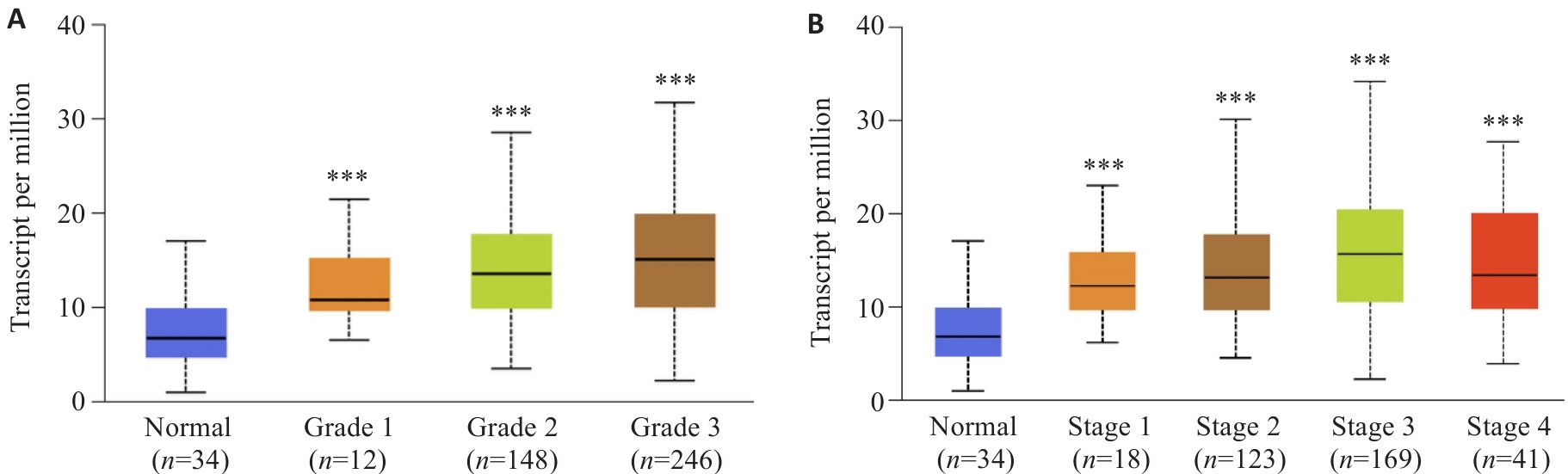
图2 YEATS2高表达与胃癌患者肿瘤分级和分期相关
Fig.2 High YEATS2 expression is significantly associated with tumor grading and staging in gastric cancer patients. A: Correlation between YEATS2 TPM and tumor grading in gastric cancer. B: Correlation between YEATS2 TPM and tumor staging in gastric cancer. ***P<0.001 vs adjacent tissue.
| Factor | n | YEATS2 expression | χ2 | P | ||
|---|---|---|---|---|---|---|
| Low (n=50) | High (n=50) | |||||
| Gender | Female | 19 | 6 (31.58%) | 13 (68.42%) | 3.184 | 0.074 |
| Male | 81 | 44 (54.32%) | 37 (45.68%) | |||
| Age (year) | ˂60 | 35 | 18 (51.43%) | 17 (48.57%) | 0.044 | 0.834 |
| ≥60 | 65 | 32 (49.23%) | 33 (50.77%) | |||
| Cancer cell type | Adenocarcinoma | 79 | 42 (53.16%) | 37 (46.84%) | 1.507 | 0.220 |
| Other | 21 | 8 (38.10%) | 13 (61.90%) | |||
| CEA (μg/L) | ˂5 | 35 | 24 (68.57%) | 11 (31.43%) | 7.429 | 0.006 |
| ≥5 | 65 | 26 (40.00%) | 39 (60.00%) | |||
| CA19-9 (kU/L) | ˂37 | 44 | 27 (61.36%) | 17 (38.64%) | 4.058 | 0.044 |
| ≥37 | 56 | 23 (41.07%) | 33 (58.93%) | |||
| Tumor size (cm) | ˂5 | 49 | 27 (55.10%) | 22 (44.90%) | 1.000 | 0.317 |
| ≥5 | 51 | 23 (45.10%) | 28 (54.90%) | |||
| T stage | T1-T2 | 36 | 25 (69.44%) | 11 (30.56%) | 8.507 | 0.004 |
| T3-T4 | 64 | 25 (39.06%) | 39 (60.94%) | |||
| N stage | N0-N1 | 43 | 27 (62.79%) | 16 (37.21%) | 4.937 | 0.026 |
| N2-N3 | 57 | 23 (40.35%) | 34 (59.65%) | |||
表1 胃癌组织中YEATS2表达水平与胃癌恶性进展参数间的关系
Tab.1 Relationship between YEATS2 expression level and parameters of malignant progression in gastric cancer tissues (n=50)
| Factor | n | YEATS2 expression | χ2 | P | ||
|---|---|---|---|---|---|---|
| Low (n=50) | High (n=50) | |||||
| Gender | Female | 19 | 6 (31.58%) | 13 (68.42%) | 3.184 | 0.074 |
| Male | 81 | 44 (54.32%) | 37 (45.68%) | |||
| Age (year) | ˂60 | 35 | 18 (51.43%) | 17 (48.57%) | 0.044 | 0.834 |
| ≥60 | 65 | 32 (49.23%) | 33 (50.77%) | |||
| Cancer cell type | Adenocarcinoma | 79 | 42 (53.16%) | 37 (46.84%) | 1.507 | 0.220 |
| Other | 21 | 8 (38.10%) | 13 (61.90%) | |||
| CEA (μg/L) | ˂5 | 35 | 24 (68.57%) | 11 (31.43%) | 7.429 | 0.006 |
| ≥5 | 65 | 26 (40.00%) | 39 (60.00%) | |||
| CA19-9 (kU/L) | ˂37 | 44 | 27 (61.36%) | 17 (38.64%) | 4.058 | 0.044 |
| ≥37 | 56 | 23 (41.07%) | 33 (58.93%) | |||
| Tumor size (cm) | ˂5 | 49 | 27 (55.10%) | 22 (44.90%) | 1.000 | 0.317 |
| ≥5 | 51 | 23 (45.10%) | 28 (54.90%) | |||
| T stage | T1-T2 | 36 | 25 (69.44%) | 11 (30.56%) | 8.507 | 0.004 |
| T3-T4 | 64 | 25 (39.06%) | 39 (60.94%) | |||
| N stage | N0-N1 | 43 | 27 (62.79%) | 16 (37.21%) | 4.937 | 0.026 |
| N2-N3 | 57 | 23 (40.35%) | 34 (59.65%) | |||
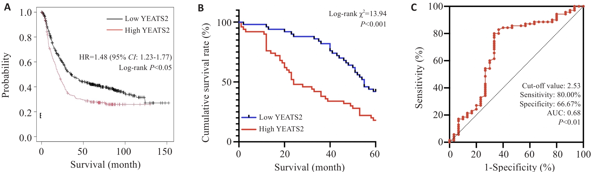
图3 YEATS2对胃癌患者预后的影响
Fig.3 Effect of YEATS2 on prognosis of patients with gastric cancer. A: Kaplan-Meier analysis. B: Survival curves. C: Predictive value of YEATS2 for 5-year survival after radical gastrectomy.
| Factor | Univariate analysis | Multivariate analysis | ||||
|---|---|---|---|---|---|---|
| Log-rank χ2 | P | HR | 95% CI | P | ||
| Gender (male vs female) | <0.001 | 0.989 | ||||
| Age (˂60 years vs ≥60 years ) | 0.014 | 0.907 | ||||
| Cancer cell type (adenocarcinoma vs other) | 0.539 | 0.463 | ||||
| CEA (˂5 μg/L vs ≥5 μg/L) | 24.128 | <0.001 | 2.760 | 1.480-5.147 | 0.001 | |
| CA19-9 (˂37kU/L vs ≥37 kU/L) | 30.316 | <0.001 | 2.600 | 1.480-4.569 | <0.001 | |
| Tumor size (˂5 cm vs ≥5 cm) | 1.041 | 0.308 | ||||
| T stage (T1-T2 vs T3-T4) | 15.477 | <0.001 | 2.015 | 1.129-3.593 | 0.018 | |
| N stage (N0-N1 vs N2-N3) | 25.115 | <0.001 | 2.238 | 1.275-3.928 | 0.005 | |
| YEATS2 expression (high vs low) | 13.939 | <0.001 | 1.675 | 1.013-2.771 | 0.045 | |
表2 胃癌患者术后5年生存期的影响因素分析
Tab.2 Analysis of risk factors affecting 5-year survival rate of gastric cancer patients after radical gastrectomy
| Factor | Univariate analysis | Multivariate analysis | ||||
|---|---|---|---|---|---|---|
| Log-rank χ2 | P | HR | 95% CI | P | ||
| Gender (male vs female) | <0.001 | 0.989 | ||||
| Age (˂60 years vs ≥60 years ) | 0.014 | 0.907 | ||||
| Cancer cell type (adenocarcinoma vs other) | 0.539 | 0.463 | ||||
| CEA (˂5 μg/L vs ≥5 μg/L) | 24.128 | <0.001 | 2.760 | 1.480-5.147 | 0.001 | |
| CA19-9 (˂37kU/L vs ≥37 kU/L) | 30.316 | <0.001 | 2.600 | 1.480-4.569 | <0.001 | |
| Tumor size (˂5 cm vs ≥5 cm) | 1.041 | 0.308 | ||||
| T stage (T1-T2 vs T3-T4) | 15.477 | <0.001 | 2.015 | 1.129-3.593 | 0.018 | |
| N stage (N0-N1 vs N2-N3) | 25.115 | <0.001 | 2.238 | 1.275-3.928 | 0.005 | |
| YEATS2 expression (high vs low) | 13.939 | <0.001 | 1.675 | 1.013-2.771 | 0.045 | |
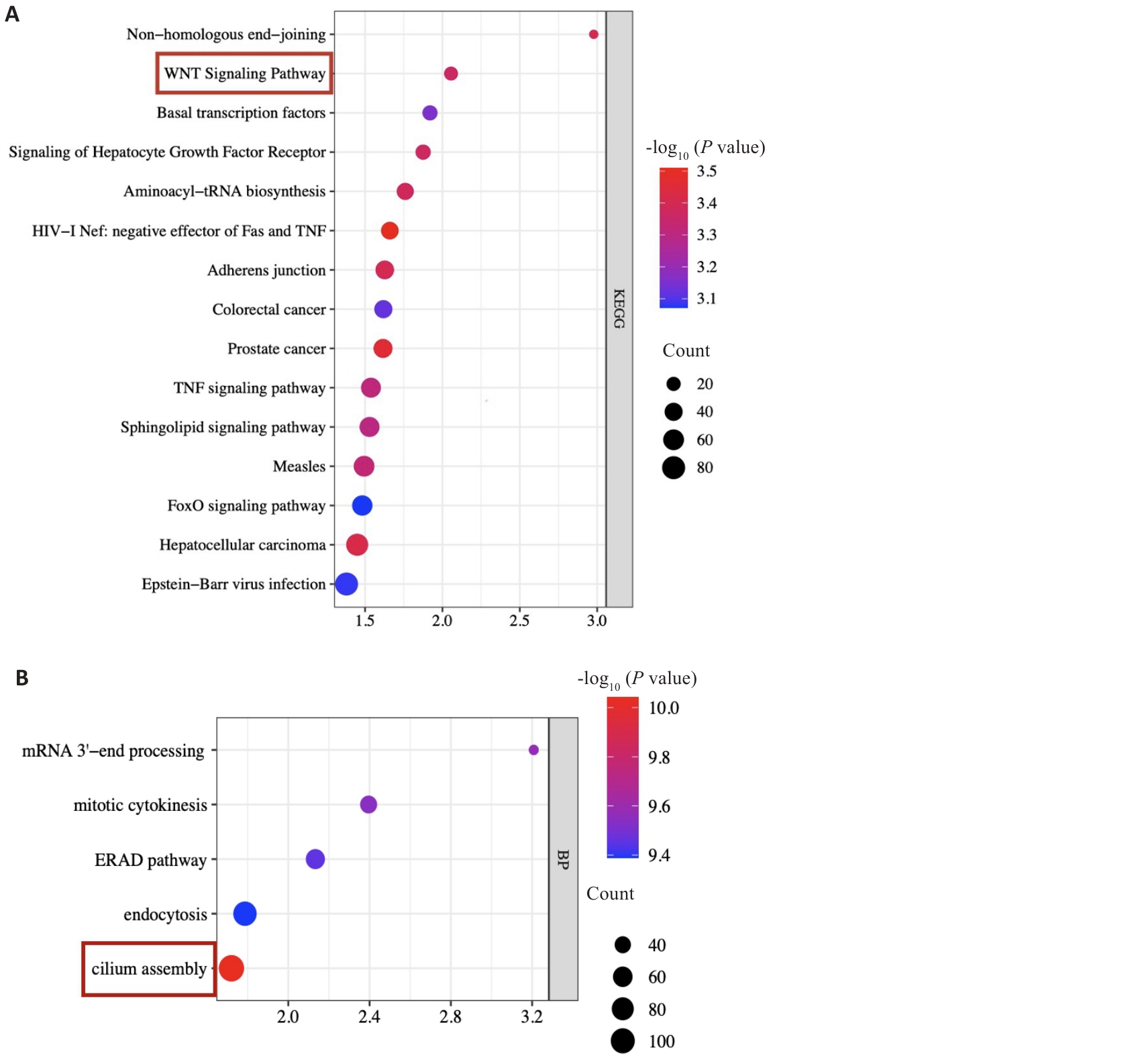
图4 YEATS2的KEGG和GO富集分析
Fig.4 KEGG and GO enrichment analysis of YEATS2. A: KEGG enrichment analysis results. B: GO enrichment analysis results.

图5 YEATS2促进胃癌细胞的迁移
Fig.5 YEATS2 promotes migration of gastric cancer cells. A-D: Validation of YEATS2 knockdown and overexpression in gastric cancer cells. E, F: High expression of YEATS2 promotes HGC-27 cell migration. G, H: High expression of YEATS2 promotes AGS cell migration. n=3, *P<0.05 vs shNC or vs Vector.

图6 YEATS2促进胃癌细胞的迁移和侵袭
Fig.6 YEATS2 promotes migration and invasion of gastric cancer cells. A-D: High expression of YEATS2 promotes HGC-27 cell migration and invasion. E-H: High expression of YEATS2 promotes AGS cell migration and invasion. n=3, *P<0.05 vs shNC or vs Vector.
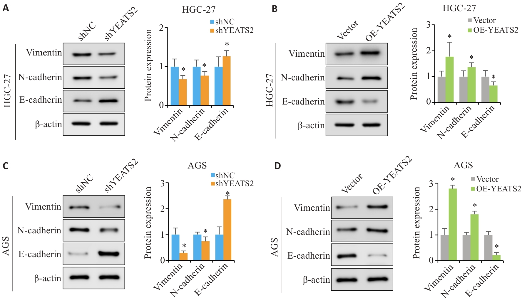
图7 YEATS2促进胃癌细胞的EMT进程
Fig.7 YEATS2 promotes EMT process in gastric cancer cells. A, B : Expressions of key EMT-related proteins in HGC27 cells. C, D: Expressions of key EMT-related proteins in AGS cells. n=3, *P<0.05 vs shNC or vs Vector.

图8 胃癌中YEATS2的表达升高激活Wnt/β-catenin信号通路
Fig.8 Elevated expression of YEATS2 in gastric cancer cells activates the Wnt/β-catenin signaling pathway. A, B: Expressions of Wnt, active β-catenin and c-myc in HGC-27 cells. C, D: Expressions of Wnt, active β-catenin and c-myc in AGS cells. E: Expressions of key EMT-related proteins in HGC27 cells. F: Expressions of key EMT-related proteins in AGS cells. n=3, *P<0.05 vs shNC or vs Vector or vs OE-YEATS2.
| [1] | Smyth EC, Nilsson M, Grabsch HI, et al. Gastric cancer[J]. Lancet, 2020, 396(10251): 635-48. doi:10.1016/s0140-6736(20)31288-5 |
| [2] | Thrift AP, Wenker TN, El-Serag HB. Global burden of gastric cancer: epidemiological trends, risk factors, screening and prevention[J]. Nat Rev Clin Oncol, 2023, 20(5): 338-49. doi:10.1038/s41571-023-00747-0 |
| [3] | Bray F, Laversanne M, Sung H, et al. Global cancer statistics 2022: GLOBOCAN estimates of incidence and mortality worldwide for 36 cancers in 185 countries[J]. CA Cancer J Clin, 2024, 74(3): 229-63. doi:10.3322/caac.21834 |
| [4] | Cho H, Yamada M, Sekine S, et al. Gastric cancer is highly prevalent in Lynch syndrome patients with atrophic gastritis[J]. Gastric Cancer, 2021, 24(2): 283-91. doi:10.1007/s10120-020-01113-0 |
| [5] | Wei J, Lu XF, Liu Q, et al. Neoadjuvant sintilimab in combination with concurrent chemoradiotherapy for locally advanced gastric or gastroesophageal junction adenocarcinoma: a single-arm phase 2 trial[J]. Nat Commun, 2023, 14(1): 4904. doi:10.1038/s41467-023-40480-x |
| [6] | Zhang CM, Tang RY, Zhu HL, et al. Comparison of treatment strategies and survival of early-onset gastric cancer: a population-based study[J]. Sci Rep, 2022, 12(1): 6288. doi:10.1038/s41598-022-10156-5 |
| [7] | Markouli M, Strepkos D, Basdra EK, et al. Prominent role of histone modifications in the regulation of tumor metastasis[J]. Int J Mol Sci, 2021, 22(5): 2778. doi:10.3390/ijms22052778 |
| [8] | Nopour R. Prediction of five-year survival among esophageal cancer patients using machine learning[J]. Heliyon, 2023, 9(12): e22654. doi:10.1016/j.heliyon.2023.e22654 |
| [9] | Huang CM, Liu H, Hu YF, et al. Laparoscopic vs open distal gastrectomy for locally advanced gastric cancer: five-year outcomes from the CLASS-01 randomized clinical trial[J]. JAMA Surg, 2022, 157(1): 9-17. doi:10.1001/jamasurg.2021.7583 |
| [10] | Zhou Q, Wu XY, Wang XF, et al. The reciprocal interaction between tumor cells and activated fibroblasts mediated by TNF-α/IL-33/ST2L signaling promotes gastric cancer metastasis[J]. Oncogene, 2020, 39(7): 1414-28. doi:10.1038/s41388-019-1078-x |
| [11] | 左芦根, 王 炼, 杨 子, 等. 高表达CAMSAP2通过上调TGF-β信号促进胃癌细胞的侵袭和转移[J]. 南方医科大学学报, 2023, 43(9): 1460-8. doi:10.12122/j.issn.1673-4254.2023.09.02 |
| [12] | Xia X, Zhang ZZ, Zhu CC, et al. Neutrophil extracellular traps promote metastasis in gastric cancer patients with postoperative abdominal infectious complications[J]. Nat Commun, 2022, 13: 1017. doi:10.1038/s41467-022-28492-5 |
| [13] | Wang ZH, Liu ZW, Lv MX, et al. Novel histone modifications and liver cancer: emerging frontiers in epigenetic regulation[J]. Clin Epigenetics, 2025, 17(1): 30. doi:10.1186/s13148-025-01838-8 |
| [14] | Li CX, Hou SN, Ma XY, et al. Epigenetic regulation of virulence and the transcription of ribosomal protein genes involves a YEATS family protein in Cryptococcus deneoformans [J]. FEMS Yeast Res, 2021, 21(1): foab001. doi:10.1093/femsyr/foab001 |
| [15] | Zhai YF, Zhang FY, Shi XY, et al. YEATS2 promotes malignant phenotypes of esophageal squamous cell carcinoma via H3K27ac activated-IL6ST[J]. Front Cell Dev Biol, 2025, 13: 1497290. doi:10.3389/fcell.2025.1497290 |
| [16] | Wu Q, Zheng Q, Yuan L, et al. Repression of YEATS2 induces cellular senescence in hepatocellular carcinoma and inhibits tumor growth[J]. Cell Cycle, 2024, 23(4): 478-94. doi:10.1080/15384101.2024.2342714 |
| [17] | Sheng H, Zheng F, Lan T, et al. YEATS2 regulates the activation of TAK1/NF-κB pathway and is critical for pancreatic ductal adenocarcinoma cell survival[J]. Cell Biol Toxicol, 2023, 39(3): 1-16. doi:10.1007/s10565-021-09671-4 |
| [18] | Alipour M, Moghanibashi M, Naeimi S, et al. LINC00894 YEATS2-AS1 and SUGP2 genes as novel biomarkers for N0 status of lung adenocarcinoma[J]. Sci Rep, 2025, 15(1): 10628. doi:10.1038/s41598-024-84640-5 |
| [19] | 李世超, 许文娟, 王玉兰. YEATS2在肝细胞癌中的表达及临床意义[J]. 中国癌症防治杂志, 2020, 12(3): 297-302. |
| [20] | Ren XL, Zhou Y, Xue ZY, et al. Histone benzoylation serves as an epigenetic mark for DPF and YEATS family proteins[J]. Nucleic Acids Res, 2021, 49(1): 114-26. doi:10.1093/nar/gkaa1130 |
| [21] | Yeewa R, Chaiya P, Jantrapirom S, et al. Multifaceted roles of YEATS domain-containing proteins and novel links to neurological diseases[J]. Cell Mol Life Sci, 2022, 79(3): 183. doi:10.1007/s00018-022-04218-0 |
| [22] | Liu X, Hu Y, Li CR, et al. Overexpression of YEATS2 remodels the extracellular matrix to promote hepatocellular carcinoma progression via the PI3K/AKT pathway[J]. Cancers (Basel), 2023, 15(6): 1850. doi:10.3390/cancers15061850 |
| [23] | Roy J, Kumar A, Chakravarty S, et al. Dynamic interaction of MYC enhancer RNA with YEATS2 protein regulates MYC gene transcription in pancreatic cancer[J]. EMBO Rep, 2025, 26(10): 2519-44. doi:10.1038/s44319-025-00446-0 |
| [24] | Zeng ZR, Lei S, He ZW, et al. YEATS2 is a target of HIF1α and promotes pancreatic cancer cell proliferation and migration[J]. J Cell Physiol, 2021, 236(3): 2087-98. doi:10.1002/jcp.29995 |
| [25] | Du N, Yi LL, Wang JM, et al. High expression of YEATS2 as a predictive factor of poor prognosis in patients with hepatocellular carcinoma[J]. Sci Rep, 2024, 14(1): 17246. doi:10.1038/s41598-024-68348-0 |
| [26] | Lan T, Chen HF, Zheng F, et al. Cinobufacini retards progression of pancreatic ductal adenocarcinoma through targeting YEATS2/TAK1/NF-κB axis[J]. Phytomedicine, 2023, 109: 154564. doi:10.1016/j.phymed.2022.154564 |
| [27] | Manfioletti G, Fedele M. Epithelial-mesenchymal transition (EMT)[J]. Int J Mol Sci, 2023, 24(14): 11386. doi:10.3390/ijms241411386 |
| [28] | Akrida I, Mulita F, Plachouri KM, et al. Epithelial to mesenchymal transition (EMT) in metaplastic breast cancer and Phyllodes breast tumors[J]. Med Oncol, 2023, 41(1): 20. doi:10.1007/s12032-023-02259-4 |
| [29] | 张文静, 张 诺, 杨 子, 等. BZW1高表达促进胃癌细胞的侵袭和转移: 基于调控Wnt//β-catenin通路和促进上皮间质转化[J]. 南方医科大学学报, 2024, 44(2): 354-62. |
| [30] | Zhang J, Hu ZM, Horta CA, et al. Regulation of epithelial-mesenchymal transition by tumor microenvironmental signals and its implication in cancer therapeutics[J]. Semin Cancer Biol, 2023, 88: 46-66. doi:10.1016/j.semcancer.2022.12.002 |
| [1] | 王莹, 李静, 王伊迪, 华明钰, 胡玮彬, 张晓智. 原发性肝癌患者的临床结局与治疗反应预测模型:基于失巢凋亡和免疫基因[J]. 南方医科大学学报, 2025, 45(9): 1967-1979. |
| [2] | 张瑜, 李海涛, 潘玉卿, 曹杰贤, 翟丽, 张曦. MZB1基因在泛癌中的表达及其与免疫浸润及预后的关系[J]. 南方医科大学学报, 2025, 45(9): 2006-2018. |
| [3] | 王子良, 陈孝华, 杨晶晶, 严晨, 张志郅, 黄炳轶, 赵萌, 刘嵩, 葛思堂, 左芦根, 陈德利. 高表达SURF4通过抑制紧密连接蛋白表达促进胃癌细胞的恶性生物学行为[J]. 南方医科大学学报, 2025, 45(8): 1732-1742. |
| [4] | 陈鑫源, 吴成挺, 李瑞迪, 潘雪芹, 张耀丹, 陶俊宇, 林才志. 双术汤通过P53/SLC7A11/GPX4通路诱导胃癌细胞铁死亡[J]. 南方医科大学学报, 2025, 45(7): 1363-1371. |
| [5] | 庞金龙, 赵新丽, 张振, 王豪杰, 周星琦, 杨玉梅, 李姗姗, 常小强, 李锋, 李娴. 皮肤黑色素瘤中MMRN2高表达促进肿瘤细胞的侵袭和迁移并与不良预后相关[J]. 南方医科大学学报, 2025, 45(7): 1479-1489. |
| [6] | 吴璇, 方家敏, 韩玮玮, 陈琳, 孙菁, 金齐力. 高表达PRELID1促进胃癌细胞上皮间质转化并与不良预后相关[J]. 南方医科大学学报, 2025, 45(7): 1535-1542. |
| [7] | 王康, 李海宾, 余靖, 孟源, 张虹丽. ELFN1高表达是结肠癌的预后生物标志物并促进结肠癌细胞的增殖转移[J]. 南方医科大学学报, 2025, 45(7): 1543-1553. |
| [8] | 莫艳秀, 舒洋, 莫钰兰, 刘峻彤, 徐欧欧, 邓华菲, 王岐本. 敲除CDC20可明显抑制宫颈癌细胞的增殖及侵袭转移[J]. 南方医科大学学报, 2025, 45(6): 1200-1211. |
| [9] | 侯鑫睿, 张振东, 曹明远, 杜予心, 王小平. 红景天苷靶向miR-1343-3p-OGDHL/PDHB糖代谢轴抑制胃癌细胞的体内外增殖[J]. 南方医科大学学报, 2025, 45(6): 1226-1239. |
| [10] | 张毅, 沈昱, 万志强, 陶嵩, 柳亚魁, 王栓虎. CDKN3高表达促进胃癌细胞的迁移和侵袭:基于调控p53/NF-κB信号通路和抑制胃癌细胞凋亡[J]. 南方医科大学学报, 2025, 45(4): 853-861. |
| [11] | 黄晴晴, 张文静, 张小凤, 王炼, 宋雪, 耿志军, 左芦根, 王月月, 李静, 胡建国. 高表达MYO1B促进胃癌细胞增殖、迁移和侵袭并与患者的不良预后有关[J]. 南方医科大学学报, 2025, 45(3): 622-631. |
| [12] | 李华莉, 宋婷, 刘嘉雯, 李永宝, 姜兆静, 窦文, 周凌宏. 预后导向的肺癌调强放疗计划优化新方法[J]. 南方医科大学学报, 2025, 45(3): 643-649. |
| [13] | 宋雪, 陈悦, 张敏, 张诺, 左芦根, 李静, 耿志军, 张小凤, 王月月, 王炼, 胡建国. GPSM2在胃癌组织中高表达并通过促进肿瘤细胞的增殖影响患者预后[J]. 南方医科大学学报, 2025, 45(2): 229-238. |
| [14] | 唐天威, 李路安, 陈源汉, 张丽, 徐丽霞, 李志莲, 冯仲林, 张辉林, 华瑞芳, 叶智明, 梁馨苓, 李锐钊. 高血清胱抑素C水平是IgA肾病不良预后的独立危险因素[J]. 南方医科大学学报, 2025, 45(2): 379-386. |
| [15] | 宋博文, 周仁杰, 徐盈, 施金冉, 张志郅, 李静, 耿志军, 宋雪, 王炼, 王月月, 左芦根. TMCO1在胃癌中高表达与患者不良预后相关并通过抑制调亡促进肿瘤恶性进展[J]. 南方医科大学学报, 2025, 45(11): 2385-2393. |
| 阅读次数 | ||||||
|
全文 |
|
|||||
|
摘要 |
|
|||||