南方医科大学学报 ›› 2025, Vol. 45 ›› Issue (11): 2427-2436.doi: 10.12122/j.issn.1673-4254.2025.11.15
易明1( ), 罗烨2, 吴露1, 吴泽衡3, 蒋翠平1, 陈史钰3, 柯晓3()
收稿日期:2025-05-09
出版日期:2025-11-20
发布日期:2025-11-28
通讯作者:
柯晓
E-mail:mingming8909@163.com;xiaokehospital@126.com
作者简介:易 明,硕士,主治医师,E-mail: mingming8909@163.com
基金资助:
Ming YI1(), Ye LUO2, Lu WU1, Zeheng WU3, Cuiping JIANG1, Shiyu CHEN3, Xiao KE3()
Received:2025-05-09
Online:2025-11-20
Published:2025-11-28
Contact:
Xiao KE
E-mail:mingming8909@163.com;xiaokehospital@126.com
摘要:
目的 探究黄芪甲苷Ⅳ(AS-IV)通过PINK1/Parkin信号通路,调控细胞线粒体自噬水平减轻D-半乳糖(D-GAL)内皮细胞(HUVEC)衰老的作用机制。 方法 将体外培养的人脐静脉内皮细胞随机分为5组:空白对照组(NC组)、D-GAL(40 g/L D-GAL)组、AS-IV(200 μmol/L)组、D-GAL+AS-IV(40 g/L D-GAL+200 μmol/L)组,D-GAL+AS-IV+MTK458(40 g/L D-GAL+200 μmol/L+25 μmol/L)组,干预48 h。评估细胞增殖、迁移和血管生成能力;检测细胞凋亡、活性氧水平、线粒体膜电位,以及检测自噬相关蛋白(LC3-II/LC3-I)和PINK1/Parkin通路蛋白的表达。 结果 经AS-IV干预后,D-GAL对HUVEC细胞活力的抑制作用显著降低(P<0.05),AS-IV有效缓解D-GAL诱导的HUVEC管状结构形成障碍,促进血管生成(P<0.05)、恢复细胞的迁移能力(P<0.05),D-GAL诱导的HUVEC细胞中衰老相关β-半乳糖苷酶(SA-β-Gal)染色阳性率也显著降低(P<0.05),并抑制衰老相关基因P21和P53的表达。AS-IV恢复D-GAL诱导的线粒体膜电位,降低细胞内活性氧水平(P<0.05);并抑制D-GAL诱导的HUVEC细胞中自噬体与溶酶体的融合,阻止自噬流的完成。当加入线粒体自噬激动剂MTK458(25μmol/L)后,与D-GAL+AS-IV组相比,D-GAL+AS-IV+MTK458组中细胞黄色斑点明显增加(P<0.05),P21、P53、PINK1、Parkin、LC3、Beclin等蛋白表达上升(P<0.05)。 结论 AS-IV通过抑制PINK1/Parkin通路调控线粒体自噬,从而减轻D-GAL诱导的内皮细胞衰老。
易明, 罗烨, 吴露, 吴泽衡, 蒋翠平, 陈史钰, 柯晓. 黄芪甲苷通过抑制PINK1/Parkin通路调控细胞线粒体自噬减轻D-半乳糖诱导的内皮细胞衰老[J]. 南方医科大学学报, 2025, 45(11): 2427-2436.
Ming YI, Ye LUO, Lu WU, Zeheng WU, Cuiping JIANG, Shiyu CHEN, Xiao KE. Astragaloside IV alleviates D-GAL-induced endothelial cell senescence by promoting mitochondrial autophagy via inhibiting the PINK1/Parkin pathway[J]. Journal of Southern Medical University, 2025, 45(11): 2427-2436.
| Gene | Primer sequences (5'-3') |
|---|---|
| human-GAPDH | F:GGAGCGAGATCCCTCCAAAAT |
| R:GGCTGTTGTCATACTTCTCATGG | |
| human-TP53 (P53) | F:CAGCACATGACGGAGGTTGT |
| R:TCATCCAAATACTCCACACGC | |
| human-p21 | F:TGTCCGTCAGAACCCATGC |
| R:AAAGTCGAAGTTCCATCGCTC | |
| Human-PINK1 | F: GCCTCATCGAGGAAAAACAGG |
| R: GTCTCGTGTCCAACGGGTC | |
| Human-parkin | F:GTGTTTGTCAGGTTCAACTCCA |
| R:GAAAATCACACGCAACTGGTC | |
| Human-p62 | F:GCACCCCAATGTGATCTGC |
| R:CGCTACACAAGTCGTAGTCTGG | |
| Human-Baclin1 | F:CCATGCAGGTGAGCTTCGT |
| R:GAATCTGCGAGAGACACCATC |
表1 qRT-PCR引物序列
Tab.1 Primers sequences for qRT-PCR
| Gene | Primer sequences (5'-3') |
|---|---|
| human-GAPDH | F:GGAGCGAGATCCCTCCAAAAT |
| R:GGCTGTTGTCATACTTCTCATGG | |
| human-TP53 (P53) | F:CAGCACATGACGGAGGTTGT |
| R:TCATCCAAATACTCCACACGC | |
| human-p21 | F:TGTCCGTCAGAACCCATGC |
| R:AAAGTCGAAGTTCCATCGCTC | |
| Human-PINK1 | F: GCCTCATCGAGGAAAAACAGG |
| R: GTCTCGTGTCCAACGGGTC | |
| Human-parkin | F:GTGTTTGTCAGGTTCAACTCCA |
| R:GAAAATCACACGCAACTGGTC | |
| Human-p62 | F:GCACCCCAATGTGATCTGC |
| R:CGCTACACAAGTCGTAGTCTGG | |
| Human-Baclin1 | F:CCATGCAGGTGAGCTTCGT |
| R:GAATCTGCGAGAGACACCATC |

图1 AS-IV对D-GAL诱导HUVEC细胞活力的影响
Fig.1 Effect of AS-IV on viability of D-GAL-induced human umbilical vein endothelial cells (HUVECs). A: Effect of different concentrations of D-GAL on HUVEC viability. B: Viability of HUVECs with different treatments. ***P<0.001, ****P<0.0001 vs NC group; ####P<0.0001 vs D-gal group.
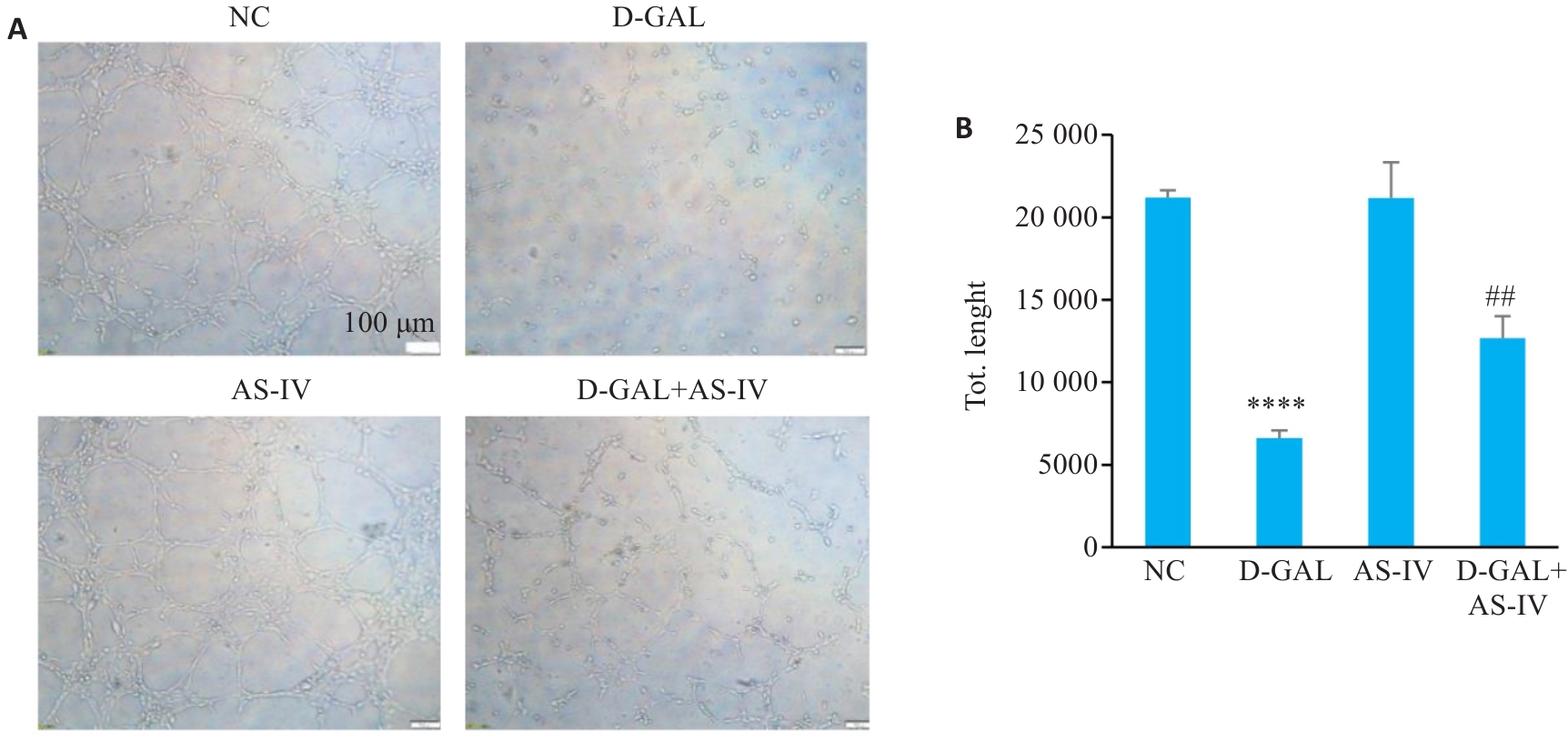
图2 AS-IV对D-GAL诱导的HUVEC管状结构形成能力的影响
Fig.2 Effect of AS-IV on tube formation ability of HUVEC induced by D-GAL. A: Tubule formation assay images of HUVEC cells after treatment with various groups. B: Statistical graph of the tubule formation assay (Scale bar=100 μm), ****P<0.0001 vs NC group, ##P<0.01 vs D-GAL group.

图3 AS-IV对D-GAL诱导的HUVEC细胞迁移能力影响
Fig.3 Effect of AS-IV on migration ability of D-GAL-induced HUVECs. A: Observation of migration of HUVECs after different treatments (Scale bar=200 μm). B: Wound healing rates of HUVECs with different treatments. ****P<0.0001 vs NC group; ###P<0.001 vs D-GAL group.
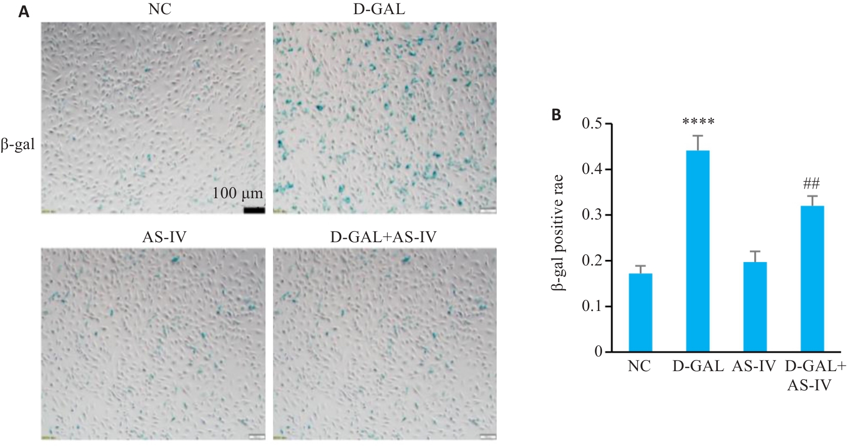
图4 AS-IV对D-GAL诱导的HUVEC细胞半乳糖染色阳性率影响
Fig.4 Effect of AS-IV on SA‑β‑Gal staining positivity rate in D-GAL-induced HUVECs. A: SA‑β-Gal staining of HUVEC cells with different treatments (Scale bar=100 μm). B: SA-β-Gal staining positiving rates of the cells. ****P<0.0001 vs NC group; ##P<0.01 vs D-GAL group.
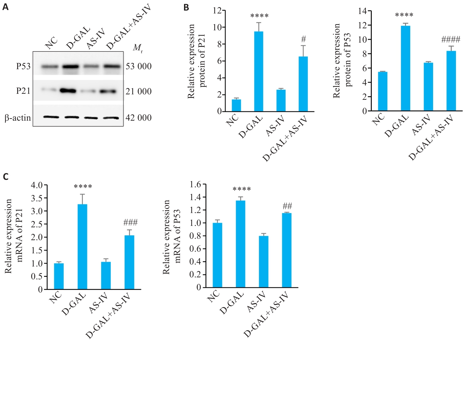
图5 AS-IV对D-GAL诱导的HUVEC细胞中P21和P53的表达影响
Fig.5 Effect of AS-IV on the Expression of P21 and P53 in D-GAL-Induced HUVEC Cells. A: Protein band diagram of P53 and P21 in four groups of cells. B, C: Statistical graph of P21 and P53 gene and protein expression.****P<0.0001 vs NC group; #P<0.05, ##P<0.01, ###P<0.001, ####P<0.0001 vs D-GAL group.
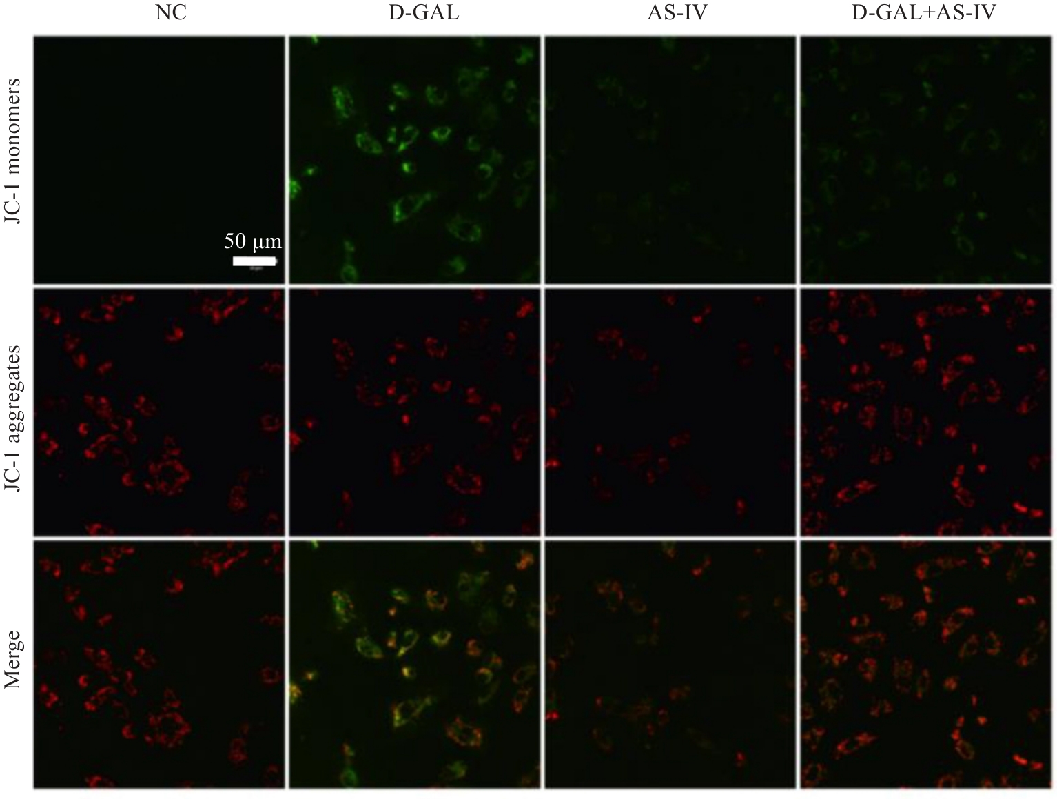
图6 AS-IV对D-GAL诱导的HUVEC细胞中线粒体膜电位影响
Fig.6 Effect of AS-IV on mitochondrial membrane potential in D-GAL-induced HUVECs (Scale bar=50 μm).
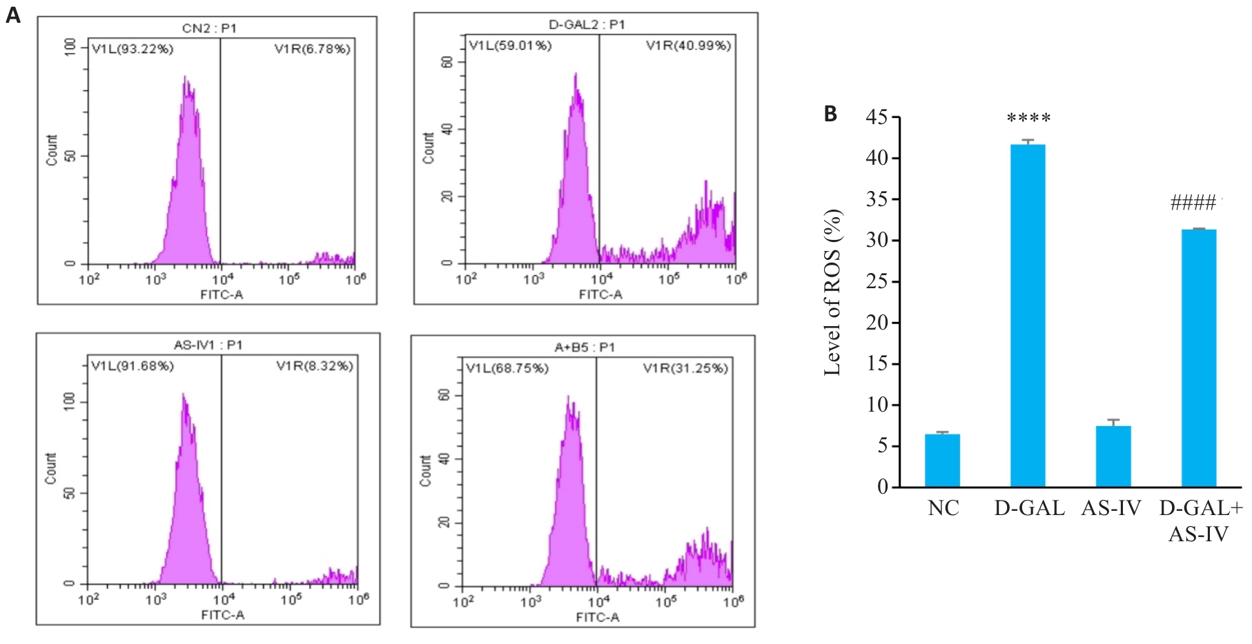
图7 AS-IV对D-GAL诱导的HUVEC细胞中ROS阳性细胞百分比影响
Fig.7 Effect of AS-IV on percentage of ROS-positive HUVECs induced by D-GAL. A: ROS levels in HUVEC cells with different treatments. B: Statistical graph of ROS levels. ****P<0.0001 vs NC group; ####P<0.0001 vs D-gal group.

图8 AS-IV对D-GAL诱导的HUVEC细胞中自噬体与溶酶体的融合及自噬流的影响
Fig.8 Effect of AS-IV on autophagosome-lysosome fusion and autophagy flux in D-GAL-induced HUVECs. A: Autophagy flux assay of HUVECs with different treatments (Scale bar=50 μm). B: Statistical graph of puncta counts. ****P<0.0001 vs control group; ####P<0.0001 vs D-GAL group.

图9 AS-IV对D-GAL诱导的HUVEC细胞中PINK1/Parkin通路及细胞自噬相关蛋白表达的影响
Fig.9 Effect of AS-IV on expression of PINK1/Parkin pathway and autophagy-related proteins in D-GAL-induced HUVECs. A: Protein bands of P62, LC3II/I, Beclin, PINK1, and Parkin in the 4 groups. B: Relative mRNA expressions of P62, Beclin, PINK, and Parkin. C: Statistical graph of grayscale values of the protein bands of P62, LC3II/I, Beclin, PINK1, and Parkin. *P<0.05, **P<0.01, ***P<0.001, ****P<0.0001 vs NC group; #P<0.05, ###P<0.001, ####P<0.0001 vs D-gal group.

图10 MTK458对D-GAL诱导的HUVEC细胞中自噬体与溶酶体的融合及自噬流的影响
Fig.10 Effect of MTK458 on autophagosome-lysosome fusion and autophagy flux in D-GAL-induced HUVECs. A: Autophagy flux assay of HUVECs with different treatments (Scale bar=50 μm). B: Statistical graph of puncta counts. ****P<0.0001 vs NC group; ####P<0.0001 vs D-gal group; △△△△P<0.0001 vs D-gal+AS-IV group.
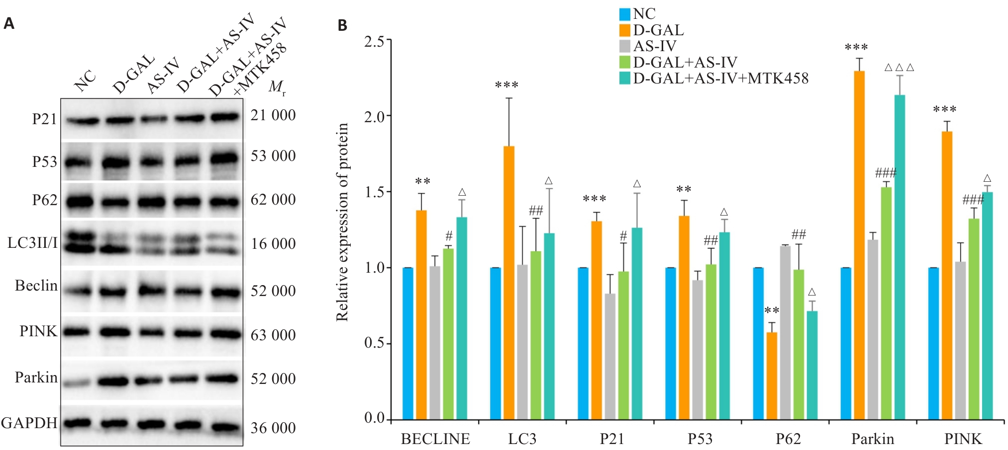
图11 MTK458对D-GAL诱导的HUVEC细胞中PINK1/Parkin通路及细胞衰老、自噬相关蛋白表达的影响
Fig.11 Effect of MTK458 on the PINK1/Parkin pathway and expressions of proteins related to cellular senescence and autophagy in D-GAL-induced HUVECs. A: Protein bands in the 5 groups of cells. B: Statistical graph of grayscale values of the protein bands. **P<0.01, ***P<0.001 vs NC group; #P<0.05, ##P<0.01, ###P<0.001 vs D-gal group; △P<0.05, △△△P<0.001 vs D-gal+AS-IV group.
| [1] | Campisi J, Andersen JK, Kapahi P, et al. Cellular senescence: a link between cancer and age-related degenerative disease[J]? Semin Cancer Biol, 2011, 21(6): 354-9. |
| [2] | Minamino T, Miyauchi H, Yoshida T, et al. Endothelial cell senescence in human atherosclerosis: role of telomere in endothelial dysfunction[J]. Circulation, 2002, 105(13): 1541-4. doi:10.1161/01.cir.0000013836.85741.17 |
| [3] | Bu LL, Yuan HH, Xie LL, et al. New dawn for atherosclerosis: vascular endothelial cell senescence and death[J]. Int J Mol Sci, 2023, 24(20): 15160. doi:10.3390/ijms242015160 |
| [4] | Bloom SI, Islam MT, Lesniewski LA, et al. Mechanisms and consequences of endothelial cell senescence[J]. Nat Rev Cardiol, 2023, 20(1): 38-51. doi:10.1038/s41569-022-00739-0 |
| [5] | Li AQ, Gao M, Liu BL, et al. Mitochondrial autophagy: molecular mechanisms and implications for cardiovascular disease[J]. Cell Death Dis, 2022, 13(5): 444. doi:10.1038/s41419-022-04906-6 |
| [6] | Narendra DP, Youle RJ. The role of PINK1-Parkin in mitochondrial quality control[J]. Nat Cell Biol, 2024, 26(10): 1639-51. doi:10.1038/s41556-024-01513-9 |
| [7] | 齐苗苗. 黄芪甲苷通过PINK1/Parkin介导线粒体自噬改善心肌细胞氧化应激损伤的研究[D]. 兰州: 兰州大学, 2021. |
| [8] | Yi SL, Zheng B, Zhu Y, et al. Melatonin ameliorates excessive PINK1/Parkin-mediated mitophagy by enhancing SIRT1 expression in granulosa cells of PCOS[J]. Am J Physiol Endocrinol Metab, 2020, 319(1): E91-E101. doi:10.1152/ajpendo.00006.2020 |
| [9] | Sun KY, Yang PY, Zhao R, et al. Matrine attenuates D-galactose-induced aging-related behavior in mice via inhibition of cellular senescence and oxidative stress[J]. Oxid Med Cell Longev, 2018, 2018: 7108604. doi:10.1155/2018/7108604 |
| [10] | Zeng M, He YL, Yang YL, et al. Mesenchymal stem cell-derived extracellular vesicles relieve endothelial cell senescence via recovering CTRP9 upon repressing miR-674-5p in atherosclerosis[J]. Regen Ther, 2024, 27: 354-64. doi:10.1016/j.reth.2024.03.027 |
| [11] | Chu QQ, Li YJ, Wu JC, et al. Oxysterol sensing through GPR183 triggers endothelial senescence in hypertension[J]. Circ Res, 2024, 135(7): 708-21. doi:10.1161/circresaha.124.324722 |
| [12] | Liu MM, Wang DN, Qi CY, et al. Brain ischemia causes systemic Notch1 activity in endothelial cells to drive atherosclerosis[J]. Immunity, 2024, 57(9): 2157-72. e7. doi:10.1016/j.immuni.2024.07.002 |
| [13] | Wang SS, Zhang X, Ke ZZ, et al. D-galactose-induced cardiac ageing: a review of model establishment and potential interventions[J]. J Cell Mol Med, 2022, 26(21): 5335-59. doi:10.1111/jcmm.17580 |
| [14] | Kumari R, Jat P. Mechanisms of cellular senescence: cell cycle arrest and senescence associated secretory phenotype[J]. Front Cell Dev Biol, 2021, 9: 645593. doi:10.3389/fcell.2021.645593 |
| [15] | Zhang WJ, Frei B. Astragaloside IV inhibits NF‑κB activation and inflammatory gene expression in LPS-treated mice[J]. Mediators Inflamm, 2015, 2015: 274314. doi:10.1155/2015/274314 |
| [16] | Xu CH, Tang FT, Lu ML, et al. Pretreatment with Astragaloside IV protects human umbilical vein endothelial cells from hydrogen peroxide induced oxidative stress and cell dysfunction via inhibiting ENOS uncoupling and NADPH oxidase-ROS-NF‑κB pathway[J]. Can J Physiol Pharmacol, 2016, 94(11): 1132-40. doi:10.1139/cjpp-2015-0572 |
| [17] | Sun YX, Ma YH, Sun FY, et al. Astragaloside IV attenuates lipopolysaccharide induced liver injury by modulating Nrf2-mediated oxidative stress and NLRP3-mediated inflammation[J]. Heliyon, 2023, 9(4): e15436. doi:10.1016/j.heliyon.2023.e15436 |
| [18] | Xiao XL, Zheng YL, Mo Y, et al. Astragaloside IV alleviates oxidative stress-related damage via inhibiting NLRP3 infla-mmasome in a MAPK signaling dependent pathway in human lens epithelial cells[J]. Drug Dev Res, 2022, 83(4): 1016-23. doi:10.1002/ddr.21929 |
| [19] | Qu CY, Tan XY, Hu QC, et al. A systematic review of astragaloside IV effects on animal models of diabetes mellitus and its complications[J]. Heliyon, 2024, 10(5): e26863. doi:10.1016/j.heliyon.2024.e26863 |
| [20] | Cheng SY, Zhang XX, Feng Q, et al. Astragaloside IV exerts angiogenesis and cardioprotection after myocardial infarction via regulating PTEN/PI3K/Akt signaling pathway[J]. Life Sci, 2019, 227: 82-93. doi:10.1016/j.lfs.2019.04.040 |
| [21] | Shen Q, Fang J, Guo HJ, et al. Astragaloside IV attenuates podocyte apoptosis through ameliorating mitochondrial dysfunction by up-regulated Nrf2-ARE/TFAM signaling in diabetic kidney disease[J]. Free Radic Biol Med, 2023, 203: 45-57. doi:10.1016/j.freeradbiomed.2023.03.022 |
| [22] | Liang YT, Chen BQ, Liang D, et al. Pharmacological effects of astragaloside IV: a review[J]. Molecules, 2023, 28(16): 6118. doi:10.3390/molecules28166118 |
| [23] | Wang SL, Long HJ, Hou LJ, et al. The mitophagy pathway and its implications in human diseases[J]. Signal Transduct Target Ther, 2023, 8(1): 304. doi:10.1038/s41392-023-01503-7 |
| [24] | Ajoolabady A, Chiong M, Lavandero S, et al. Mitophagy in cardiovascular diseases: molecular mechanisms, pathogenesis, and treatment[J]. Trends Mol Med, 2022, 28(10): 836-49. doi:10.1016/j.molmed.2022.06.007 |
| [25] | Xue A, Zhao DP, Zhao CY, et al. Study on the neuroprotective effect of Zhimu-Huangbo extract on mitochondrial dysfunction in HT22 cells induced by D-galactose by promoting mitochondrial autophagy[J]. J Ethnopharmacol, 2024, 318: 117012. doi:10.1016/j.jep.2023.117012 |
| [26] | Zhou XH, Tan BW, Gui WW, et al. IGF2 deficiency promotes liver aging through mitochondrial dysfunction and upregulated CEBPB signaling in D-galactose-induced aging mice[J]. Mol Med, 2023, 29(1): 161. doi:10.1186/s10020-023-00752-0 |
| [27] | Li H, Guan KF, Wang RC, et al. Synergistic effects of MFG-E8 and whey protein on mitigating d-galactose-induced sarcopenia through PI3K/AKT/PGC-1α and MAPK/ERK signaling pathways[J]. J Dairy Sci, 2024, 107(1): 9-23. doi:10.3168/jds.2023-23637 |
| [28] | Yang L, Shi J, Wang XW, et al. Curcumin alleviates D-galactose-induced cardiomyocyte senescence by promoting autophagy via the SIRT1/AMPK/mTOR pathway[J]. Evid Based Complement Alternat Med, 2022, 2022: 2990843. doi:10.1155/2022/2990843 |
| [29] | Li HJ, Xu JL, Zhang YN, et al. Astragaloside IV alleviates senescence of vascular smooth muscle cells through activating Parkin-mediated mitophagy[J]. Hum Cell, 2022, 35(6): 1684-96. doi:10.1007/s13577-022-00758-6 |
| [30] | Li J, Yang DM, Li ZP, et al. PINK1/Parkin-mediated mitophagy in neurodegenerative diseases[J]. Ageing Res Rev, 2023, 84: 101817. doi:10.1016/j.arr.2022.101817 |
| [31] | Cao YH, Chen X, Pan FQ, et al. Xinmaikang-mediated mitophagy attenuates atherosclerosis via the PINK1/Parkin signaling pathway[J]. Phytomedicine, 2023, 119: 154955. doi:10.1016/j.phymed.2023.154955 |
| [32] | Zhao XP, Wang Z, Wang LJ, et al. The PINK1/Parkin signaling pathway-mediated mitophagy: a forgotten protagonist in myocardial ischemia/reperfusion injury[J]. Pharmacol Res, 2024, 209: 107466. doi:10.1016/j.phrs.2024.107466 |
| [33] | He HL, Huang WY, Xiong LD, et al. FUNDC1-mediated mitophagy regulates photodamage independently of the PINK1/Parkin-dependent pathway[J]. Free Radic Biol Med, 2024, 225: 630-40. doi:10.1016/j.freeradbiomed.2024.10.272 |
| [34] | Chen CY, Bu XL, Deng LP, et al. Astragaloside IV as a promising therapeutic agent for liver diseases: current landscape and future perspectives[J]. Front Pharmacol, 2025, 16: 1574154. doi:10.3389/fphar.2025.1574154 |
| [35] | Liu T, Ai L, Jiang AB, et al. Astragaloside IV suppresses the proliferation and inflammatory response of human epidermal keratinocytes and ameliorates imiquimod-induced psoriasis-like skin damage in mice[J]. Allergol Immunopathol (Madr), 2024, 52(5): 44-50. doi:10.15586/aei.v52i5.1140 |
| [36] | Xia DQ, Li WJ, Tang C, et al. Astragaloside IV, as a potential anticancer agent[J]. Front Pharmacol, 2023, 14: 1065505. doi:10.3389/fphar.2023.1065505 |
| [37] | Deng LL. Astragaloside IV as potential antioxidant against diabetic ketoacidosis in juvenile mice through activating JNK/Nrf2 signaling pathway[J]. Arch Med Res, 2020, 51(7): 654-63. doi:10.1016/j.arcmed.2020.06.013 |
| [1] | 张玉, 胡音琦, 李佩佩, 时潇, 徐伟, 胡建鹏. 脑络欣通通过激活HIF-1α/VEGF信号通路促进氧糖剥夺/复氧复糖损伤后大鼠的脑微血管内皮细胞增殖[J]. 南方医科大学学报, 2025, 45(9): 1980-1988. |
| [2] | 常笑语, 张瀚文, 曹红亭, 侯玲, 孟鑫, 陶虹, 罗彦, 李光华. 热应激对大鼠胸主动脉内皮细胞生物钟基因 Bmal1和细胞周期蛋白表达水平的影响[J]. 南方医科大学学报, 2025, 45(7): 1353-1362. |
| [3] | 董妍妍, 张可敬, 储俊, 储全根. 抵当汤含药血清通过PI3K/Akt/mTOR信号通路增强高糖诱导的大鼠肾小球内皮细胞自噬[J]. 南方医科大学学报, 2025, 45(3): 461-469. |
| [4] | 廖茗, 钟文华, 张冉, 梁娟, 徐文陶睿, 万文珺, 吴超, 李曙. 源自蛇毒的蛋白C激活剂通过调控HIF-1α抑制BNIP3活性氧生成保护人脐静脉内皮细胞免受缺氧-复氧损伤[J]. 南方医科大学学报, 2025, 45(3): 614-621. |
| [5] | 郭克磊, 李颖利, 宣晨光, 侯紫君, 叶松山, 李林运, 陈丽平, 韩立, 卞华. 益气养阴化浊通络方通过调控miR-21a-5p/FoxO1/PINK1介导的线粒体自噬减轻糖尿病肾病小鼠的足细胞损伤[J]. 南方医科大学学报, 2025, 45(1): 27-34. |
| [6] | 展俊平, 黄硕, 孟庆良, 范围, 谷慧敏, 崔家康, 王慧莲. 缺氧微环境下补阳还五汤通过抑制BNIP3-PI3K/Akt通路抑制类风湿关节炎滑膜成纤维细胞的线粒体自噬[J]. 南方医科大学学报, 2025, 45(1): 35-42. |
| [7] | 廖阅, 马雪, 邓三春, 陈苏衡, 李玉兰. 过劳诱导小鼠血管内皮屏障功能障碍[J]. 南方医科大学学报, 2024, 44(9): 1814-1820. |
| [8] | 程瑶, 王远迎, 姚飞扬, 胡盼, 陈铭勰, 吴宁. 黄芩苷通过调控PI3K/AKT信号通路抑制登革病毒感染诱导的人静脉内皮细胞的自噬[J]. 南方医科大学学报, 2024, 44(7): 1272-1283. |
| [9] | 白钰婷, 刚保才, 张梦洁, 万子雨, 刘国权, 顾 玮. FAK抑制剂PF-562271减轻老化血小板诱导的人脐静脉内皮细胞损伤[J]. 南方医科大学学报, 2024, 44(2): 252-259. |
| [10] | 姜诚诚, 李洋洋, 段可欣, 詹婷婷, 陈子龙, 王永雪, 赵蕊, 马彩云, 郭俣, 刘长青. Parkin通过介导PINK 1/Parkin线粒体自噬信号通路加速小鼠帕金森病发展及加剧神经炎症发生[J]. 南方医科大学学报, 2024, 44(12): 2359-2366. |
| [11] | 苏晓飞, 李霖, 戴靖榕, 肖宝, 金子琦, 刘斌. PAD4抑制剂GSK484通过抑制H3Cit表达减轻小鼠脓毒症肺损伤后内皮功能障碍的发生[J]. 南方医科大学学报, 2024, 44(12): 2396-2403. |
| [12] | 王磊, 卞芬兰, 马飞扬, 方舒, 凌梓涵, 刘梦然, 孙红燕, 付程文, 倪诗垚, 赵晓阳, 冯心茹, 孙正宇, 卢国庆, 康品方, 吴士礼. 激活ALDH2通过上调SIRT1/PGC-1α信号通路减轻小鼠缺氧性肺动脉高压[J]. 南方医科大学学报, 2024, 44(10): 1955-1964. |
| [13] | 罗 瑞, 田龙海, 杨永曜. 高良姜素通过下调lncRNA H19的表达抑制ox-LDL诱导的人源正常主动脉内皮细胞血管生成活性[J]. 南方医科大学学报, 2024, 44(1): 52-59. |
| [14] | 于佳池, 李芮冰, 夏 天, 王佳楠, 金家丞, 袁漫秋, 李绵洋. 沉默PDCD4表达可减轻脓毒症血管内皮细胞损伤:基于改善线粒体动力学[J]. 南方医科大学学报, 2024, 44(1): 25-35. |
| [15] | 李洪涛, 邓 宇, 王添乐, 黄克勇, 于传沛, 陈朝俊. 丹参新醌乙减轻ox-LDL诱导的内皮细胞损伤:基于抑制NF-κB/NLRP3信号通路介导的细胞焦亡[J]. 南方医科大学学报, 2023, 43(8): 1425-1431. |
| 阅读次数 | ||||||
|
全文 |
|
|||||
|
摘要 |
|
|||||