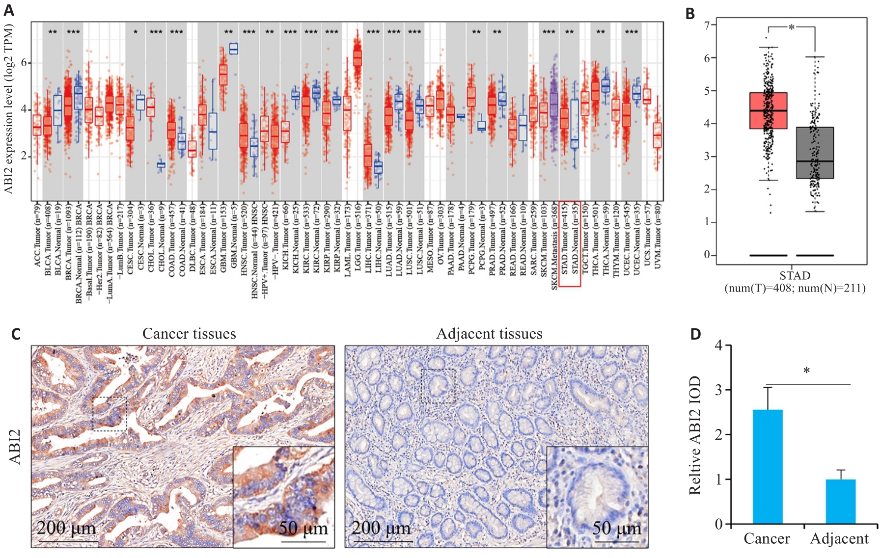
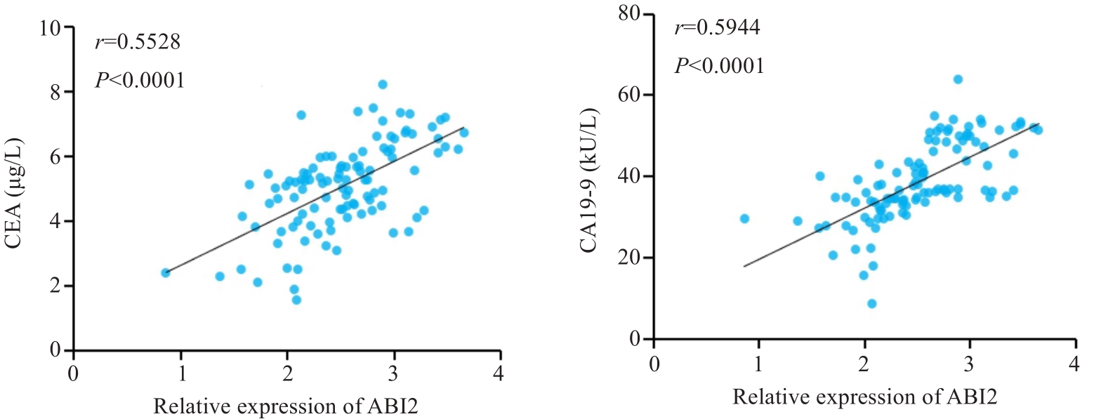
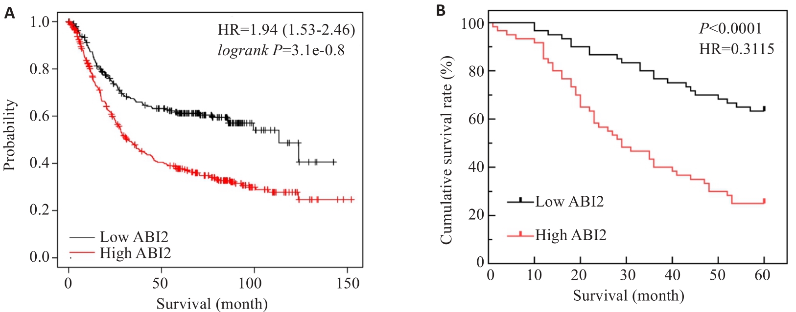
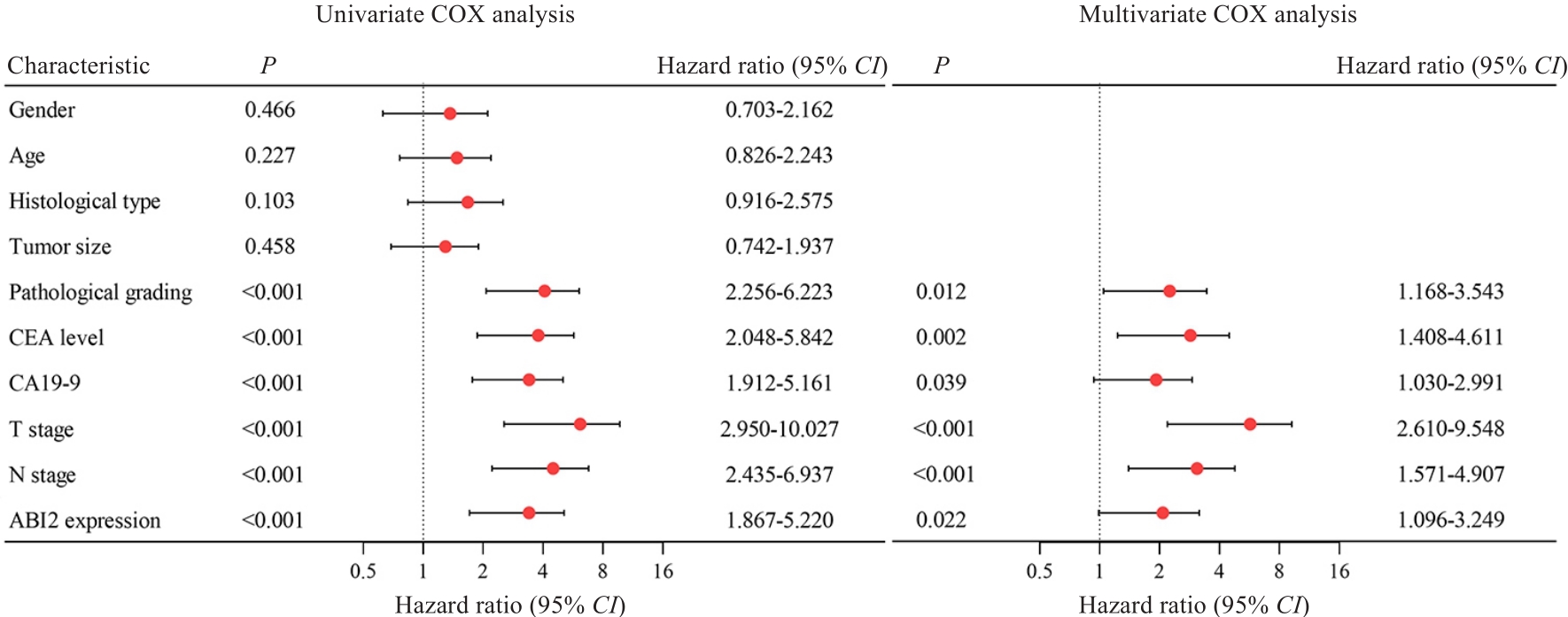
南方医科大学学报 ›› 2024, Vol. 44 ›› Issue (9): 1653-1661.doi: 10.12122/j.issn.1673-4254.2024.09.04
陈孝华1(), 鲁辉1, 王子良1, 王炼1, 夏勇生1, 耿志军2,4, 张小凤2,4, 宋雪2,4, 王月月3,4, 李静3,4, 胡建国3,4, 左芦根1,4(
)
收稿日期:
2024-04-22
出版日期:
2024-09-20
发布日期:
2024-09-30
通讯作者:
左芦根
E-mail:chenxiaohua9116@163.com;zuolugen@126.com
作者简介:
陈孝华,硕士,E-mail: chenxiaohua9116@163.com
基金资助:
Xiaohua CHEN1(), Hui LU1, Ziliang WANG1, Lian WANG1, Yongsheng XIA1, Zhijun GENG2,4, Xiaofeng ZHANG2,4, Xue SONG2,4, Yueyue WANG3,4, Jing LI3,4, Jianguo HU3,4, Lugen ZUO1,4(
)
Received:
2024-04-22
Online:
2024-09-20
Published:
2024-09-30
Contact:
Lugen ZUO
E-mail:chenxiaohua9116@163.com;zuolugen@126.com
摘要:
目的 探讨Abelson相互作用因子2(ABI2)在胃癌进展和预后中的作用及机制。 方法 通过TIMER2.0和GEPIA数据库分析ABI2在泛癌和胃癌中的表达,采用Kaplan-Meier Plotter数据库分析ABI2表达水平与胃癌预后的关系,通过DAVID数据库预测ABI2在生物系统中的功能。纳入在我院于2016年1月~2018年10月接受胃癌根治术治疗的120例胃癌患者,检测胃癌及癌旁组织中ABI2的表达水平,分析其与疾病进展的联系,并通过Cox单因素和多因素分析影响患者预后的危险因素。构建胃癌细胞系(MGC-803)ABI2干扰及过表达模型并构建裸鼠移植瘤模型,联合CCK-8、划痕、Transwell和免疫印迹实验分析ABI2对胃癌细胞增殖、迁移和侵袭的影响。结合体内外研究分析ABI2影响胃癌细胞恶性生物学行为的可能分子机理。 结果 生信分析和本院胃癌组织检测结果表明ABI2在胃癌组织中高表达(P<0.05)。Kaplan-Meier Plotter数据库分析表明ABI2低表达患者的术后生存率优于高表达患者(P<0.0001)。本院患者的生存分析结果显示,胃癌组织中ABI2高表达患者术后5年生存率降低(P<0.0001),Cox单因素和多因素生存分析进一步表明,ABI2高表达是影响胃癌患者预后的独立危险因素(P=0.022,HR=1.887,95% CI:1.096~3.249)。富集分析提示,ABI2的作用可能与Wnt信号通路相关。上调ABI2促进裸鼠移植瘤和胃癌细胞的增殖能力(P<0.05),并增加Vimentin和N-cadherin的表达,同时降低E-cadherin的表达(P<0.05);下调ABI2则相反(P<0.05)。机制分析发现上调ABI2促进移植瘤组织和胃癌细胞中Wnt2和β-catenin的表达(P<0.05),下调ABI2则相反(P<0.05)。 结论 ABI2在胃癌中表达升高并影响疾病远期预后,其可能与调控Wnt信号通路进而促进胃癌细胞增殖、迁移和侵袭能力有关。
陈孝华, 鲁辉, 王子良, 王炼, 夏勇生, 耿志军, 张小凤, 宋雪, 王月月, 李静, 胡建国, 左芦根. ABI2在胃癌进展和预后中的作用及其调控机制[J]. 南方医科大学学报, 2024, 44(9): 1653-1661.
Xiaohua CHEN, Hui LU, Ziliang WANG, Lian WANG, Yongsheng XIA, Zhijun GENG, Xiaofeng ZHANG, Xue SONG, Yueyue WANG, Jing LI, Jianguo HU, Lugen ZUO. Role of Abelson interactor 2 in progression and prognosis of gastric cancer and its regulatory mechanisms[J]. Journal of Southern Medical University, 2024, 44(9): 1653-1661.
图1 ABI2在胃癌组织中表达水平升高
Fig.1 ABI2 is highly expressed in gastric cancer. A: Pan-cancer expression analysis. B: Expression of ABI2 in gastric and adjacent tissues. C, D: Immunohistochemical staining of ABI2 in gastric and adjacent tissues. *P<0.05, **P<0.01, ***P<0.001.
Factors | n | ABI2 expression (n, %) | χ2 | Ρ | ||
---|---|---|---|---|---|---|
Low (n=60) | High (n=60) | |||||
Gender | Male | 95 | 47 (49.47%) | 48 (50.53%) | 0.051 | 0.822 |
Female | 25 | 13 (52.00%) | 12 (48.00%) | |||
Age (year) | <60 | 49 | 27 (55.10%) | 22 (44.90%) | 0.862 | 0.353 |
≥60 | 71 | 33 (46.48%) | 38 (53.52%) | |||
CEA (μg/L) | <5 | 57 | 35 (61.40%) | 22 (38.60%) | 5.647 | 0.017 |
≥5 | 63 | 25 (39.68%) | 38 (60.32%) | |||
CA19-9 (kU/L) | <37 | 67 | 44 (65.67%) | 23 (34.33%) | 14.903 | <0.001 |
≥37 | 53 | 16 (30.19%) | 37 (69.81%) | |||
Tumor size (cm) | <5 | 64 | 33 (51.56%) | 31 (48.44%) | 0.134 | 0.714 |
≥5 | 56 | 27 (48.21%) | 29 (51.79%) | |||
Cancer cell type | Adenocarcinoma | 89 | 46 (51.69%) | 43 (48.31%) | 0.391 | 0.532 |
Other | 31 | 14 (45.16%) | 17 (54.84%) | |||
Pathological grading | G1-G2 | 66 | 41 (62.12%) | 25 (37.88%) | 8.620 | 0.003 |
G3-G4 | 54 | 19 (35.19%) | 35 (64.81%) | |||
T stage | T1-T2 | 51 | 33 (64.71%) | 18 (35.29%) | 7.673 | 0.006 |
T3-T4 | 69 | 27 (39.13%) | 42 (60.87%) | |||
N stage | N0-N1 | 60 | 36 (60.00%) | 24 (40.00%) | 4.800 | 0.028 |
N2-N3 | 60 | 24 (40.00%) | 36 (60.00%) |
表1 胃癌组织中ABI2的表达量与胃癌患者临床及病理参数间的关系
Tab.1 Correlation of ABI2 expression level with clinicopathological parameters of gastric cancer patients
Factors | n | ABI2 expression (n, %) | χ2 | Ρ | ||
---|---|---|---|---|---|---|
Low (n=60) | High (n=60) | |||||
Gender | Male | 95 | 47 (49.47%) | 48 (50.53%) | 0.051 | 0.822 |
Female | 25 | 13 (52.00%) | 12 (48.00%) | |||
Age (year) | <60 | 49 | 27 (55.10%) | 22 (44.90%) | 0.862 | 0.353 |
≥60 | 71 | 33 (46.48%) | 38 (53.52%) | |||
CEA (μg/L) | <5 | 57 | 35 (61.40%) | 22 (38.60%) | 5.647 | 0.017 |
≥5 | 63 | 25 (39.68%) | 38 (60.32%) | |||
CA19-9 (kU/L) | <37 | 67 | 44 (65.67%) | 23 (34.33%) | 14.903 | <0.001 |
≥37 | 53 | 16 (30.19%) | 37 (69.81%) | |||
Tumor size (cm) | <5 | 64 | 33 (51.56%) | 31 (48.44%) | 0.134 | 0.714 |
≥5 | 56 | 27 (48.21%) | 29 (51.79%) | |||
Cancer cell type | Adenocarcinoma | 89 | 46 (51.69%) | 43 (48.31%) | 0.391 | 0.532 |
Other | 31 | 14 (45.16%) | 17 (54.84%) | |||
Pathological grading | G1-G2 | 66 | 41 (62.12%) | 25 (37.88%) | 8.620 | 0.003 |
G3-G4 | 54 | 19 (35.19%) | 35 (64.81%) | |||
T stage | T1-T2 | 51 | 33 (64.71%) | 18 (35.29%) | 7.673 | 0.006 |
T3-T4 | 69 | 27 (39.13%) | 42 (60.87%) | |||
N stage | N0-N1 | 60 | 36 (60.00%) | 24 (40.00%) | 4.800 | 0.028 |
N2-N3 | 60 | 24 (40.00%) | 36 (60.00%) |
图2 胃癌组织中ABI2表达水平与外周血CEA和CA19-9水平呈正相关
Fig.2 ABI2 expression level in gastric cancer tissue is positively correlated with peripheral blood levels of CEA and CA19-9.
图3 胃癌组织中ABI2表达水平影响胃癌患者预后
Fig.3 ABI2 expression level in gastric cancer tissues affects prognosis of the patients. A: ABI2 expression level affects overall survival of gastric cancer patients. B: Kaplan-Meier survival analysis of 5-year survival rate of the patients after radical gastrectomy.
图5 ABI2的KEGG和GO富集分析结果
Fig.5 Results of KEGG and GO enrichment analysis. A: KEGG enrichment analysis. B-D: Biological processes, cellular component and molecular function in GO enrichment analysis.
图6 ABI2在体外实验中促进胃癌细胞增殖、迁移、侵袭和上皮间充质转化
Fig.6 ABI2 overexpression promotes proliferation, migration, invasion, and epithelial-mesenchymal transition (EMT) of gastric cancer cells in vitro. A, B: Lentiviral transfection efficiency. C, D: Results of CCK-8 assay (presented as A values) from day 1 to day 5. E, F: Expression of EMT markers in MGC803 cells. G, H: Wound-healing assay of the cells. I-L: Migration and invasion of MGC803 cells. Si: siRNA; LV: overexpression. *P<0.05 vs Control (n=3).
图7 ABI2在体内实验中促进胃癌细胞增殖和上皮间充质转化
Fig.7 ABI2 overexpression promotes proliferation and epithelial-mesenchymal transition of gastric cancer cells in nude mice. A: Representative image of the transplanted tumors. B, C: Transplanted tumor volume from day 5 to day 25. D, E: Expression of EMT markers in the transplanted tumor. *P<0.05 vs Control (n=3).
图8 ABI2对胃癌细胞恶性生物学行为的影响可能与Wnt通路相关
Fig.8 The effect of ABI2 on malignant biological behaviors of gastric cancer cells is related to the Wnt signaling pathway. A, B: Expression of Wnt2 and β-catenin in MGC803 cells. C, D: Expression of Wnt2 and β-catenin in the transplanted tumors. *P<0.05 vs Control (n=3).
1 | Smyth EC, Nilsson M, Grabsch HI, et al. Gastric cancer[J]. Lancet, 2020, 396(10251): 635-48. |
2 | 郑荣寿, 张思维, 孙可欣, 等. 2016年中国恶性肿瘤流行情况分析[J]. 中华肿瘤杂志, 2023, 45(3): 212-20. DOI: 10.3760/cma.j.cn112152-20220922-00647 |
3 | Thrift AP, El-Serag HB. Burden of gastric cancer[J]. Clin Gastroenterol Hepatol, 2020, 18(3): 534-42. |
4 | Chia NY, Tan P. Molecular classification of gastric cancer[J]. Ann Oncol, 2016, 27(5): 763-9. |
5 | Jiang PX, Tang SN, Hudgins H, et al. The Abl/Abi signaling links WAVE regulatory complex to Cbl E3 ubiquitin ligase and is essential for breast cancer cell metastasis[J]. Neoplasia, 2022, 32: 100819. |
6 | Ichigotani Y, Fujii K, Hamaguchi M, et al. In search of a function for the E3B1/Abi2/Argbp1/NESH family (Review)[J]. Int J Mol Med, 2002, 9(6): 591-5. |
7 | Zipfel PA, Bunnell SC, Witherow DS, et al. Role for the Abi/wave protein complex in T cell receptor-mediated proliferation and cytoskeletal remodeling[J]. Curr Biol, 2006, 16(1): 35-46. |
8 | Courtney KD, Grove M, Vandongen H, et al. Localization and phosphorylation of Abl-interactor proteins, Abi-1 and Abi-2, in the developing nervous system[J]. Mol Cell Neurosci, 2000, 16(3): 244-57. |
9 | Hirao N, Sato S, Gotoh T, et al. NESH (Abi-3) is present in the Abi/WAVE complex but does not promote c-Abl-mediated phosphorylation[J]. FEBS Lett, 2006, 580(27): 6464-70. |
10 | Li YZ, Clough N, Sun XL, et al. Bcr-Abl induces abnormal cytoskeleton remodeling, beta1 integrin clustering and increased cell adhesion to fibronectin through the Abl interactor 1 pathway[J]. J Cell Sci, 2007, 120(Pt 8): 1436-46. |
11 | Ryu JR, Echarri A, Li R, et al. Regulation of cell-cell adhesion by Abi/Diaphanous complexes[J]. Mol Cell Biol, 2009, 29(7): 1735-48. |
12 | Stradal T, Courtney KD, Rottner K, et al. The Abl interactor proteins localize to sites of actin polymerization at the tips of lamellipodia and filopodia[J]. Curr Biol, 2001, 11(11): 891-5. |
13 | Jensen CC, Clements AN, Liou H, et al. PIM1 phosphorylates ABI2 to enhance actin dynamics and promote tumor invasion[J]. J Cell Biol, 2023, 222(6): e202208136. |
14 | Chen JD, Li HZ, Zhang B, et al. ABI2-mediated MEOX2/KLF4-NANOG axis promotes liver cancer stem cell and drives tumour recurrence[J]. Liver Int, 2022, 42(11): 2562-76. |
15 | Guan WL, He Y, Xu RH. Gastric cancer treatment: recent progress and future perspectives[J]. J Hematol Oncol, 2023, 16(1): 57. |
16 | Wang B, Mysliwiec T, Krainc D, et al. Identification of ArgBP1, an Arg protein tyrosine kinase binding protein that is the human homologue of a CNS-specific Xenopus gene[J]. Oncogene, 1996, 12(9): 1921-9. |
17 | Dai Z, Pendergast AM. Abi-2, a novel SH3-containing protein interacts with the c-Abl tyrosine kinase and modulates c-Abl transforming activity[J]. Genes Dev, 1995, 9(21): 2569-82. |
18 | Grove M, Demyanenko G, Echarri A, et al. ABI2-deficient mice exhibit defective cell migration, aberrant dendritic spine morphogenesis, and deficits in learning and memory[J]. Mol Cell Biol, 2004, 24(24): 10905-22. |
19 | Chen ZC, Borek D, Padrick SB, et al. Structure and control of the actin regulatory WAVE complex[J]. Nature, 2010, 468(7323): 533-8. |
20 | Shami Shah A, Batrouni AG, Kim D, et al. PLEKHA4/kramer attenuates dishevelled ubiquitination to modulate Wnt and planar cell polarity signaling[J]. Cell Rep, 2019, 27(7): 2157-70. e8. |
21 | Nusse R, Clevers H. Wnt/β-catenin signaling, disease, and emerging therapeutic modalities[J]. Cell, 2017, 169(6): 985-99. |
22 | Zhao H, Ming TQ, Tang S, et al. Wnt signaling in colorectal cancer: pathogenic role and therapeutic target[J]. Mol Cancer, 2022, 21(1): 144. |
23 | Li HJ, Ke FY, Lin CC, et al. ENO1 promotes lung cancer metastasis via HGFR and WNT signaling-driven epithelial-to-mesenchymal transition[J]. Cancer Res, 2021, 81(15): 4094-109. |
24 | Hiremath IS, Goel A, Warrier S, et al. The multidimensional role of the Wnt/β‑catenin signaling pathway in human malignancies[J]. J Cell Physiol, 2022, 237(1): 199-238. |
25 | Rim EY, Clevers H, Nusse R. The Wnt pathway: from signaling mechanisms to synthetic modulators[J]. Annu Rev Biochem, 2022, 91: 571-98. |
26 | Albrecht LV, Tejeda-Muñoz N, de Robertis EM. Cell biology of canonical Wnt signaling[J]. Annu Rev Cell Dev Biol, 2021, 37: 369-89. |
27 | Cheng XX, Wang ZC, Chen XY, et al. Frequent loss of membranous E-cadherin in gastric cancers: a cross-talk with Wnt in determining the fate of beta-catenin[J]. Clin Exp Metastasis, 2005, 22(1): 85-93. |
28 | Guo Q, Xu J, Huang Z, et al. ADMA mediates gastric cancer cell migration and invasion via Wnt/β‑catenin signaling pathway[J]. Clin Transl Oncol, 2021, 23(2): 325-34. |
29 | Wang J, Cai H, Liu QL, et al. Cinobufacini inhibits colon cancer invasion and metastasis via suppressing Wnt/β‑catenin signaling pathway and EMT[J]. Am J Chin Med, 2020, 48(3): 703-18. |
30 | Park JK, Song JH, He TC, et al. Overexpression of Wnt-2 in colorectal cancers[J]. Neoplasma, 2009, 56(2): 119-23. |
31 | Shi YH, He B, Kuchenbecker KM, et al. Inhibition of Wnt-2 and galectin-3 synergistically destabilizes beta-catenin and induces apoptosis in human colorectal cancer cells[J]. Int J Cancer, 2007, 121(6): 1175-81. |
32 | Vider BZ, Zimber A, Chastre E, et al. Evidence for the involvement of the Wnt 2 gene in human colorectal cancer[J]. Oncogene, 1996, 12(1): 153-8. |
33 | Katoh M. WNT2 and human gastrointestinal cancer (review)[J]. Int J Mol Med, 2003, 12(5): 811-6. |
34 | Kramer N, Schmöllerl J, Unger C, et al. Autocrine WNT2 signaling in fibroblasts promotes colorectal cancer progression[J]. Oncogene, 2017, 36(39): 5460-72. |
35 | Lei L, Wang Y, Li ZH, et al. PHLDA3 promotes lung adenocarcinoma cell proliferation and invasion via activation of the Wnt signaling pathway[J]. Lab Invest, 2021, 101(9): 1130-41. |
36 | Pu P, Zhang Z, Kang C, et al. Downregulation of Wnt2 and beta-catenin by siRNA suppresses malignant glioma cell growth[J]. Cancer Gene Ther, 2009, 16(4): 351-61. |
[1] | 薛良军, 谈秋瑜, 许静文, 冯璐, 李文锦, 颜亮, 李玉磊. MiR-6838-5p过表达下调DDR1基因表达抑制乳腺癌MCF-7细胞的增殖[J]. 南方医科大学学报, 2024, 44(9): 1677-1684. |
[2] | 叶梦楠, 武鸿美, 梅琰, 张庆玲. CREM在胃癌中高表达并与患者的不良预后相关[J]. 南方医科大学学报, 2024, 44(9): 1776-1782. |
[3] | 耿志军, 杨晶晶, 牛民主, 刘馨悦, 施金冉, 刘亦珂, 姚新宇, 张雨路, 张小凤, 胡建国. 桑黄酮G通过调控PI3K/AKT/mTOR通路抑制胃癌细胞的生长、迁移和侵袭[J]. 南方医科大学学报, 2024, 44(8): 1476-1484. |
[4] | 庞一丹, 刘雅, 陈思嫒, 张荆雷, 曾今, 潘元明, 安娟. SPAG5在胃癌细胞恶性增殖中的生物学作用[J]. 南方医科大学学报, 2024, 44(8): 1497-1507. |
[5] | 从小凡, 陈腾, 李硕, 王媛媛, 周龙云, 李小龙, 张配, 孙小锦, 赵素容. 双氢青蒿素通过促进活性氧的产生增强鼻咽癌细胞对顺铂诱导凋亡的敏感性[J]. 南方医科大学学报, 2024, 44(8): 1553-1560. |
[6] | 柯志勇, 黄子城, 何若琳, 张倩, 陈思旭, 崔忠凯, 丁晶. 抑制Hmga2促进小鼠脂肪间充质干细胞成骨分化并加速骨缺损修复[J]. 南方医科大学学报, 2024, 44(7): 1227-1235. |
[7] | 郑孟冬, 刘妍, 刘娇娇, 康巧珍, 王婷. 蛋白4.1R对肝细胞HL-7702增殖、凋亡以及糖酵解的影响[J]. 南方医科大学学报, 2024, 44(7): 1355-1360. |
[8] | 何华星, 刘璐琳, 刘颖茵, 陈纳川, 孙素霞. 丁酸钠与索拉非尼可能通过YAP诱导铁死亡协同抑制肝癌细胞增殖[J]. 南方医科大学学报, 2024, 44(7): 1425-1430. |
[9] | 房锦存, 刘立威, 林俊豪, 陈逢生. CDHR2过表达通过抑制PI3K/Akt通路抑制乳腺癌细胞增殖[J]. 南方医科大学学报, 2024, 44(6): 1117-1125. |
[10] | 崔芝, 马萃娇, 王倩茹, 陈金豪, 严子阳, 杨建林, 吕亚丰, 曹春雨. 表达 TGF-βⅡ受体的腺相关病毒载体抑制小鼠三阴性乳腺癌4T1细胞的增殖和肺转移[J]. 南方医科大学学报, 2024, 44(5): 818-826. |
[11] | 刘鹏程, 娄丽娟, 刘霞, 王建, 姜颖. M2巨噬细胞特征基因风险评分能准确预测HBV相关肝细胞癌患者的预后[J]. 南方医科大学学报, 2024, 44(5): 827-840. |
[12] | 夏勇生, 王炼, 陈孝华, 张雨路, 孙奥飞, 陈德利. 过表达TSR2通过下调PI3K/AKT信号通路抑制胃癌细胞的增殖和侵袭[J]. 南方医科大学学报, 2024, 44(5): 913-919. |
[13] | 杨晶晶, 殷丽霞, 段婷, 牛民主, 何震东, 陈心蕊, 张小凤, 李静, 耿志军, 左芦根. 胃癌组织中高表达ATP5A1与患者术后的不良预后和肿瘤细胞的糖代谢有关[J]. 南方医科大学学报, 2024, 44(5): 974-980. |
[14] | 高志强, 林 洁, 洪 鹏, 胡再宏, 董军君, 石秦林, 田小毛, 刘 丰, 魏光辉. 基于高通量 RNA 测序分析 Wilms 瘤中关键基因对预后及免疫应答的影响[J]. 南方医科大学学报, 2024, 44(4): 727-738. |
[15] | 黄秋虎, 周 建, 王子珍, 杨 堃, 陈政纲. miR-26b-3p 靶向 CREB1 调控神经胶质瘤细胞的增殖、迁移及侵袭[J]. 南方医科大学学报, 2024, 44(3): 578-584. |
阅读次数 | ||||||
全文 |
|
|||||
摘要 |
|
|||||