南方医科大学学报 ›› 2024, Vol. 44 ›› Issue (7): 1227-1235.doi: 10.12122/j.issn.1673-4254.2024.07.02
柯志勇1( ), 黄子城1(), 何若琳1, 张倩1, 陈思旭1, 崔忠凯1(), 丁晶2()
收稿日期:2024-05-31
出版日期:2024-07-20
发布日期:2024-07-25
通讯作者:
崔忠凯,丁晶
E-mail:kezhy@smu.edu.cn;huangzicheng2@qq.com;zhongkaicui@smu.edu.cn;doctor2049@sina.com
作者简介:柯志勇,硕士,高级实验师,E-mail: kezhy@smu.edu.cn基金资助:
Zhiyong KE1(), Zicheng HUANG1(), Ruolin HE1, Qian ZHANG1, Sixu CHEN1, Zhong-Kai CUI1(), Jing DING2()
Received:2024-05-31
Online:2024-07-20
Published:2024-07-25
Contact:
Zhong-Kai CUI, Jing DING
E-mail:kezhy@smu.edu.cn;huangzicheng2@qq.com;zhongkaicui@smu.edu.cn;doctor2049@sina.com
Supported by:摘要:
目的 探讨高迁移率族蛋白A2(HMGA2)在脂肪间充质干细胞(ADSCs)成骨分化进程中的作用及其在骨缺损修复中的应用。 方法 通过GEO数据库和Rstudio软件,挖掘出在ADSCs“成脂-成骨”分化平衡中的关键节点因子HMGA2,并通过在线蛋白互作网络分析工具String和绘图软件Cytoscape,绘制HMGA2在成骨分化中的互作关系网络,预测其下游作用靶点。设计Hmga2 siRNA并转染小鼠原代脂肪间充质干细胞(mADSCs),诱导其体外成骨分化,在不同时间点(Day 3,Day 7,Day 14)收集样本,通过碱性磷酸酶染色和茜素红染色评估成骨分化能力,并通过RT-qPCR和Western blotting检测成骨特异性标志物Runt相关转录因子2(RUNX2)、骨桥蛋白(OPN)和骨钙素(OCN)的表达。将敲低 Hmga2 的mADSCs移植至小鼠不可自愈合颅骨缺损处,术后6周通过μCT扫描、骨组织学染色检测成骨标志物,评价骨缺损修复效果。 结果 GEO数据库分析结果显示HMGA2在ADSCs成脂分化进程中表达上调。蛋白互作网络分析提示在ADSCs成骨分化中,HMGA2的潜在作用靶点包括SMAD7、CDH1、CDH2、SNAI1、SMAD9、IGF2BP3、ALDH1A1。抑制Hmga2后,mADSCs中成骨分化相关标志物RUNX2、OPN和OCN的表达显著上调,且碱性磷酸酶的表达和钙结节的形成增加(P<0.05)。在小鼠颅骨缺损模型中,敲低Hmga2促进了骨缺损部位的新骨形成(P<0.05)。 结论 HMGA2是调控ADSCs成骨分化的重要因子,抑制Hmga2能显著促进ADSCs成骨分化,并加速体内骨缺损的修复。
柯志勇, 黄子城, 何若琳, 张倩, 陈思旭, 崔忠凯, 丁晶. 抑制Hmga2促进小鼠脂肪间充质干细胞成骨分化并加速骨缺损修复[J]. 南方医科大学学报, 2024, 44(7): 1227-1235.
Zhiyong KE, Zicheng HUANG, Ruolin HE, Qian ZHANG, Sixu CHEN, Zhong-Kai CUI, Jing DING. Hmga2 knockdown enhances osteogenic differentiation of adipose-derived mesenchymal stem cells and accelerates bone defect healing in mice[J]. Journal of Southern Medical University, 2024, 44(7): 1227-1235.
| Gene | Sequence | |
|---|---|---|
| Hmga2 | Forward | CCGGTAGAGGCAGTGGTAGC |
| Reverse | GGTTGTTCCCTGGGCTGATGT | |
| Alp | Forward | GAGCAGGAACAGAAGTTTGC |
| Reverse | GTTGCAGGGTCTGGAGAGTA | |
| Osx | Forward | GCCGCTTTGTGCCTTTGAAATG |
| Reverse | CGTTATGCTCTTCCCAGACTCC | |
| Runx2 | Forward | CCGCACGACAACCGCACCAT |
| Reverse | CGCTCCGGCCCACAAATCTC | |
| Opn | Forward | CCCTCGATGTCATCCCTGTT |
| Reverse | CCCTTTCCGTTGTTGTCCTG | |
| Ocn | Forward | AGCTCAACCCCAATTGTGAC |
| Reverse | AGCTGTGCCGTCCATACTTT | |
| Dlk1 | Forward | GCGGGAACGCAACAACATC |
| Reverse | GTCACTGGTCAACTCCAGCAC | |
| Pparγ | Forward | GTGATGGAAGACCACTCGCATT |
| Reverse | CCATGAGGGAGTTAGAAGGTTC |
表1 RT-qPCR 所用引物序列
Tab.1 Primer sequences for RT-qPCR
| Gene | Sequence | |
|---|---|---|
| Hmga2 | Forward | CCGGTAGAGGCAGTGGTAGC |
| Reverse | GGTTGTTCCCTGGGCTGATGT | |
| Alp | Forward | GAGCAGGAACAGAAGTTTGC |
| Reverse | GTTGCAGGGTCTGGAGAGTA | |
| Osx | Forward | GCCGCTTTGTGCCTTTGAAATG |
| Reverse | CGTTATGCTCTTCCCAGACTCC | |
| Runx2 | Forward | CCGCACGACAACCGCACCAT |
| Reverse | CGCTCCGGCCCACAAATCTC | |
| Opn | Forward | CCCTCGATGTCATCCCTGTT |
| Reverse | CCCTTTCCGTTGTTGTCCTG | |
| Ocn | Forward | AGCTCAACCCCAATTGTGAC |
| Reverse | AGCTGTGCCGTCCATACTTT | |
| Dlk1 | Forward | GCGGGAACGCAACAACATC |
| Reverse | GTCACTGGTCAACTCCAGCAC | |
| Pparγ | Forward | GTGATGGAAGACCACTCGCATT |
| Reverse | CCATGAGGGAGTTAGAAGGTTC |
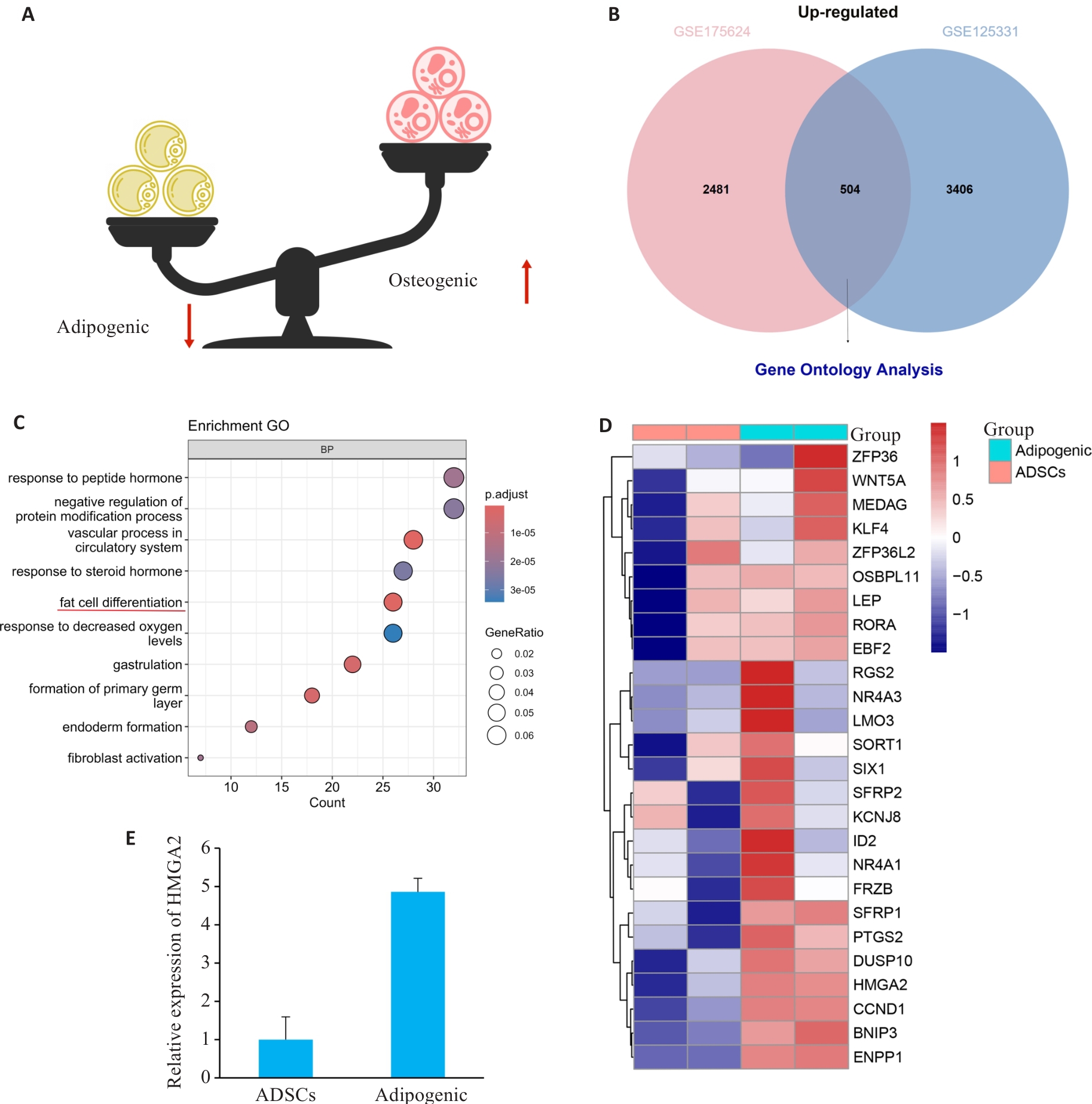
图1 hADSCs成脂分化进程中的差异基因表达情况
Fig.1 Differential gene expression profile during adipogenic differentiation of human ADSCs. A: Schematic illustration of the balance between adipogenic and osteogenic differentiation. B: Venn plot of up-regulated genes in the GSE175624 and GSE125331 datasets. C: Bubble chart of GO pathway enrichment analysis of the differentially expressed genes. D: Heatmap of the differentially expressed genes under the "fat cell differentiation" term. E: Expression of HMGA2 in GSE175614 dataset.

图2 抑制Hmga2在体外促进ADSCs成骨分化
Fig.2 Hmga2 knockdown promotes osteogenic differentiation of mouse ADSCs in vitro. A: si-Hmga2 inhibits Hmga2 mRNA expression in ADSCs. B-E: ALP staining and activity assay of mouse ADSCs on day 3 (B, C) and 7 (D, E) of osteogenic induction. F, G: ARS staining and quantification of calcium deposition in ADSCs on day 14 of osteogenic induction. *P<0.05, ***P<0.001 vs si-NC group. Scale bar: 500 μm.

图3 抑制Hmga2对ADSCs成骨成脂分化标志的影响
Fig.3 Effects of Hmga2 knockdown on expressions of osteogenic and adipogenic markers of ADSCs. A-C: Relative expression levels of Osx, Pparγ, and Dlk1 mRNAs in ADSCs on day 3 of osteogenic induction. D-F:Expressions of Runx2 and Opn mRNA and proteins on day 7. G: Expressions of OCN protein on day 7. *P<0.05, **P<0.01, ***P<0.001, ****P <0.0001 vs si-NC group.

图4 HMGA2影响ADSCs成骨分化的作用机制图
Fig.4 Mechanistic illustration of the role of HMGA2 in osteogenic differentiation of ADSCs.

图5 术后6周小鼠颅骨μCT三维重建图及骨计量学分析
Fig.5 Three-dimensional micro-CT reconstruction and histomorphometric analysis bone defect repair in mice at 6 weeks after surgery. A: Reconstructed micro-CT images. B: Morphometric analyses of bone regeneration in calvarial defects by assessing relative bone growth surface area, bone volume/tissue volume (BV/TV%), trabecular number (Tb.N) and trabecular thickness (Tb.Th). **P<0.01, ***P<0.001, ****P<0.0001 vs Blank group.

图6 术后6周小鼠颅骨缺损骨再生的组织学HE和Masson染色
Fig.6 HE and Masson's trichrome staining of bone regeneration in the calvarial defects at 6 weeks after surgery. A, C: HE staining overview image (scale bar: 500 μm) and magnified image (scale bar: 100 μm). B, D: Masson's trichrome staining overview image (scale bar: 500 μm) and magnified image (scale bar: 100 μm).

图7 HMGA2的蛋白互作网络分析
Fig.7 Protein-protein interaction (PPI) network analysis of HMGA2. A: PPI networks after cluster analyses. B: Predicted HMGA2 direct interacting proteins.
| Gene | Combined score | Experimentally determined interaction | Coexpression |
|---|---|---|---|
| IGF2BP3 0.909 0.292 0.257 | |||
| SMAD9 | 0.816 | 0.294 | 0.067 |
| SNAI1 | 0.728 | 0 | 0.042 |
| SMAD7 | 0.68 | 0.045 | 0.055 |
| CDH1 | 0.593 | 0.059 | 0 |
| ALDH1A1 | 0.569 | 0 | 0 |
| CDH2 | 0.538 | 0.095 | 0.097 |
表2 HMGA2互作蛋白评分
Tab.2 HMGA2-interacting protein possibility score
| Gene | Combined score | Experimentally determined interaction | Coexpression |
|---|---|---|---|
| IGF2BP3 0.909 0.292 0.257 | |||
| SMAD9 | 0.816 | 0.294 | 0.067 |
| SNAI1 | 0.728 | 0 | 0.042 |
| SMAD7 | 0.68 | 0.045 | 0.055 |
| CDH1 | 0.593 | 0.059 | 0 |
| ALDH1A1 | 0.569 | 0 | 0 |
| CDH2 | 0.538 | 0.095 | 0.097 |
| 1 | Karalashvili L, Kakabadze A, Uhryn M, et al. Bone grafts for reconstruction of bone defects (review)[J]. Georgian Med News, 2018(282): 44-9. |
| 2 | Marx RE. Bone and bone graft healing[J]. Oral Maxillofac Surg Clin North Am, 2007, 19(4): 455-66, v. |
| 3 | Bläsius F, Delbrück H, Hildebrand F, et al. Surgical treatment of bone sarcoma[J]. Cancers, 2022, 14(11): 2694. |
| 4 | Myeroff C, Archdeacon M. Autogenous bone graft: donor sites and techniques[J]. J Bone Joint Surg Am, 2011, 93(23): 2227-36. |
| 5 | Tabrizi R, Shafiei S, Moslemi H, et al. Impact of osteoporosis on autogenous bone graft resorption[J]. J Oral Maxillofac Surg, 2024,[Online ahead of print]. |
| 6 | Shimada Y, Ishikawa T, Endo J, et al. Treatment of atypical ulnar fractures associated with long-term bisphosphonate therapy for osteoporosis: autogenous bone graft with internal fixation[J]. Case Rep Orthop, 2017, 2017: 8602573. |
| 7 | Chou LB, Mann RA, Coughlin MJ, et al. Stress fracture as a complication of autogenous bone graft harvest from the distal tibia[J]. Foot Ankle Int, 2007, 28(2): 199-201. |
| 8 | Bharadwaz A, Jayasuriya AC. Recent trends in the application of widely used natural and synthetic polymer nanocomposites in bone tissue regeneration[J]. Mater Sci Eng C Mater Biol Appl, 2020, 110: 110698. |
| 9 | Moghaddam A, Bahrami M, Mirzadeh M, et al. Recent trends in bone tissue engineering: a review of materials, methods, and structures[J]. Biomed Mater, 2024, 19(4): 1088. |
| 10 | 茹江英, 牛云飞, 刘雅克, 等. 骨不连、骨缺损治疗新材料、新技术的 基础研究及临床应用[Z]. 2016. |
| 11 | El-Rashidy AA, Roether JA, Harhaus L, et al. Regenerating bone with bioactive glass scaffolds: a review of in vivo studies in bone defect models[J]. Acta Biomater, 2017, 62: 1-28. |
| 12 | 吴展羽, 叶 川. 干细胞在骨科多种疾病治疗中的应用: 问题及前景[J]. 中国组织工程研究, 2018, 22(17): 2775-82. DOI: 10.3969/j.issn.2095-4344.0513 |
| 13 | Gou YN, Huang YR, Luo WP, et al. Adipose-derived mesenchymal stem cells (MSCs) are a superior cell source for bone tissue engineering[J]. Bioact Mater, 2024, 34: 51-63. |
| 14 | Gaur S, Agnihotri R. Application of adipose tissue stem cells in regenerative dentistry: a systematic review[J]. J Int Soc Prev Community Dent, 2021, 11(3): 266-71. |
| 15 | Dziedzic DSM, Mogharbel BF, Ferreira PE, et al. Transplantation of adipose-derived cells for periodontal regeneration: a systematic review[J]. Curr Stem Cell Res Ther, 2019, 14(6): 504-18. |
| 16 | James AW. Review of signaling pathways governing MSC osteogenic and adipogenic differentiation[J]. Scientifica, 2013, 2013: 684736. |
| 17 | Hammond SM, Sharpless NE. HMGA2, microRNAs, and stem cell aging[J]. Cell, 2008, 135(6): 1013-6. |
| 18 | West RC, McWhorter ES, Ali A, et al. HMGA2 is regulated by LIN28 and BRCA1 in human placental cells[J]. Biol Reprod, 2019, 100(1): 227-38. |
| 19 | Yan JJ, Yang YL, Liu YR, et al. MicroRNA let-7g links foam cell formation and adipogenic differentiation: a key regulator of Paeonol treating atherosclerosis-osteoporosis[J]. Phytomedicine, 2024, 126: 155447. |
| 20 | Ligon AH, Moore SD, Parisi MA, et al. Constitutional rearrangement of the architectural factor HMGA2: a novel human phenotype including overgrowth and lipomas[J]. Am J Hum Genet, 2005, 76(2): 340-8. |
| 21 | Erickson-Johnson MR, Seys AR, Roth CW, et al. Carboxypeptidase M: a biomarker for the discrimination of well-differentiated liposarcoma from lipoma[J]. Mod Pathol, 2009, 22(12): 1541-7. |
| 22 | Kalomoiris S, Cicchetto AC, Lakatos K, et al. Fibroblast growth factor 2 regulates high mobility group A2 expression in human bone marrow-derived mesenchymal stem cells[J]. J Cell Biochem, 2016, 117(9): 2128-37. |
| 23 | Zhang L, Xie HQ, Li SL. LncRNA LOXL1-AS1 controls osteogenic and adipocytic differentiation of bone marrow mesenchymal stem cells in postmenopausal osteoporosis through regulating the miR-196a-5p/Hmga2 axis[J]. J Bone Miner Metab, 2020, 38(6): 794-805. |
| 24 | Zhang Y, Liu Y, Wu M, et al. MicroRNA-664a-5p promotes osteogenic differentiation of human bone marrow-derived mesenchymal stem cells by directly downregulating HMGA2[J]. Biochem Biophys Res Commun, 2020, 521(1): 9-14. |
| 25 | Wei JF, Li HL, Wang SH, et al. Let-7 enhances osteogenesis and bone formation while repressing adipogenesis of human stromal/mesenchymal stem cells by regulating HMGA2[J]. Stem Cells Dev, 2014, 23(13): 1452-63. |
| 26 | Gao XL, Cao MG, Ai GG, et al. MiR-98 reduces the expression of HMGA2 and promotes osteogenic differentiation of mesenchymal stem cells[J]. Eur Rev Med Pharmacol Sci, 2018, 22(11): 3311-7. |
| 27 | Zhao HQ, Yang YX, Wang Y, et al. MicroRNA-497-5p stimulates osteoblast differentiation through HMGA2-mediated JNK signaling pathway[J]. J Orthop Surg Res, 2020, 15(1): 515. |
| 28 | 曹振宇, 冶 怡, 马建武, 等. MiR-98-5p靶向HMGA2通过PI3K/Akt/GSK-3β通路调控骨再生的机制研究[J]. 解放军医药杂志, 2021, 33(8): 40-4, 61. DOI: 10.3969/j.issn.2095-140X.2021.08.008 |
| 29 | Tian Z, Zhou HZ, Xu YB, et al. MicroRNA-495 inhibits new bone regeneration via targeting high mobility group AT-hook 2 (HMGA2)[J]. Med Sci Monit, 2017, 23: 4689-98. |
| 30 | Negishi T, Mihara N, Chiba T, et al. High mobility group AT-hook 2 regulates osteoblast differentiation and facial bone development[J]. Biochem Biophys Res Commun, 2022, 590: 68-74. |
| 31 | Cui ZK, Kim S, Baljon JJ, et al. Microporous methacrylated glycol chitosan-montmorillonite nanocomposite hydrogel for bone tissue engineering[J]. Nat Commun, 2019, 10(1): 3523. |
| 32 | Dimitriou R, Jones E, McGonagle D, et al. Bone regeneration: current concepts and future directions[J]. BMC Med, 2011, 9: 66. |
| 33 | Nauth A, Schemitsch E, Norris B, et al. Critical-size bone defects: is there a consensus for diagnosis and treatment[J]? J Orthop Trauma, 2018, 32(): S7-S11. |
| [1] | 陈梓锋, 李胜发, 张祐鸣, 杨婉雯, 王 婷. 脂质运载蛋白2自限性抑制间充质干细胞的成骨细胞分化[J]. 南方医科大学学报, 2023, 43(8): 1339-1344. |
| [2] | 刘 屿, 曾 莲, 王卫红, 杨艳玲, 王 洲, 刘建启, 李 卫, 孙婧宇, 余晓宏. 人骨髓间充质干细胞外泌体来源的miR-335-5p促进人牙周膜干细胞的成骨分化:基于下调DKK1表达[J]. 南方医科大学学报, 2023, 43(3): 420-427. |
| [3] | 金晓丽, 许 嘉, 陈煊威, 陈 瑾, 黄 慧, 张 婷, 任 军, 许 健. 冬凌草甲素逆转硫代乙酰胺对破骨和成骨细胞分化的机制研究[J]. 南方医科大学学报, 2023, 43(11): 1892-1900. |
| [4] | 路晓淼, 田瑞雪, 刘姗姗, 徐锦程. 神经生长因子联合牙髓干细胞可促进大鼠种植体周围骨结合[J]. 南方医科大学学报, 2021, 41(9): 1304-1309. |
| [5] | 罗玉婷, 杨正艳, 李 蒙, 赵曼竹, 温秀杰, 周 智. Mage-D1与活化后的p75神经营养因子受体结合可正向调节大鼠外胚间充质干细胞的矿化[J]. 南方医科大学学报, 2021, 41(10): 1547-1553. |
| [6] | 陆进,张浩轩,俞鹏,龚义凤,龚喜旺,范强强,杨月. miR-144-3p在大鼠骨髓间充质干细胞成骨分化过程中的表达及其靶向调控作用[J]. 南方医科大学学报, 2018, 38(09): 1083-. |
| [7] | 刘松,吴建群,胡稷杰,王簕,王钊,孟欢,卓灵剑,郑健雄. 神经肽Y Y1受体拮抗剂促进大鼠BMSCs成骨分化和股骨缺损修复[J]. 南方医科大学学报, 2018, 38(06): 669-. |
| [8] | 叶明侠,俞凌,王淑芳,范文生,孟元光. 经γ射线辐照后的脂肪间充质干细胞对大鼠薄型子宫内膜的治疗作用[J]. 南方医科大学学报, 2017, 37(05): 575-. |
| [9] | 杜婷婷,刘娜,张维,石海刚,张彤. 年龄因素对牙周膜干细胞增殖和分化能力的影响[J]. 南方医科大学学报, 2017, 37(03): 360-. |
| [10] | 马强,杨俊杰,周浩,张颖,陈韵岱. 艾塞那肽通过SDF-1/CXCR-4/Rho GTPase通路增强脂肪来源间充质干细胞的趋化性迁移[J]. 南方医科大学学报, 2016, 36(08): 1034-. |
| [11] | 李理,覃永保,马刚,李兵. 氯化锂复合磷酸钙骨水泥可促进大鼠胫骨骨缺损修复[J]. 南方医科大学学报, 2016, 36(06): 824-. |
| [12] | 谭咏梅,侯晋,杨小军,梁悦娥,张琰,赵望泓. 侵入细胞内的牙龈卟啉单胞菌影响人牙周膜细胞的增殖及成骨分化[J]. 南方医科大学学报, 2016, 36(04): 525-. |
| [13] | 路博闻,刘娜,徐璐璐,石海刚,张洋,张维. 人脱落乳牙牙髓干细胞与人恒牙牙髓干细胞成骨分化及破骨能力的差异[J]. 南方医科大学学报, 2016, 36(02): 180-. |
| [14] | 胡稷杰,刘亚伟,何敏毅,余斌,王钢. LASP1下调和ferritin上调在rhBMP-2 诱导的比格犬BMSCs成骨分化中起重要作用[J]. 南方医科大学学报, 2013, 33(08): 1207-. |
| [15] | 黄懿文,杨锐,陈思,孙嘉,陈容平,黄震. PPARγ通路对模拟微重力条件下大鼠骨髓间充质干细胞向成骨细胞分化的影响[J]. 南方医科大学学报, 2013, 33(04): 573-. |
| 阅读次数 | ||||||
|
全文 |
|
|||||
|
摘要 |
|
|||||