南方医科大学学报 ›› 2024, Vol. 44 ›› Issue (11): 2192-2200.doi: 10.12122/j.issn.1673-4254.2024.11.16
蔡祺晖1( ), 蓝海强2, 冼柏俊1, 刘连3, 王楠1, 黄晓蕾1, 牛晓璐1, 胡欣雨1, 李辰1, 谢俊毅1, 廖钊宏1,2()
收稿日期:2024-05-16
出版日期:2024-11-20
发布日期:2024-11-29
通讯作者:
廖钊宏
E-mail:m19802076244@163.com;liao1219315353@163.com
作者简介:蔡祺晖,在读本科生,E-mail: m19802076244@163.com
基金资助:
Qihui CAI1(), Haiqiang LAN2, Bojun XIAN1, Lian LIU3, Nan WANG1, Xiaolei HUANG1, Xiaolu NIU1, Xinyu HU1, Chen LI1, Junyi XIE1, Zhaohong LIAO1,2()
Received:2024-05-16
Online:2024-11-20
Published:2024-11-29
Contact:
Zhaohong LIAO
E-mail:m19802076244@163.com;liao1219315353@163.com
摘要:
目的 探究骨骼肌E2信号对Cardiotoxin (CTX)诱导的小鼠急性损伤骨骼肌内巨噬细胞胞葬作用的影响。 方法 选择野生 C57BL/6雌鼠150只、C57BL/6雄鼠33只,对一部分雌鼠进行卵巢去势(OVX)操作。CTX胫骨前肌注射诱导小鼠急性肌损伤,后给予β-雌二醇(β-Estradiol)、4-他莫昔芬(4-OHT)等试剂肌注,分为Control(Female)组、Male组、Control+β-Estradiol组、Control+4-OHT组、OVX组、OVX+β-Estradiol组。采用ELISA法比较各组小鼠损伤肌内血清中E2表达。采用免疫荧光、流式细胞术等方法比较损伤肌内炎性渗出巨噬细胞数量、表型、胞葬作用与肌再生修复的差异。紫外照射法体外诱导细胞凋亡。体外分化培养C2C12细胞,分化为成熟肌管后,将之于炎性环境中与巨噬细胞、凋亡细胞共培养,且给予β-Estradiol、4-OHT等处理,主要分为Control(C2C12+HS+Mac+ACs)组、C2C12+HS+IFN-γ+Mac+ACs组、C2C12+HS+IFN-γ+β-Estradiol+Mac+ACs组、C2C12+HS+IFN-γ+4-OHT+Mac+ACs组、C2C12+HS+IFN-γ+β-Estradiol+4-OHT+Mac+ACs组。采用免疫荧光、流式细胞术等方法比较各组巨噬细胞胞葬作用的差异。 结果 较之于Control组,OVX鼠损伤肌内炎性单核巨噬细胞渗出增加,M1细胞比例增加(P<0.05),但M2细胞比例、胞葬作用下调(P<0.05),肌纤维再生修复延迟。体外炎性环境中,β-Estradiol共培养体系中的M2巨噬细胞数目、巨噬细胞胞葬较之于Control组均上调(P<0.05),而4-OHT组的趋势则相反(P<0.05)。 结论 骨骼肌纤维E2信号通过促使损伤肌内M1向M2的转变,以促进损伤肌内巨噬细胞胞葬作用,继而促进炎症撤退与肌再生修复。
蔡祺晖, 蓝海强, 冼柏俊, 刘连, 王楠, 黄晓蕾, 牛晓璐, 胡欣雨, 李辰, 谢俊毅, 廖钊宏. 肌纤维E2信号促进小鼠急性损伤骨骼肌内的巨噬细胞胞葬[J]. 南方医科大学学报, 2024, 44(11): 2192-2200.
Qihui CAI, Haiqiang LAN, Bojun XIAN, Lian LIU, Nan WANG, Xiaolei HUANG, Xiaolu NIU, Xinyu HU, Chen LI, Junyi XIE, Zhaohong LIAO. E2 signaling in myofibers promots macrophage efferocytosis in mouse skeletal muscles with cardiotoxin-induced acute injury[J]. Journal of Southern Medical University, 2024, 44(11): 2192-2200.
| Gene | Primer sequence (5'-3') |
|---|---|
| ERα | For:ACTGGCCAATCTTTCTCTGC Rev:CAATTCATCCCCAAAGACATGGAC |
| ERβ | For:TCACTTCTGCGCTGTCTGCAGCG Rev:CCTGGGTCGCTGTGCCAAG |
| GAPDH | For:CAATGTGTCCGTCGTGGATCT Rev:GTCCTCAGTGTAGCCCAAGATG |
表1 qRT-PCR的引物序列
Tab.1 Primer sequence for qRT-PCR
| Gene | Primer sequence (5'-3') |
|---|---|
| ERα | For:ACTGGCCAATCTTTCTCTGC Rev:CAATTCATCCCCAAAGACATGGAC |
| ERβ | For:TCACTTCTGCGCTGTCTGCAGCG Rev:CCTGGGTCGCTGTGCCAAG |
| GAPDH | For:CAATGTGTCCGTCGTGGATCT Rev:GTCCTCAGTGTAGCCCAAGATG |
| Gene | Primer sequence(5'-3') |
|---|---|
| IL-1β | For:GCCCATCCTCTGTGACTC Rev:TGTGCCGTCTTTCATTAC |
| IL-10 | For:TTTCAAACAAAGGACCAG Rev:GGATCATTTCCGATAAGG |
| iNOS | For:CTTCCGGGCAGCCTGTGAGACG Rev:ATCCCCAGGTGTTCCCCAGGTAGG |
| TNF-α | For:GCTGTCTCCCCCGAAAGATG Rev:AGGCAGGTGTAGATGTTGTGG |
| Arg1 | For:CTCCAAGCCAAAGTCCTTAGAG Rev:AGGAGCTGTCATTAGGGACA |
| Mrc1 | For:CTCTGTTCAGCTATTGGACGC Rev:TGGCACTCCCAAACATAATTTGA |
| GAPDH | For:CAATGTGTCCGTCGTGGATCT Rev:GTCCTCAGTGTAGCCCAAGATG |
表2 qRT-PCR反应因子的引物序列
Tab.2 Primer sequence for qRT-PCR
| Gene | Primer sequence(5'-3') |
|---|---|
| IL-1β | For:GCCCATCCTCTGTGACTC Rev:TGTGCCGTCTTTCATTAC |
| IL-10 | For:TTTCAAACAAAGGACCAG Rev:GGATCATTTCCGATAAGG |
| iNOS | For:CTTCCGGGCAGCCTGTGAGACG Rev:ATCCCCAGGTGTTCCCCAGGTAGG |
| TNF-α | For:GCTGTCTCCCCCGAAAGATG Rev:AGGCAGGTGTAGATGTTGTGG |
| Arg1 | For:CTCCAAGCCAAAGTCCTTAGAG Rev:AGGAGCTGTCATTAGGGACA |
| Mrc1 | For:CTCTGTTCAGCTATTGGACGC Rev:TGGCACTCCCAAACATAATTTGA |
| GAPDH | For:CAATGTGTCCGTCGTGGATCT Rev:GTCCTCAGTGTAGCCCAAGATG |
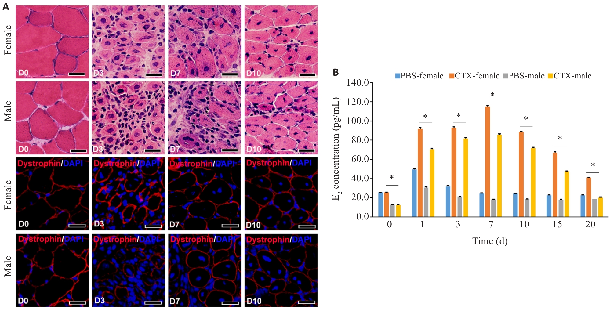
图1 肌纤维E2信号对急性骨骼肌炎症的影响
Fig.1 Effect of E2 in myofiber signaling on acute skeletal myositis in mice. A: HE and immunofluorescence staining for detecting inflammation in the tibialis anterior muscle (TA) after CTX injury (Scale bar=50 μm). B: ELISA for detection of serum E2 in male and female mice at different time points after muscle injury (*P<0.05).

图2 急性骨骼肌炎中雌激素的受体表达
Fig.2 Expression of estrogen receptors in acute skeletal myositis. A: Western blotting of ERα and ERβ expression level in mature myotubes derived from C2C12 cells. B: qRT-PCR analysis of ERα and ERβ mRNA levels in the damaged muscle of female mice. C: Immunofluorescence staining of ERβ expression level in injured mouse muscle (Scale bar=50 μm). *P<0.05, **P<0.01.

图3 肌纤维E2信号对急性骨骼肌炎症损伤肌内单核巨噬细胞渗出影响
Fig.3 Effect of E2 signaling in myofibers on exudation of intramuscular mononuclear macrophages after acute skeletal muscle injury. A: ELISA for detecting serum E2 level in female mice after different treatments. B: ELISA for detecting serum E2 levels in female mice at different time points after OVX treatment. C: HE staining for detecting muscular inflammation in female mice with different treatments after CTX injury. D: Immunofluorescence staining for detecting muscular inflammation in female mice after CTX injury with different treatments. *P<0.05, **P<0.01; Scale bar=50 μm.

图4 肌纤维E2信号对急性骨骼肌炎症损伤肌内巨噬细胞表型影响
Fig.4 Effect of E2 signaling in myofibers on phenotype of intramuscular macrophages in mice after acute skeletal muscle injury. A: Flow cytometric analysis of mononuclear macrophages in the damaged muscle of female mice after OVX treatment. B: Flow cytometric analysis of the phenotype of macrophages in the injured muscles of female mice after OVX treatment. *P<0.05, **P<0.01.

图5 肌纤维E2信号对急性骨骼肌炎症损伤肌内炎症因子的影响
Fig.5 Effect of E2 signaling in myofibers on intramuscular inflammatory cytokines after acute skeletal muscle injury. A: qRT-PCR analysis of inflammatory cytokines in female mice after OVX treatment. B: qRT-PCR analysis of inflammatory cytokines in macrophages from the injured muscles isolated by flow cytometry in female mice after OVX treatment. *P<0.05, **P<0.01.

图6 肌纤维E2信号驱动急性骨骼肌炎症损伤肌内巨噬细胞胞葬作用
Fig.6 E2 signaling in myofibers drives intramuscular macrophage efferocytosis in acute skeletal myositis. A: Immunofluorescence detection of intramuscular macrophage efferocytosis in the damaged muscle. (scale bar=50 μm). B: Flow cytometry of intramuscular macrophage efferocytosis in the damaged muscle (*P<0.05).

图7 体外共培养体系中E2信号对巨噬细胞表型与胞葬作用的影响
Fig.7 Effect of E2 signaling on macrophage phenotypes and efferocytosis in an in vitro co-culture system. A: Immunofluorescence detection of macrophage phenotypes in the in vitro co-culture system. B: Immunofluorescence detection and flow cytometry analysis of macrophage efferocytosis in the in vitro co-culture system. *P<0.05; Scale bar=50 μm.

图8 肌纤维E2信号调控损伤骨骼肌内肌纤维再生修复
Fig.8 E2 signaling in myofibers regulates regeneration and repair of intramuscular myofibers in injured mouse skeletal muscles of female mice after different treatments (Immunofluorescence staining, scale bar=50 μm; *P<0.05).
| 1 | Zhong HH, Sian V, Johari M, et al. Revealing myopathy spectrum: integrating transcriptional and clinical features of human skeletal muscles with varying health conditions[J]. Commun Biol, 2024, 7(1): 438. |
| 2 | Zeller J, Peter K, Eisenhardt SU. Intravital imaging of leukocyte-endothelial interaction in hindlimb ischemia/reperfusion injury by intravital multiphoton microscopy[J]. Methods Mol Biol, 2024, 2711: 89-104. |
| 3 | Zhang H, Qi G, Wang K, et al. Oxidative stress: Roles in skeletal muscle atrophy [J]. Biochem Pharmacol, 2023, 214: 115664-77. |
| 4 | Al-Kharashi L, Attia H, Alsaffi A, et al. Pentoxifylline and thiamine ameliorate rhabdomyolysis-induced acute kidney injury in rats via suppressing TLR4/NF‑κB and NLRP-3/caspase-1/gasdermin mediated-pyroptosis[J]. Toxicol Appl Pharmacol, 2023, 461: 116387. |
| 5 | Boudhabhay I, Poillerat V, Grunenwald A, et al. Complement activation is a crucial driver of acute kidney injury in rhabdomyolysis[J]. Kidney Int, 2021, 99(3): 581-97. |
| 6 | Geraci A, Calvani R, Ferri E, et al. Sarcopenia and menopause: the role of estradiol[J]. Front Endocrinol, 2021, 12: 682012. |
| 7 | Ferreira J, Carneiro A, Vila I, et al. Inflammation and loss of skeletal muscle mass in chronic limb threatening ischemia[J]. Ann Vasc Surg, 2023, 88: 164-73. |
| 8 | Petrocelli JJ, McKenzie AI, de Hart NMMP, et al. Disuse-induced muscle fibrosis, cellular senescence, and senescence-associated secretory phenotype in older adults are alleviated during re-ambulation with metformin pre-treatment[J]. Aging Cell, 2023, 22(11): e13936. |
| 9 | Livshits G, Kalinkovich A. Restoration of epigenetic impairment in the skeletal muscle and chronic inflammation resolution as a therapeutic approach in sarcopenia[J]. Ageing Res Rev, 2024, 96: 102267. |
| 10 | Aluganti Narasimhulu C, Singla DK. Amelioration of diabetes-induced inflammation mediated pyroptosis, sarcopenia, and adverse muscle remodelling by bone morphogenetic protein-7[J]. J Cachexia Sarcopenia Muscle, 2021, 12(2): 403-20. |
| 11 | Antuña E, Cachán-Vega C, Bermejo-Millo JC, et al. Inflammaging: implications in sarcopenia[J]. Int J Mol Sci, 2022, 23(23): 15039. |
| 12 | Ji Y, Lin J, Liu R, et al. Celecoxib attenuates hindlimb unloading-induced muscle atrophy via suppressing inflammation, oxidative stress and ER stress by inhibiting STAT3[J]. Inflammo-pharmacology, 2024, 32(2): 1633-46. |
| 13 | Fischer V, Ragipoglu D, Diedrich J, et al. Mast cells trigger disturbed bone healing in osteoporotic mice[J]. J Bone Miner Res, 2022, 37(1): 137-51. |
| 14 | Zhang Y, Chang YC, Han ZW, et al. Estrogen protects against renal ischemia-reperfusion injury by regulating Th17/treg cell immune balance[J]. Dis Markers, 2022, 2022: 7812099. |
| 15 | Kurmann L, Okoniewski M, Dubey RK. Estradiol inhibits human brain vascular pericyte migration activity: a functional and transcriptomic analysis[J]. Cells, 2021, 10(9): 2314. |
| 16 | Rodríguez-Benítez E, López-García K, Xelhuantzi N, et al. Shift from pro-to anti-inflammatory phase in pelvic floor muscles at postpartum matches histological signs of regeneration in multiparous rabbits[J]. Medicina, 2024, 60(4): 675. |
| 17 | Luo ZW, Qi BJ, Sun YY, et al. Engineering bioactive M2 macrophage-polarized, anti-inflammatory, miRNA-based liposomes for functional muscle repair: from exosomal mechanisms to biomaterials[J]. Small, 2022, 18(34): e2201957. |
| 18 | Guimarães-Pinto K, Maia EP, Ferreira JRM, et al. Efferocytosis in lung mucosae: implications for health and disease[J]. Immunol Lett, 2022, 248: 109-18. |
| 19 | Doran AC, Yurdagul A Jr, Tabas I. Efferocytosis in health and disease[J]. Nat Rev Immunol, 2020, 20(4): 254-67. |
| 20 | 吴泽锴, 黄 涛, 廖钊宏, 等. 肌纤维转化生长因子- β信号激活与急性肌损伤炎症反应的相关性研究[J]. 中华创伤骨科杂志, 2021, 23(3): 254-61. DOI: 10.3760/cma.j.cn115530-20210107-00010 |
| 21 | Xiao JW, Huang JW, Jian XT, et al. IRE1α arm of unfolded protein response in muscle-specific TGF-β signaling-mediated regulation of muscle cell immunological properties[J]. Cell Mol Biol Lett, 2023, 28(1): 15. |
| 22 | Hoffman DB, Raymond-Pope CJ, Sorensen JR, et al. Temporal changes in the muscle extracellular matrix due to volumetric muscle loss injury[J]. Connect Tissue Res, 2022, 63(2): 124-37. |
| 23 | Navi A, Patel H, Xu SW, et al. Role of toll-like receptor 4 in skeletal muscle damage in chronic limb-threatening ischemia[J]. JVS Vasc Sci, 2024, 5: 100194. |
| 24 | Hirtz A, Rech F, Dubois-Pot-Schneider H, et al. Estrogen signaling in healthy and tumor brain[J]. Steroids, 2023, 199: 109285. |
| 25 | Geraci A, Calvani R, Ferri E, et al. Sarcopenia and Menopause: The Role of Estradiol[J]. Front Endocrinol (Lausanne), 2021, 12: 682012-16. |
| 26 | Chaiyasing R, Sugiura A, Ishikawa T, et al. Estrogen modulates the skeletal muscle regeneration process and myotube morphogenesis: morphological analysis in mice with a low estrogen status[J]. J Vet Med Sci, 2021, 83(12): 1812-9. |
| 27 | Lou YY, Fu ZJ, Tian Y, et al. Estrogen-sensitive activation of SGK1 induces M2 macrophages with anti-inflammatory properties and a Th2 response at the maternal-fetal interface[J]. Reprod Biol Endocrinol, 2023, 21(1): 50. |
| 28 | Bauerschmitz G, Hüchel S, Gallwas J, et al. Inhibition of increased invasiveness of breast cancer cells with acquired tamoxifen resistance by suppression of CYR61[J]. Cancer Genomics Proteomics, 2023, 20(6): 531-8. |
| 29 | Valero-Breton M, Tacchi F, Abrigo J, et al. Angiotensin-(1-7) improves skeletal muscle regeneration[J]. Eur J Transl Myol, 2023, 33(4): 12037. |
| 30 | Landeros RV, Jobe SO, Aranda-Pino G, et al. Convergent ERK1/2, p38 and JNK mitogen activated protein kinases (MAPKs) signalling mediate catecholoestradiol-induced proliferation of ovine uterine artery endothelial cells[J]. J Physiol, 2017, 595(14): 4663-76. |
| 31 | Mitchnick KA, Mendell AL, Wideman CE, et al. Dissociable involvement of estrogen receptors in perirhinal cortex-mediated object-place memory in male rats[J]. Psychoneuroendocrinology, 2019, 107: 98-108. |
| [1] | 范正媛, 沈子涵, 李亚, 沈婷婷, 李高峰, 李素云. 补肺益肾方对香烟烟雾提取物诱导的人支气管上皮细胞损伤的保护作用及其机制[J]. 南方医科大学学报, 2025, 45(7): 1372-1379. |
| [2] | 王立明, 陈宏睿, 杜燕, 赵鹏, 王玉洁, 田燕歌, 刘新光, 李建生. 益气滋肾方通过抑制PI3K/Akt/NF-κB通路改善小鼠慢性阻塞性肺疾病的炎症反应[J]. 南方医科大学学报, 2025, 45(7): 1409-1422. |
| [3] | 夏冰, 彭进, 丁九阳, 王杰, 唐国伟, 刘国杰, 王沄, 万昌武, 乐翠云. ATF3通过NF-κB信号通路调控动脉粥样硬化斑块内的炎症反应[J]. 南方医科大学学报, 2025, 45(6): 1131-1142. |
| [4] | 王心恒, 邵小涵, 李童童, 张璐, 杨勤军, 叶卫东, 童佳兵, 李泽庚, 方向明. 平喘宁方通过调控HMGB1/Beclin-1轴介导的自噬改善患寒哮证大鼠的气道炎症[J]. 南方医科大学学报, 2025, 45(6): 1153-1162. |
| [5] | 牛民主, 殷丽霞, 乔通, 尹林, 张可妮, 胡建国, 宋传旺, 耿志军, 李静. 旱莲苷A通过调控JAK2/STAT3通路抑制M1型巨噬细胞极化改善葡聚糖硫酸钠诱导的小鼠结肠炎[J]. 南方医科大学学报, 2025, 45(6): 1297-1306. |
| [6] | 杨洋, 王凯, 柳鉴修, 周志谟, 贾雯, 吴思谋, 李金星, 何方, 程如越. 生命早期两歧双歧杆菌BD-1干预可缓解注意缺陷多动障碍雌性大鼠幼年期的多动行为[J]. 南方医科大学学报, 2025, 45(4): 702-710. |
| [7] | 朱正望, 王琳琳, 赵静涵, 马瑞雪, 余雨春, 蔡庆春, 王兵, 朱平生, 苗明三. 退黄合剂通过调控法尼醇X受体抑制NLRP3炎症小体改善α-萘异硫氰酸酯诱导的大鼠胆汁淤积[J]. 南方医科大学学报, 2025, 45(4): 718-724. |
| [8] | 储菲, 陈孝华, 宋博文, 杨晶晶, 左芦根. 苏荠宁黄酮通过抑制PI3K/AKT信号通路拮抗肠上皮细胞凋亡改善小鼠实验性结肠炎[J]. 南方医科大学学报, 2025, 45(4): 819-828. |
| [9] | 张金水, 李硕, 魏栋栋, 成昕, 邓云, 张有志. 石墨烯发热膜热疗改善小鼠冻伤的作用及机制[J]. 南方医科大学学报, 2025, 45(3): 522-530. |
| [10] | 曹周芳, 汪元, 王梦娜, 孙玥, 刘菲菲. LINC00837/miR-671-5p/SERPINE2功能轴促进类风湿关节炎成纤维细胞样滑膜细胞的恶性病理学过程[J]. 南方医科大学学报, 2025, 45(2): 371-378. |
| [11] | 罗维, 王宇航, 刘延松, 王媛媛, 艾磊. 高糖环境通过抑制免疫反应基因1的表达诱导巨噬细胞促炎性M1型极化[J]. 南方医科大学学报, 2025, 45(1): 1-9. |
| [12] | 张玉如, 万磊, 方昊翔, 李方泽, 王丽文, 李柯霏, 闫佩文, 姜辉. miR-155-5p介导PIK3R1负调控PI3K/AKT信号通路促进原发性干燥综合征人唾液腺上皮细胞增殖[J]. 南方医科大学学报, 2025, 45(1): 65-71. |
| [13] | 左涵珺, 段兆达, 王朝, 郭涛, 石金沙, 石浩龙, 李娟娟. 天麻素经PI3K/AKT通路改善新生大鼠缺氧缺血性脑损伤后小胶质细胞介导的炎症反应[J]. 南方医科大学学报, 2024, 44(9): 1712-1719. |
| [14] | 李明远, 张玮, 华梦晴. 甲基巴多索龙通过抑制NLRP3炎症小体活化缓解小鼠急性肝损伤[J]. 南方医科大学学报, 2024, 44(9): 1662-1669. |
| [15] | 张先恒, 刘健, 韩琦, 陈一鸣, 丁香, 陈晓露. 黄芩清热除痹胶囊通过PTEN/PI3K/AKT信号通路改善痛风性关节炎大鼠的炎症反应及尿酸、脂质代谢失衡[J]. 南方医科大学学报, 2024, 44(8): 1450-1458. |
| 阅读次数 | ||||||
|
全文 |
|
|||||
|
摘要 |
|
|||||