
南方医科大学学报 ›› 2026, Vol. 46 ›› Issue (6): 1244-1255.doi: 10.12122/j.issn.1673-4254.2026.06.05
• • 上一篇
尹林1,2( ), 章雨1,2, 张可妮1,2, 乔通1,2, 张龙涛1,2, 黄菊2,3, 李静1,2, 胡建国1,2, 耿志军2,3()
收稿日期:2026-01-07
出版日期:2026-06-20
发布日期:2026-06-24
通讯作者:
耿志军
E-mail:yljykbbmu@163.com;gengzhijun1213@163.com
作者简介:尹 林,在读硕士研究生,E-mail: yljykbbmu@163.com
基金资助:
Lin YIN1,2(), Yu ZHANG1,2, Keni ZHANG1,2, Tong QIAO1,2, Longtao ZHANG1,2, Ju HUANG2,3, Jing LI1,2, Jianguo HU1,2, Zhijun GENG2,3()
Received:2026-01-07
Online:2026-06-20
Published:2026-06-24
Contact:
Zhijun GENG
E-mail:yljykbbmu@163.com;gengzhijun1213@163.com
摘要:
目的 探讨1,3-二咖啡酰奎宁酸(1,3-DA)对葡聚糖硫酸钠(DSS)诱导小鼠结肠炎的保护作用及调控磷脂酰肌醇3-激酶/蛋白激酶B(PI3K/Akt)信号通路的分子机制。 方法 体内构建DSS诱导的小鼠结肠炎模型,设置正常对照组、DSS模型组、1,3-DA干预组、PI3K抑制剂LY294002干预组及5-氨基水杨酸(5-ASA)阳性对照组,评估小鼠结肠炎病理表型,检测结肠组织炎症水平、氧化应激指标、肠上皮紧密连接蛋白及PI3K/Akt通路相关蛋白表达。体外采用过氧化氢(H2O2)诱导NCM460肠上皮细胞构建氧化应激损伤模型,设置对照组、模型组、1,3-DA干预组及1,3-DA联合PI3K激活剂740Y-P共处理组,检测细胞活性氧(ROS)生成、紧密连接蛋白与通路蛋白表达以验证调控机制。 结果 体内实验中,与DSS模型组相比,1,3-DA干预组小鼠体质量下降(P<0.001)、结肠长度缩短(P<0.001)、疾病活动指数(DAI)升高(P<0.001)及结肠黏膜病理损伤得到缓解,结肠组织促炎因子γ 干扰素(IFN-γ)(P<0.001)、髓过氧化物酶(MPO)表达显著下调(P<0.001),抗氧化酶:超氧化物歧化酶(SOD)、过氧化氢酶(CAT)、谷胱甘肽过氧化物酶(GSH-PX)活性含量明显上调(均P<0.001),脂质过氧化产物丙二醛(MDA)水平显著降低(P<0.001),肠上皮紧密连接蛋白:闭锁小带蛋白1(ZO-1)、闭合蛋白1(Claudin-1)的表达上调(P<0.001),同时磷酸化磷脂酰肌醇3-激酶(p-PI3K)与磷酸化蛋白激酶B(p-Akt)的表达上调受到显著抑制(均P<0.001)。体外实验中,与H₂O₂诱导的肠上皮细胞模型组相比,1,3-DA干预组细胞内ROS蓄积水平显著降低(P<0.001),紧密连接蛋白ZO-1、Claudin-1的表达显著上调(均P<0.001),p-PI3K(P<0.001)和p-Akt(P=0.014)的表达明显下降,且PI3K激活剂740Y-P与1,3-DA共处理后可部分逆转其对肠上皮细胞的保护作用。 结论 1,3-DA可通过抗炎、抗氧化应激、修复肠上皮屏障功能,减轻DSS诱导的小鼠结肠炎及H₂O₂介导的肠上皮细胞氧化应激损伤,其作用机制与抑制PI3K/Akt信号通路的过度异常激活密切相关。
尹林, 章雨, 张可妮, 乔通, 张龙涛, 黄菊, 李静, 胡建国, 耿志军. 1,3-二咖啡酰奎宁酸通过抑制PI3K/Akt通路减轻氧化应激缓解葡聚糖硫酸钠诱导的小鼠结肠炎[J]. 南方医科大学学报, 2026, 46(6): 1244-1255.
Lin YIN, Yu ZHANG, Keni ZHANG, Tong QIAO, Longtao ZHANG, Ju HUANG, Jing LI, Jianguo HU, Zhijun GENG. 1,3-dicaffeoylquinic acid mitigates dextran sulfate sodium-induced colitis in mice by alleviating oxidative stress via inhibiting the PI3K/Akt pathway[J]. Journal of Southern Medical University, 2026, 46(6): 1244-1255.
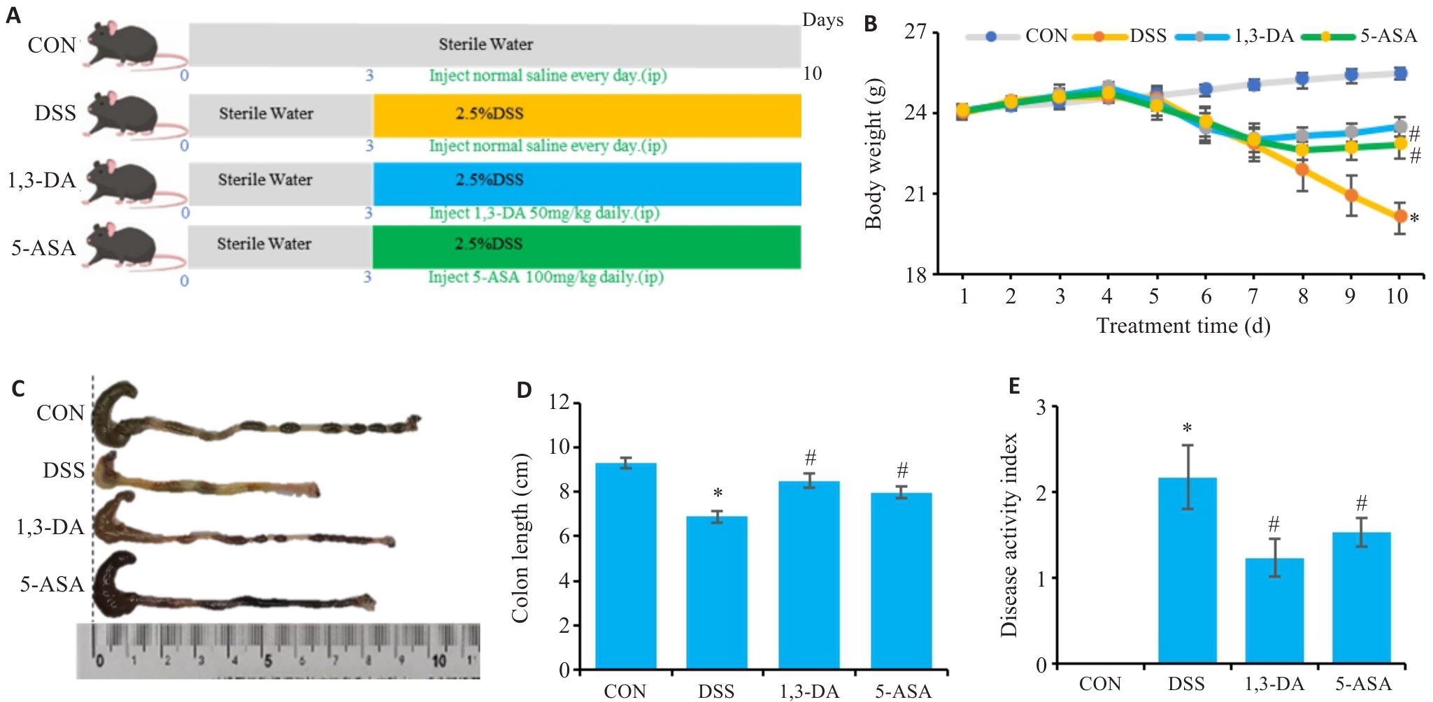
图1 1,3-DA对DSS诱导的小鼠肠炎症状的影响
Fig.1 Effect of 1,3-DA intervention on DSS-induced colitis in mice (n=10). A: Schematic of colitis modeling and treatments of the mice. B: Body weight changes of the mice in each group. C, D: Comparison of colon length of the mice among the groups. E: Disease activity index (DAI) score of the mice after different treatments. *P<0.05 vs CON group, #P<0.05 vs DSS group.
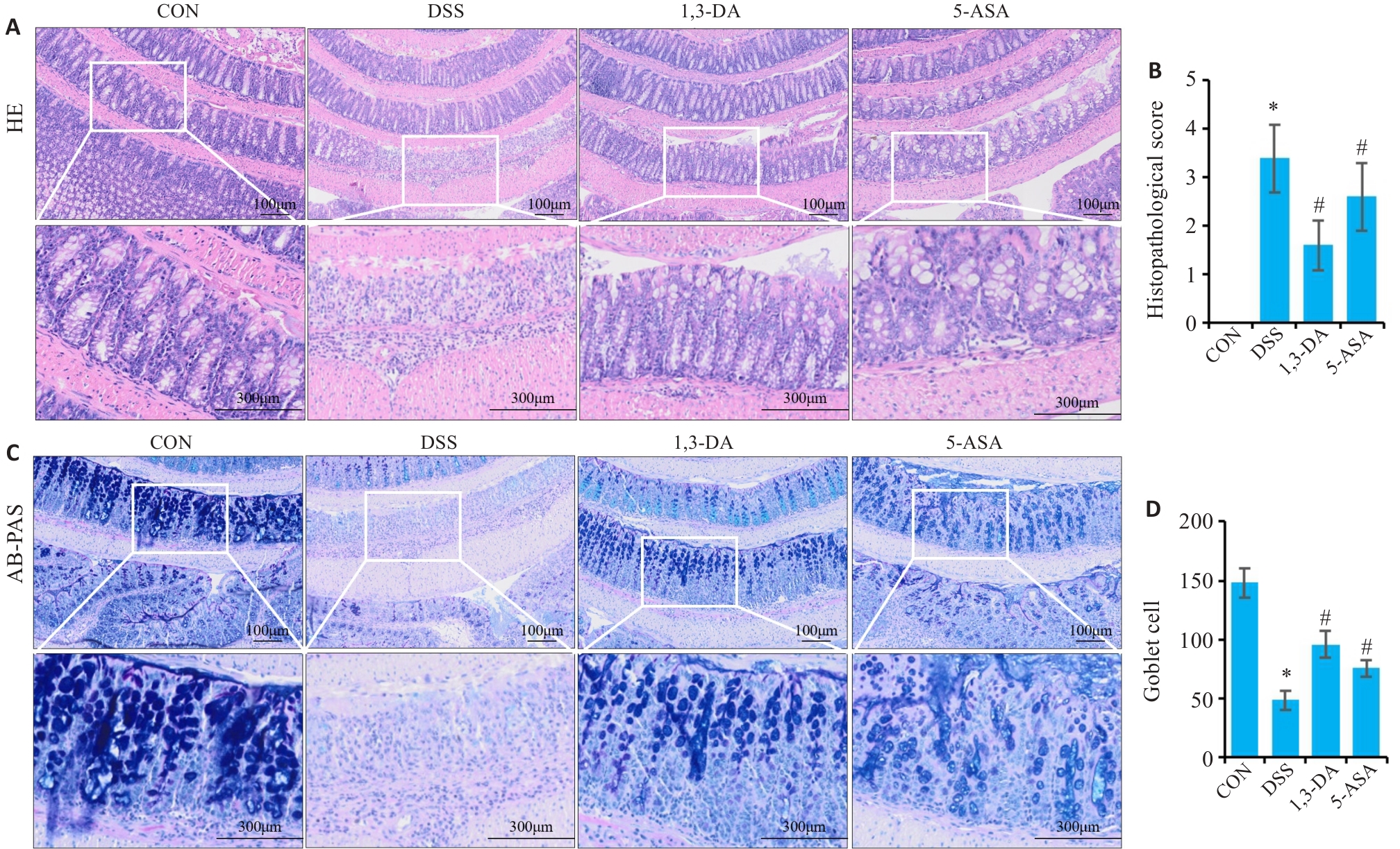
图2 1,3-DA对DSS小鼠肠组织病理学的影响
Fig.2 Effect of 1,3-DA on intestinal histopathology in DSS-treated mice (n=10). A: HE staining and inflammatory scores of intestinal tissues of the mice in each group (Original magnification: ×200). B: Histopathological scores of the mice in each group. C: AB-PAS staining of the intestinal tissues in each group (×200). D: Goblet cell counts in the visual field of each group. *P<0.05 vs CON group, #P<0.05 vs DSS group.
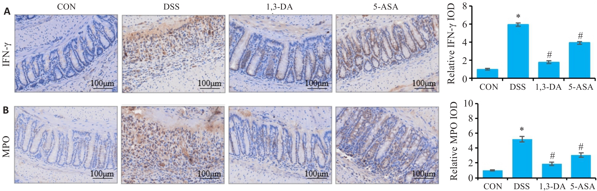
图 3 1,3-DA 对 DSS 小鼠结肠组织炎症因子的影响
Fig.3 Effect of 1,3-DA on inflammatory factors in the intestinal tissue of DSS-treated mice (n=10). A: Immunohistochemical staining for IFN-γ and relative OD values in different groups. B: Immunohistochemical staining for MPO and relative OD values in different groups (Scale bar=100 μm). *P<0.05 vs CON group, #P<0.05 vs DSS group.
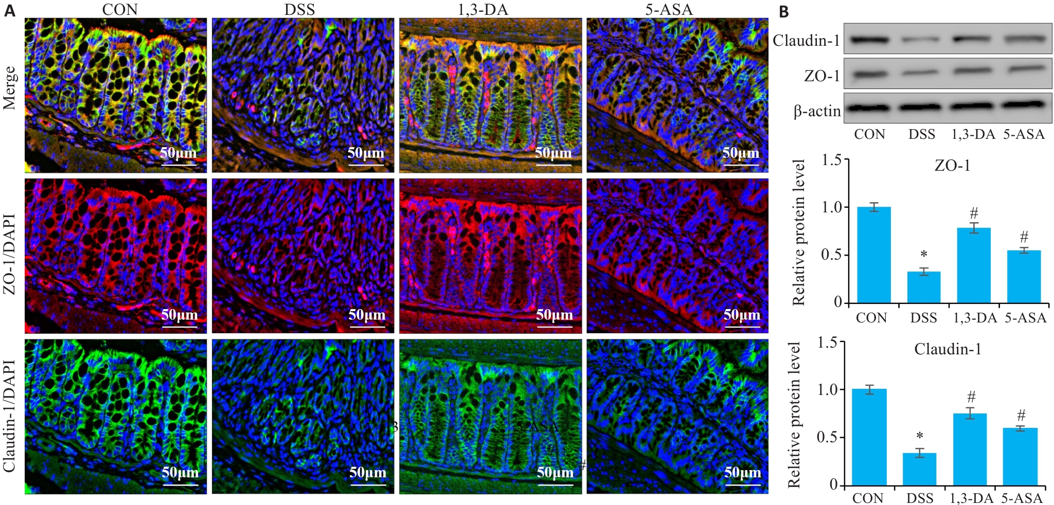
图4 1,3-DA对DSS小鼠结肠组织中紧密连接蛋白的影响
Fig.4 Effect of 1,3-DA on tight junction proteins in the colonic tissues of DSS-treated mice (n=10). A: Immunofluorescence staining in each group. B: Western blotting results in each group. *P<0.05 vs CON group, #P<0.05 vs DSS group.
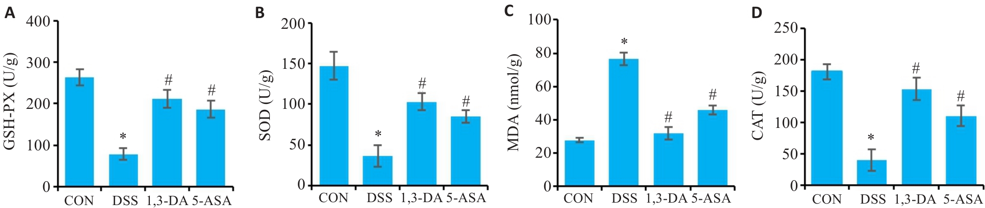
图5 1,3-DA对DSS小鼠结肠组织中氧化应激的调控作用
Fig.5 Regulatory effect of 1,3-DA on oxidative stress in the colonic tissues of DSS-treated mice (n=10). A: GSH-PX levels in each group. B: SOD levels in each group. C: MDA levels in each group. D: CAT levels in each group. *P<0.05 vs CON group, #P<0.05 vs DSS group.
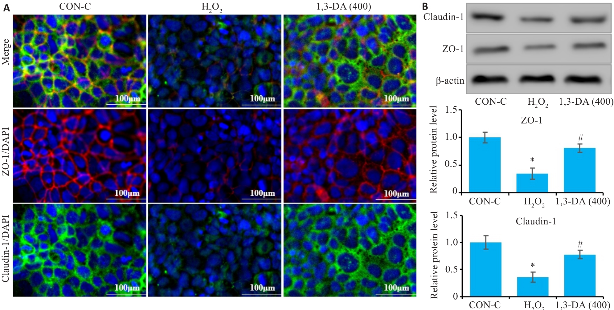
图6 1,3-DA对H₂O₂诱导的NCM460细胞肠屏障的影响
Fig.6 Effect of 1,3-DA on intestinal barrier of H2O2-induced NCM460 cells (n=6). A: Immunofluorescence staining of tight junction proteins claudin-1 and ZO-1 in the cells. B: Western blotting of claudin-1 and ZO-1 in the cells in each group. *P<0.05 vs CON-C group, #P<0.05 vs H2O2 group.
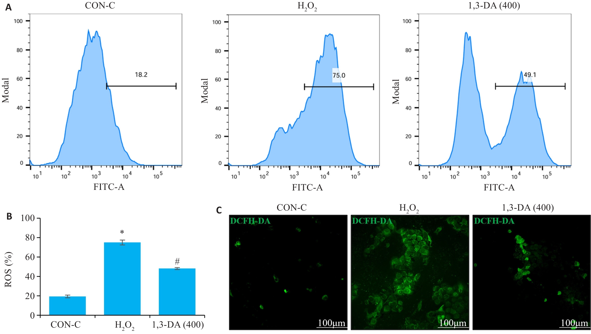
图7 1,3-DA对H2O2诱导的NCM460细胞活性氧的影响
Fig.7 Effect of 1,3-DA on ROS induced by H2O2 in NCM460 cells (n=5). A, B: Flow cytometry for analyzing ROS in the cells in each group. C: ROS staining with fluorescent probe DCFH-DA in the cells in each group. *P<0.05 vs CON-C group, #P<0.05 vs H2O2 group.
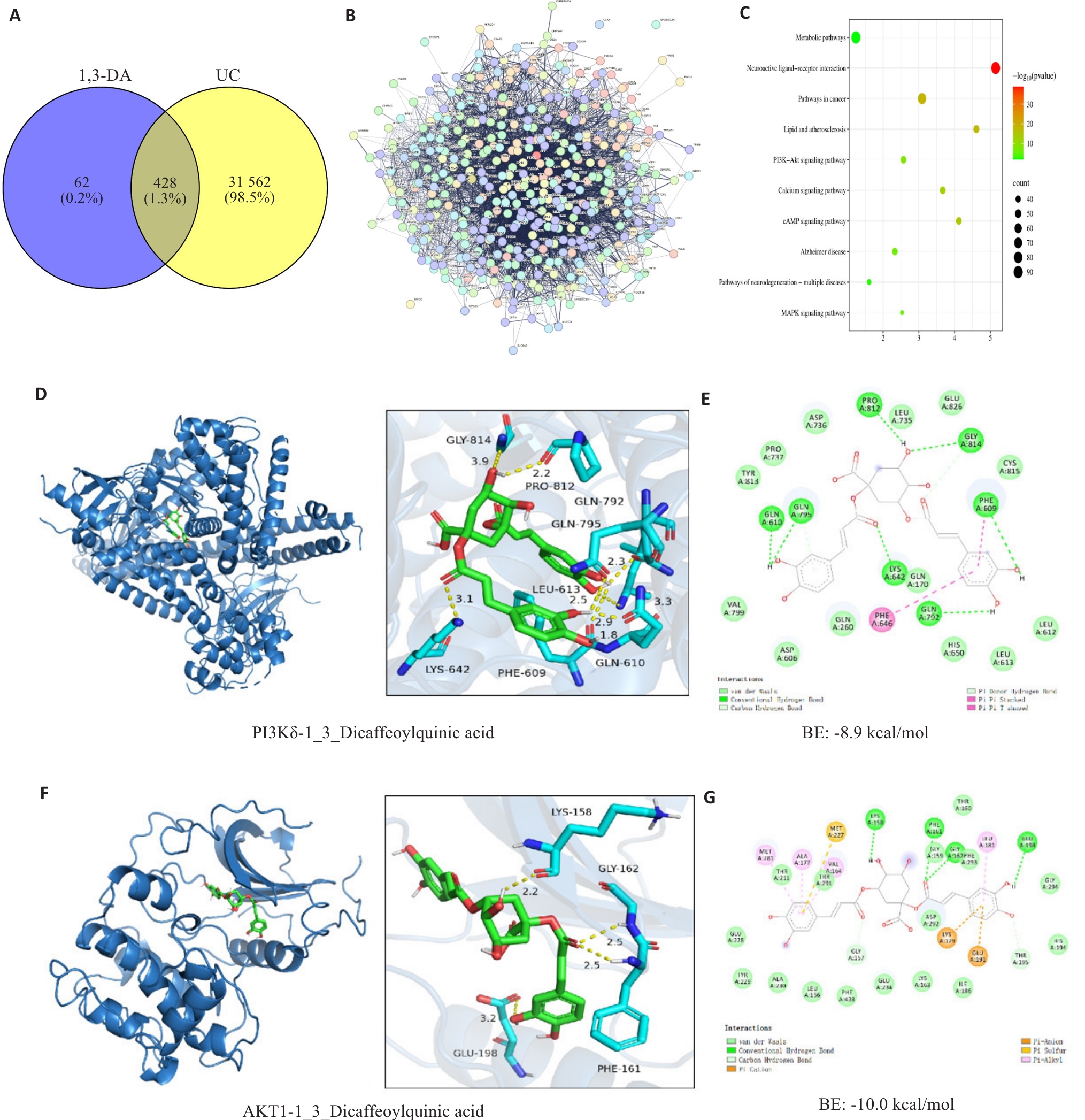
图8 1,3-DA网络药理学分析
Fig.8 1,3-DA network pharmacology. A: Venn diagram. B: PPI network diagram. C: KEGG pathway enrichment analysis. D, E: Molecular docking results of the interaction between PI3Kδ and 1,3-DA. F, G: Results of molecular docking between AKT1 and 1,3-DA.
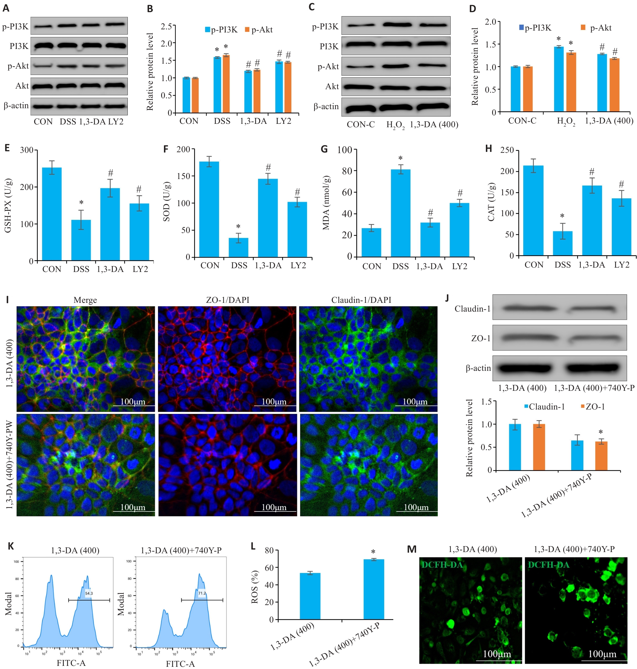
图9 1,3-DA调控PI3K/Akt通路改善结肠炎小鼠肠道氧化应激损伤
Fig.9 1,3-DA regulates the PI3K/Akt pathway to improve intestinal oxidative stress damage in mice with colitis (n=10 for in vivo experiments and n=6 for in vitro experiment). A, B: Western blotting for detecting intestinal PI3K and Akt expressions of the mice. C, D: Western blotting for detecting PI3K and Akt expressions in H2O2-induced NCM460 cells with different treatments. E-H: Intestinal oxidative stress indexes (GSH-PX, SOD, MDA, and CAT). I, J: Intestinal tight junction proteins detected by immunofluorescence staining and Western blotting. K, L: Intracellular ROS levels detected by flow cytometry (drug-treated groups vs 740Y-P activator group). M: Intracellular ROS detected using DCFH-DA staining (drug-treated vs 740Y-P activator group). *P<0.05 vs CON or CON-C group, #P<0.05 vs H2O2 or DSS group.
| [1] | Guan QD. A comprehensive review and update on the pathogenesis of inflammatory bowel disease[J]. J Immunol Res, 2019, 2019: 7247238. doi:10.1155/2019/7247238 |
| [2] | Bisgaard TH, Allin KH, Keefer L, et al. Depression and anxiety in inflammatory bowel disease: epidemiology, mechanisms and treatment[J]. Nat Rev Gastroenterol Hepatol, 2022, 19(11): 717-26. doi:10.1038/s41575-022-00634-6 |
| [3] | Sahoo DK, Heilmann RM, Paital B, et al. Oxidative stress, hormones, and effects of natural antioxidants on intestinal inflammation in inflammatory bowel disease[J]. Front Endocrinol, 2023, 14: 1217165. doi:10.3389/fendo.2023.1217165 |
| [4] | Tratenšek A, Locatelli I, Grabnar I, et al. Oxidative stress-related biomarkers as promising indicators of inflammatory bowel disease activity: a systematic review and meta-analysis[J]. Redox Biol, 2024, 77: 103380. doi:10.1016/j.redox.2024.103380 |
| [5] | Wang R, Li ZQ, Liu SJ, et al. Global, regional and national burden of inflammatory bowel disease in 204 countries and territories from 1990 to 2019: a systematic analysis based on the Global Burden of Disease Study 2019[J]. BMJ Open, 2023, 13(3): e065186. doi:10.1136/bmjopen-2022-065186 |
| [6] | Yang KQ, Zhang CH, Gong R, et al. From west to east: dissecting the global shift in inflammatory bowel disease burden and projecting future scenarios[J]. BMC Public Health, 2025, 25(1): 2696. doi:10.1186/s12889-025-24009-z |
| [7] | Tian T, Wang ZL, Zhang JH. Pathomechanisms of oxidative stress in inflammatory bowel disease and potential antioxidant therapies[J]. Oxid Med Cell Longev, 2017, 2017(1): 4535194. doi:10.1155/2017/4535194 |
| [8] | Berriche-Yahi N, Tahar A, Asselah H, et al. Effects of oral vitamin D(3) supplementation in Crohn's disease patients: Modulation of clinical active/remission phases by pro-inflammatory cytokines profile and oxidative stress[J]. Ann Biol Clin, 2022, 80(1): 29-46. doi:10.1684/abc.2021.1698 |
| [9] | Wong CH, Haque MA, Chang HC. Superoxide dismutase SOD-3 regulates redox homeostasis in the intestine[J]. MicroPubl Biol, 2023, 2023. DOI:10.17912/micropub.biology.001061 . |
| [10] | Lu Q, Jiao YQ, Wu ZD, et al. Zanthoxylum Nitidum ameliorates intestinal barrier dysfunction and inflammation in TNBS-induced colitis rats and LPS-stimulated caco-2 cells[J]. FASEB J, 2025, 39(19): e71099. doi:10.1096/fj.202501303RRRR |
| [11] | Yu T, Chen D, Qi HY, et al. Resolvins protect against diabetes-induced colonic oxidative stress, barrier dysfunction, and associated diarrhea via the HO-1 pathway[J]. Biofactors, 2024, 50(5): 967-79. doi:10.1002/biof.2049 |
| [12] | Wang YF, Zhang JQ, Zhang BH, et al. Modified Gegen Qinlian decoction ameliorated ulcerative colitis by attenuating inflammation and oxidative stress and enhancing intestinal barrier function in vivo and in vitro [J]. J Ethnopharmacol, 2023, 313: 116538. doi:10.1016/j.jep.2023.116538 |
| [13] | Hu ZY, Xu D, Meng HH, et al. Corrigendum to "4-octyl itaconate protects against oxidative stress-induced liver injury by activating the Nrf2/Sirt3 pathway through AKT and ERK1/2 phosphorylation" [Biochem. Pharmacol. 220 (2024) 115992[J]. Biochem Pharmacol, 2025, 236: 116853. doi:10.1016/j.bcp.2025.116853 |
| [14] | Yin NL, Su YG, Wang CF, et al. Farrerol alleviates microbiota dysbiosis-associated osteoporosis in IBD by inhibiting PI3K-AKT/MAPK signaling pathways[J]. Phytomedicine, 2025, 148: 157279. doi:10.1016/j.phymed.2025.157279 |
| [15] | Ni CJ, Liu HQ, Jiang HY, et al. Danthron attenuates intestinal inflammation by modulating oxidative stress via the EGFR-PI3K-AKT and Nrf2-HO-1 pathways[J]. Antioxidants, 2026, 15(2): 157. doi:10.3390/antiox15020157 |
| [16] | Huang MC, Yu QQ, Zhu YM, et al. Arabinoxylan from barley bran alleviates ulcerative colitis in mice through enhancement of mucosal barrier function and modulation of gut microbiota[J]. Int J Biol Macromol, 2025, 318(Pt 2): 144941. doi:10.1016/j.ijbiomac.2025.144941 |
| [17] | Khare V, Krnjic A, Frick A, et al. Mesalamine and azathioprine modulate junctional complexes and restore epithelial barrier function in intestinal inflammation[J]. Sci Rep, 2019, 9(1): 2842. doi:10.1038/s41598-019-39401-0 |
| [18] | Lim DW, Kim M, Yoon M, et al. 1, 3-dicaffeoylquinic acid as an active compound of Arctium lappa root extract ameliorates depressive-like behavior by regulating hippocampal nitric oxide synthesis in ovariectomized mice[J]. Antioxidants, 2021, 10(8): 1281. doi:10.3390/antiox10081281 |
| [19] | Joo SG, Kim JM, Lee HL, et al. Synurus deltoides alleviates anti-depressive like behavior dysfunction induced by chronic unpredictable mild stress via stress-related CRF/TLR pathway[J]. J Microbiol Biotechnol, 2025, 35: e2501043. doi:10.4014/jmb.2501.01043 |
| [20] | Gupta S, Kumar S, Sharma D. Natural product-based therapies for inflammatory bowel disease: targeting key signaling pathways[J]. Mini Rev Med Chem, 2025, 25(22): 1716-38. doi:10.2174/0113895575405061250923110348 |
| [21] | Wang Y, Zeng LY. Immune modulation in inflammatory bowel disease: therapeutic promise of baicalein[J]. Front Pharmacol, 2025, 16: 1723606. doi:10.3389/fphar.2025.1723606 |
| [22] | Lu XB, Sun YP, Zhang ZY, et al. Regulation of pyroptosis by natural products in ulcerative colitis: mechanisms and therapeutic potential[J]. Front Pharmacol, 2025, 16: 1573684. doi:10.3389/fphar.2025.1573684 |
| [23] | Wang SY, Yu XJ, Cheng LL, et al. Dexmedetomidine improves the circulatory dysfunction of the glymphatic system induced by sevoflurane through the PI3K/AKT/ΔFosB/AQP4 pathway in young mice[J]. Cell Death Dis, 2024, 15(6): 448. doi:10.1038/s41419-024-06845-w |
| [24] | Huang LY, Qian WW, Xu YH, et al. Mesenteric adipose tissue contributes to intestinal fibrosis in Crohn's disease through the ATX-LPA axis[J]. J Crohns Colitis, 2022, 16(7): 1124-39. doi:10.1093/ecco-jcc/jjac017 |
| [25] | Schultz M, Tonkonogy SL, Sellon RK, et al. IL-2-deficient mice raised under germfree conditions develop delayed mild focal intestinal inflammation[J]. Am J Physiol, 1999, 276(6): G1461-72. doi:10.1152/ajpgi.1999.276.6.g1461 |
| [26] | Niu SY, Zhang Y. Targeting the PI3K/Akt pathway for the treatment of ulcerative colitis: integrative regulatory features of traditional Chinese medicine[J]. Front Pharmacol, 2025, 16: 1620138. doi:10.3389/fphar.2025.1620138 |
| [27] | Hao LL, Alkry LT, Alattar A, et al. Ibrutinib attenuated DSS-induced ulcerative colitis, oxidative stress, and the inflammatory cascade by modulating the PI3K/Akt and JNK/NF‑κB pathways[J]. Arch Med Sci, 2022, 18(3): 805-15. doi:10.5114/aoms/146792 |
| [28] | Kushwaha P, Qureshi R, Khan N, et al. Revolutionizing IBD therapy: Insights into contemporary treatment strategies[J]. Int Rev Immunol, 2026, 45(1): 1-25. doi:10.1080/08830185.2025.2563522 |
| [29] | Kim A, Kim SY, Jo K, et al. Carpesium abrotanoides ethanol extract alleviates dextran sulfate sodium-induced ulcerative colitis by suppressing inflammation and apoptosis[J]. Front Pharmacol, 2025, 16: 1672111. doi:10.3389/fphar.2025.1672111 |
| [30] | Xu JG, Hu QP, Liu Y. Antioxidant and DNA-protective activities of chlorogenic acid isomers[J]. J Agric Food Chem, 2012, 60(46): 11625-30. doi:10.1021/jf303771s |
| [31] | Liang NJ, Dupuis JH, Yada RY, et al. Chlorogenic acid isomers directly interact with Keap 1-Nrf2 signaling in Caco-2 cells[J]. Mol Cell Biochem, 2019, 457(1/2): 105-18. doi:10.1007/s11010-019-03516-9 |
| [32] | Liu WY, Wang H, Xu X, et al. Natural compound rosmarinic acid displays anti-tumor activity in colorectal cancer cells by suppressing nuclear factor-kappa B signaling[J]. World J Clin Oncol, 2025, 16(5): 105341. doi:10.5306/wjco.v16.i5.105341 |
| [33] | Giera M, Lingeman H, Niessen WMA. Recent advancements in the LC- and GC-based analysis of malondialdehyde (MDA): a brief overview[J]. Chromatographia, 2012, 75(9/10): 433-40. doi:10.1007/s10337-012-2237-1 |
| [34] | Del Rio D, Stewart AJ, Pellegrini N. A review of recent studies on malondialdehyde as toxic molecule and biological marker of oxidative stress[J]. Nutr Metab Cardiovasc Dis, 2005, 15(4): 316-28. doi:10.1016/j.numecd.2005.05.003 |
| [35] | Muro P, Zhang L, Li SX, et al. The emerging role of oxidative stress in inflammatory bowel disease[J]. Front Endocrinol, 2024, 15: 1390351. doi:10.3389/fendo.2024.1390351 |
| [36] | El-Saadony MT, Saad AM, Mohammed DM, et al. Curcumin, an active component of turmeric: biological activities, nutritional aspects, immunological, bioavailability, and human health benefits- a comprehensive review[J]. Front Immunol, 2025, 16: 1603018. doi:10.3389/fimmu.2025.1603018 |
| [37] | Sato Y, Itagaki S, Kurokawa T, et al. In vitro and in vivo antioxidant properties of chlorogenic acid and caffeic acid[J]. Int J Pharm, 2011, 403(1/2): 136-8. doi:10.1016/j.ijpharm.2010.09.035 |
| [38] | Saqib U, Kelley TT, Panguluri SK, et al. Polypharmacology or promiscuity? structural interactions of resveratrol with its bandwagon of targets[J]. Front Pharmacol, 2018, 9: 1201. doi:10.3389/fphar.2018.01201 |
| [1] | 祝金超, 邵宁宁, 黄泽彧, 刘燕青, 孟祥艳, 刘子泉, 孙予杰, 董津睿, 樊毫军. 薤白超细粉通过抑制铁死亡并调控NF-κB/STAT3信号轴缓解脂多糖诱导的小鼠急性肺损伤[J]. 南方医科大学学报, 2026, 46(5): 1006-1017. |
| [2] | 张国栋, 魏思昂, 王弘, 谢艳丽, 何晖, 李嵘. 有氧运动通过氧化应激-炎症-Hippo/YAP通路轴改善心力衰竭小鼠心功能[J]. 南方医科大学学报, 2026, 46(4): 753-760. |
| [3] | 宋淇乐, 苗益恺, 冯小桐, 王一凡, 刘伟, 魏琪, 于新汝, 陈文文, 付晓艳. 硒代胱氨酸通过诱导活性氧产生启动氧化应激损伤抑制结肠癌细胞生长[J]. 南方医科大学学报, 2026, 46(3): 532-540. |
| [4] | 温贺新, 林洁, 左芦根, 刘牧林. 珠子草素通过拮抗肠上皮细胞凋亡与调控肠道Th1/Th2免疫平衡改善克罗恩病样小鼠结肠炎[J]. 南方医科大学学报, 2026, 46(3): 655-665. |
| [5] | 彭煜策, 姜毅, 马丹, 何岸, 吕鼎一, 罗明昊, 罗素新. 异牡荆素通过促进SIRT3表达减轻糖尿病小鼠的心肌氧化应激损伤[J]. 南方医科大学学报, 2026, 46(2): 316-324. |
| [6] | 李斌杰, 周晓芳, 朗晓猛, 康欣, 刘建平. 泄浊解毒方通过调节Th17/Treg免疫平衡改善大鼠溃疡性结肠炎[J]. 南方医科大学学报, 2026, 46(2): 335-344. |
| [7] | 尹林, 张可妮, 乔通, 牛民主, 殷丽霞, 刘馨悦, 耿志军, 李静, 胡建国. 藜芦酸通过激活Nrf2/HO-1信号通路减轻氧化应激缓解葡聚糖硫酸钠诱导的小鼠实验性结肠炎[J]. 南方医科大学学报, 2026, 46(2): 403-411. |
| [8] | 黄林林, 郑旺, 胡建国, 宋雪, 陶露, 耿志军, 李静, 左芦根, 葛思堂. 马鞭草苷通过抑制PI3K-AKT通路减轻肠上皮炎症改善小鼠克罗恩病样结肠炎[J]. 南方医科大学学报, 2026, 46(2): 423-433. |
| [9] | 乔通, 尹林, 张可妮, 牛民主, 黄菊, 耿志军, 李静, 胡建国. 茯苓新酸A通过调节AMPK/mTOR介导的自噬来减轻葡聚糖硫酸钠诱导的小鼠结肠炎[J]. 南方医科大学学报, 2026, 46(1): 131-140. |
| [10] | 王静娴, 任自敬, 周佩洋. S1PR5激动与过表达通过调控氧化应激增强脑微血管内皮细胞屏障功能抵抗氧糖剥夺/复氧复糖损伤[J]. 南方医科大学学报, 2025, 45(7): 1451-1459. |
| [11] | 牛民主, 殷丽霞, 乔通, 尹林, 张可妮, 胡建国, 宋传旺, 耿志军, 李静. 旱莲苷A通过调控JAK2/STAT3通路抑制M1型巨噬细胞极化改善葡聚糖硫酸钠诱导的小鼠结肠炎[J]. 南方医科大学学报, 2025, 45(6): 1297-1306. |
| [12] | 张安邦, 孙秀颀, 庞博, 吴远华, 时靖宇, 张宁, 叶涛. 电针预处理通过调节肠道-大脑轴及Nrf2/HO-1信号通路抑制铁死亡减轻大鼠脑缺血再灌注损伤[J]. 南方医科大学学报, 2025, 45(5): 911-920. |
| [13] | 储菲, 陈孝华, 宋博文, 杨晶晶, 左芦根. 苏荠宁黄酮通过抑制PI3K/AKT信号通路拮抗肠上皮细胞凋亡改善小鼠实验性结肠炎[J]. 南方医科大学学报, 2025, 45(4): 819-828. |
| [14] | 高志, 吴傲, 胡仲翔, 孙培养. 类风湿性关节炎中氧化应激与免疫浸润的生物信息学分析[J]. 南方医科大学学报, 2025, 45(4): 862-870. |
| [15] | 殷丽霞, 牛民主, 张可妮, 耿志军, 胡建国, 李江艳, 李静. 升麻素抑制MAPK通路调节辅助性T细胞免疫平衡改善小鼠克罗恩病样结肠炎[J]. 南方医科大学学报, 2025, 45(3): 595-602. |
| 阅读次数 | ||||||
|
全文 |
|
|||||
|
摘要 |
|
|||||
