
南方医科大学学报 ›› 2026, Vol. 46 ›› Issue (6): 1235-1243.doi: 10.12122/j.issn.1673-4254.2026.06.04
• • 上一篇
李瑾1,2( ), 贺一杰1,2, 毕芳芳3, 陈浚鑫1, 范桢颜1, 张宇1, 陈恩宇1, 肖鸿文1, 吴云2, 田华1(), 周宇宏1()
收稿日期:2025-12-18
出版日期:2026-06-20
发布日期:2026-06-24
通讯作者:
田华,周宇宏
E-mail:lijin960602@163.com;tianhua0912@163.com;zyh2023@xmmc.edu.cn
作者简介:李 瑾,在读博士研究生,E-mail: lijin960602@163.com
基金资助:
Jin LI1,2(), Yijie HE1,2, Fangfang BI3, Junxin CHEN1, Zhenyan FAN1, Yu ZHANG1, Enyu CHEN1, Hongwen XIAO1, Yun WU2, Hua TIAN1(), Yuhong ZHOU1()
Received:2025-12-18
Online:2026-06-20
Published:2026-06-24
Contact:
Hua TIAN, Yuhong ZHOU
E-mail:lijin960602@163.com;tianhua0912@163.com;zyh2023@xmmc.edu.cn
Supported by:摘要:
目的 探讨抑制miR-503对心肌梗死(MI)的调控作用及分子机制,明确其上游调控因子与下游效应通路,为MI治疗提供潜在靶点。 方法 通过冠状动脉结扎构建小鼠MI模型(分为假手术组、MI组、Antagomir-503组和NC组,5只/组)和原代新生大鼠心肌细胞缺氧模型(分为Control组、Hypoxia组、AMO-503组和NC组)。采用Real-time PCR检测miR-503及长链非编码RNA AK134630的表达变化;通过超声心动图评估小鼠心功能、TTC染色测定梗死面积;利用MTT法、LDH释放检测、TUNEL染色、JC-1染色及透射电镜,分别检测细胞活力、损伤、凋亡、线粒体膜电位及超微结构;采用Western blotting检测凋亡相关蛋白及关键靶点的表达水平。 结果 miR-503在MI模型(P<0.01)和缺氧模型(P<0.01)中表达均显著上调。体内抑制miR-503可改善小鼠心脏射血分数(P<0.01),并缩小梗死面积(P<0.01);同时,该干预可下调Bax、cleaved caspase-3表达,上调Apelin的表达(P<0.01)。体外抑制miR-503(AMO-503)可提高缺氧心肌细胞活力(P<0.01)、降低凋亡率(P<0.01)、恢复线粒体膜电位(P<0.01)。机制上,AK134630结合并负向调控miR-503;抑制miR-503则能解除其对下游靶基因Apelin的抑制。 结论 抑制miR-503可通过上调Apelin表达,发挥抗心肌细胞凋亡及线粒体保护作用,从而减轻MI损伤;AK134630作为miR-503的上游ceRNA参与这一调控过程。
李瑾, 贺一杰, 毕芳芳, 陈浚鑫, 范桢颜, 张宇, 陈恩宇, 肖鸿文, 吴云, 田华, 周宇宏. 靶向抑制miR-503可通过上调Apelin表达减轻小鼠心肌梗死损伤[J]. 南方医科大学学报, 2026, 46(6): 1235-1243.
Jin LI, Yijie HE, Fangfang BI, Junxin CHEN, Zhenyan FAN, Yu ZHANG, Enyu CHEN, Hongwen XIAO, Yun WU, Hua TIAN, Yuhong ZHOU. Targeted inhibition of miR-503 upregulates Apelin expression to alleviate myocardial infarction in mice[J]. Journal of Southern Medical University, 2026, 46(6): 1235-1243.
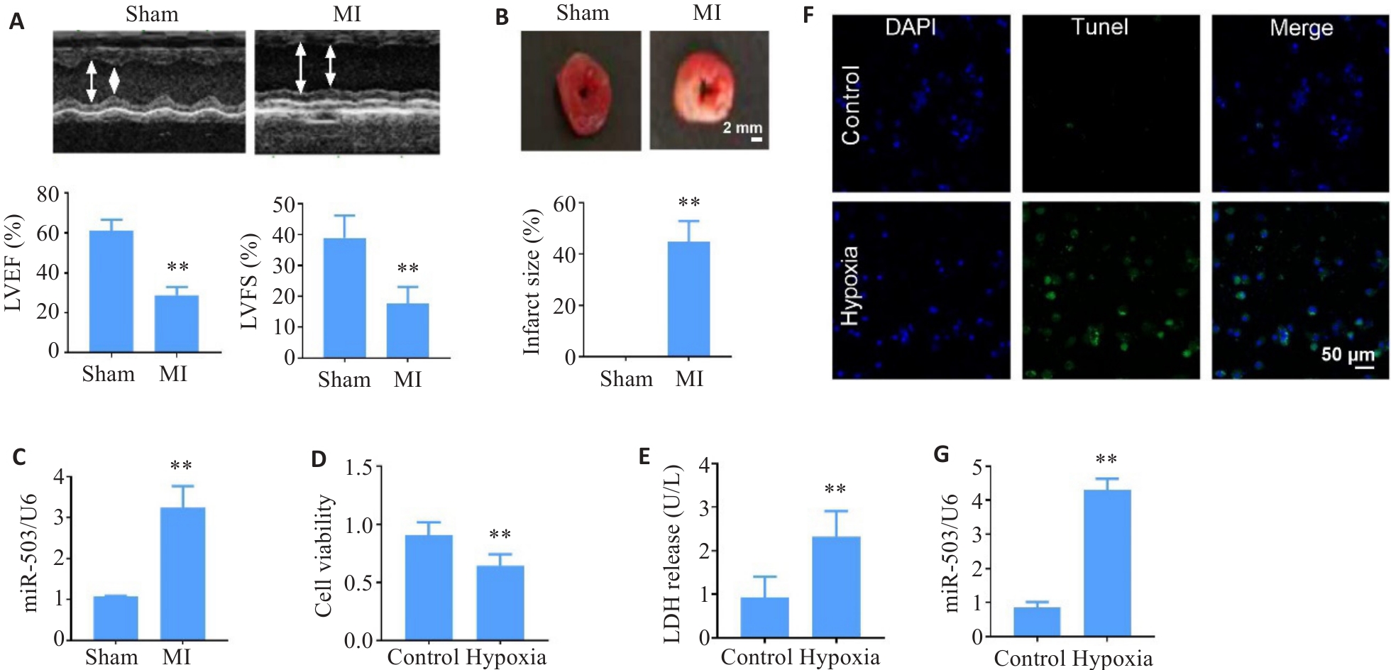
图1 MI与缺氧心肌细胞中miR-503表达上调
Fig.1 MiR-503 expression is upregulated in myocardial tissues of mice with myocardial infarction (MI) and in hypoxic primary neonatal rat cardiomyocytes. A, B: Echocardiography and TTC staining for assessing cardiac function and infarct area ion MI mice (n=5; **P<0.01 vs sham group). C: qRT-PCR showing elevated miR-503 expression in the peri-infarct zone in MI mice (n=10; **P<0.01 vs sham group). D, E: Decreased viability and increased LDH release in hypoxic primary cardiomyocytes (n=15; **P<0.01 vs control group). F: TUNEL staining showing increased apoptotic cells in the hypoxia group (Original magnification: ×100). G: Upregulated miR-503 expression in hypoxic cardiomyocytes (n=10; **P<0.01 vs control group).
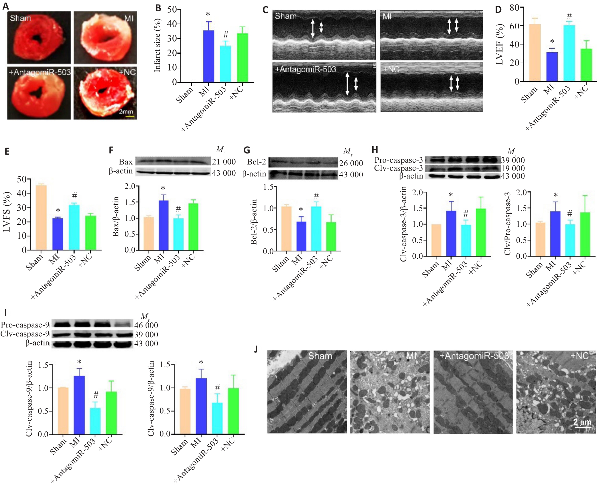
图2 AntagomiR-503减轻MI后心肌凋亡
Fig.2 AntagomiR-503 attenuates myocardial apoptosis in MI mice. A-E: TTC staining (n=4) and echocardiography (n=5) showing that antagomiR-503 improvescardiac function and reduces infarct size in MI mice (*P<0.05 vs sham group; #P<0.05 vs MI group). F, G: Western blotting showing that antagomiR-503 reduces myocardial expressions of Bax and BCl-2 in MI mice (n=6; *P<0.05 vs sham group; #P<0.05 vs MI group). H, I: Myocardial expressions of cleaved caspase-3 and cleaved caspase-9 in mice in different groups (n=4; *P<0.05 vs sham group; #P<0.05 vs MI group). J: Transmission electron microscopy showing improved mitochondrial structure after antagomiR-503 treatment in MI mice (Scale bar=2 μm).
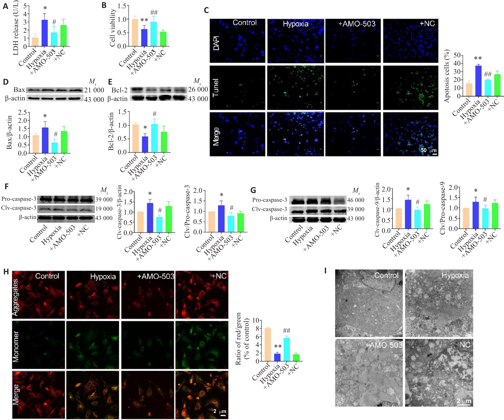
图3 AMO-503减轻缺氧诱导的心肌细胞凋亡
Fig.3 AMO-503 alleviates hypoxia-induced cardiomyocyte apoptosis. A, B: AMO-503 reduces LDH release (n=7) and increases cell viability (n=12; *P<0.05, **P<0.01 vs control group; #P<0.05,##P<0.01 vs hypoxia group). C: TUNEL staining showing that AMO-503 decreases cell apoptosis (n=5; **P<0.01 vs control group; ##P<0.01 vs hypoxia group). D-G: Western blotting showing that AMO-503 downregulates the expressions of Bax (n=5) and cleaved caspase-3/9 (n=4; *P<0.05 vs control group; #P<0.05 vs hypoxia group). H: JC-1 staining showing that AMO-503 restores mitochondrial membrane potential in hypoxic cardiomyocytes (n=6; **P<0.01 vs control group; ##P<0.01 vs hypoxia group). I: TEM images showing that AMO-503 improves mitochondrial morphology of the hypoxic cardiomyocytes (Scale bar=2 μm).
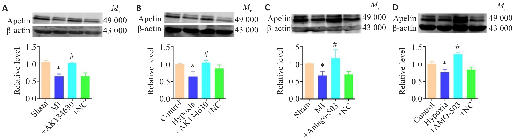
图4 AK134630/miR-503调控Apelin表达
Fig.4 Regulation of Apelin expression by AK134630/miR-503. A-D: Western blotting showed that either AK134630 overexpression or miR-503 inhibition (by Antago-503/AMO-503) upregulates Apelin expression (n=5). *P<0.05 vs sham group or hypoxia group; #P<0.05 vs MI group or hypoxia group.
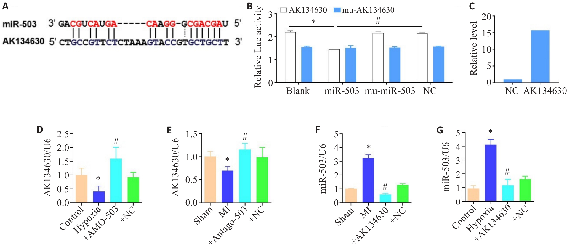
图5 miR-503与AK134630的相互作用
Fig.5 Interaction between miR-503 and AK134630. A: Schematic diagram of the predicted binding site between miR-503 and AK134630. B: Luciferase reporter assay showing that miR-503 inhibits the activity of AK134630 (n=4; *P<0.05 vs blank group; #P<0.05 vs miR-503 group). C: RNA pull-down assay showing that AK134630 bait significantly enriched miR-503 (vs. negative control NC), directly verifying their physical interaction. D-G: qRT-PCR analysis showing a negative correlation between the expression of AK134630 and miR-503 (n=5; *P<0.05 vs control group or sham group; #P<0.05 vs MI group or hypoxia group).
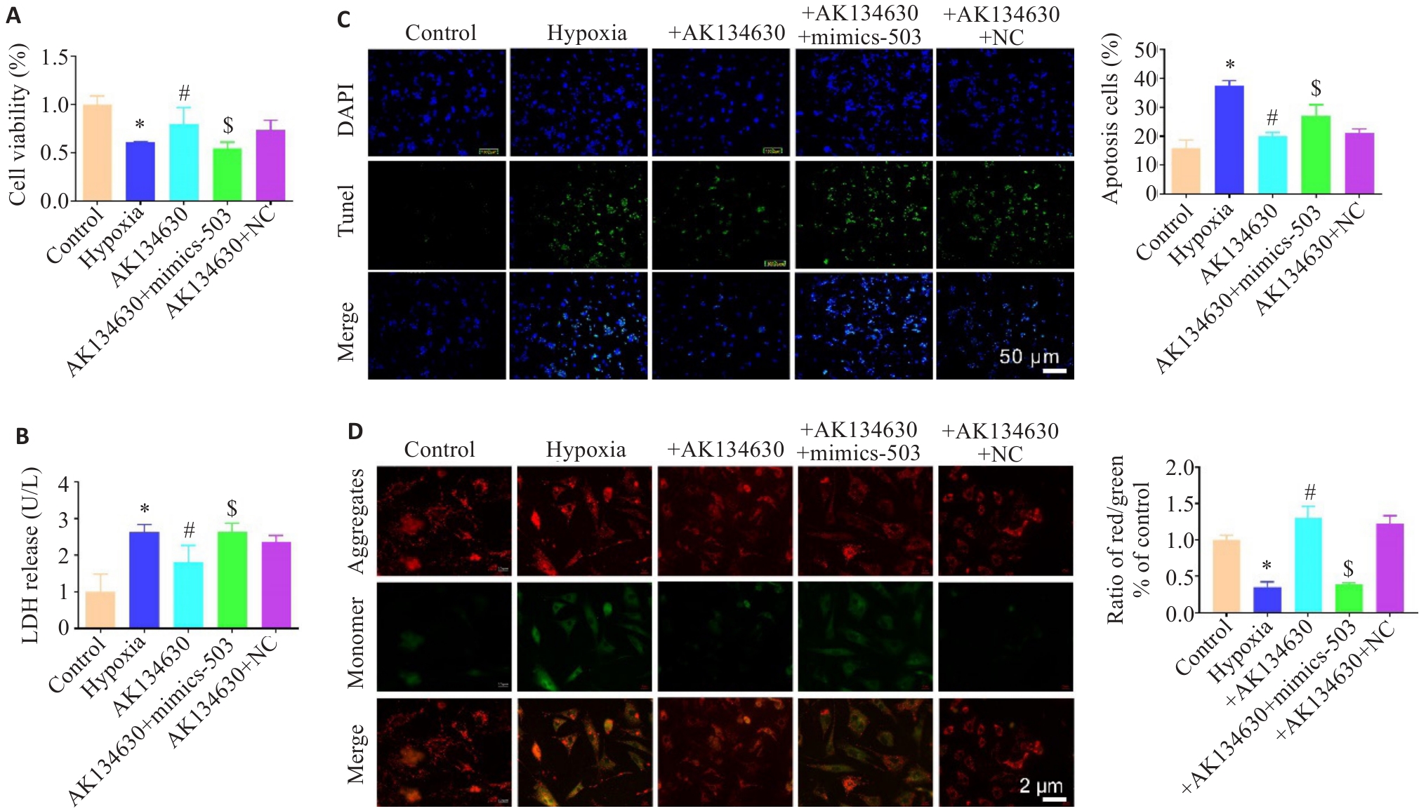
图6 AK134630通过调控miR-503减轻心肌细胞损伤
Fig.6 AK134630 alleviates cardiomyocyte injury by regulating miR-503. A, B: AK134630 overexpression increases cell viability (n=10) and decreases LDH release (n=6; *P<0.05 vs control group; #P<0.05 vs hypoxia group; $P<0.05 vs AK134630 group). C: TUNEL staining showing that AK134630 overexpression reduces hypoxic cardiomyocyte apoptosis (n=6; *P<0.05 vs control group; #P<0.05 vs hypoxia group; $P<0.05 vs AK134630 group). D: JC-1 staining showing that AK134630 overexpression restores mitochondrial membrane potential in hypoxic cardiomyocytes (n=6; *P<0.05 vs control group; #P<0.05 vs hypoxia group; $P<0.05 vs AK134630 group).
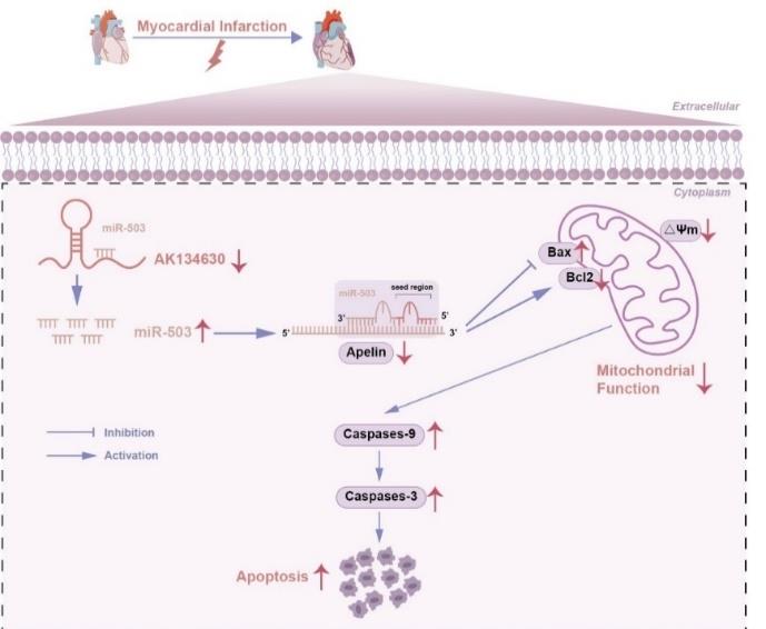
图7 机制示意图
Fig.7 Schematic diagram of the proposed mechanism. AK134630 inhibits miR-503 expression, leading to the upregulation of Apelin, which subsequently alleviates cardiomyocyte apoptosis and mitochondrial damage.
| [1] | Saito Y, Oyama K, Tsujita K, et al. Treatment strategies of acute myocardial infarction: updates on revascularization, pharmaco-logical therapy, and beyond[J]. J Cardiol, 2023, 81(2): 168-78. doi:10.1016/j.jjcc.2022.07.003 |
| [2] | Kulick N, Friede KA, Stouffer GA. Safety and efficacy of intracoronary thrombolytic agents during primary percutaneous coronary intervention for STEMI[J]. Expert Rev Cardiovasc Ther, 2023, 21(3): 165-75. doi:10.1080/14779072.2023.2184353 |
| [3] | Kim H, Lee YY, Kim VN. The biogenesis and regulation of animal microRNAs[J]. Nat Rev Mol Cell Biol, 2025, 26(4): 276-96. doi:10.1038/s41580-024-00805-0 |
| [4] | Wang ZG, Lu YJ, Yang BF. microRNAs and atrial fibrillation: new fundamentals[J]. Cardiovasc Res, 2011, 89(4): 710-21. doi:10.1093/cvr/cvq350 |
| [5] | He YJ, Cai Y, Pai PM, et al. The causes and consequences of miR-503 dysregulation and its impact on cardiovascular disease and cancer[J]. Front Pharmacol, 2021, 12: 629611. doi:10.3389/fphar.2021.629611 |
| [6] | Sun P, Wang C, Mang G, et al. Extracellular vesicle-packaged mitochondrial disturbing miRNA exacerbates cardiac injury during acute myocardial infarction[J]. Clin Transl Med, 2022, 12(4): e779. doi:10.1002/ctm2.779 |
| [7] | Wen Y, Chen R, Zhu CH, et al. miR-503 suppresses hypoxia-induced proliferation, migration and angiogenesis of endothelial progenitor cells by targeting Apelin[J]. Peptides, 2018, 105: 58-65. doi:10.1016/j.peptides.2018.05.008 |
| [8] | Sun SL, Shu YG, Tao MY. miR-503 inhibits proliferation, migration, and angiogenesis of glioma by acting on VEGFA through targeting LRIG2[J]. Cancer Manag Res, 2019, 11: 10599-608. doi:10.2147/CMAR.S222681 |
| [9] | Zhou YH, Deng L, Zhao DD, et al. microRNA-503 promotes angiotensin II-induced cardiac fibrosis by targeting Apelin-13[J]. J Cell Mol Med, 2016, 20(3): 495-505. doi:10.1111/jcmm.12754 |
| [10] | Yang Y, Zhang Y, Yang JQ, et al. Interdependent nuclear co-trafficking of ASPP1 and p53 aggravates cardiac ischemia/reperfusion injury[J]. Circ Res, 2023, 132(2): 208-22. doi:10.1161/circresaha.122.321153 |
| [11] | Bi FF, Cao M, Pan QM, et al. ITFG2, an immune-modulatory protein, targets ATP 5b to maintain mitochondrial function in myocardial infarction[J]. Biochem Pharmacol, 2024, 226: 116338. doi:10.1016/j.bcp.2024.116338 |
| [12] | Zhao PC, Zhu YQ, Sun L, et al. Circulating exosomal miR-1-3p from rats with myocardial infarction plays a protective effect on contrast-induced nephropathy via targeting ATG13 and activating the AKT signaling pathway[J]. Int J Biol Sci, 2021, 17(4): 972-85. doi:10.7150/ijbs.55887 |
| [13] | Qin W, Zhang LY, Li ZG, et al. Metoprolol protects against myocardial infarction by inhibiting miR-1 expression in rats[J]. J Pharm Pharmacol, 2020, 72(1): 76-83. doi:10.1111/jphp.13192 |
| [14] | Zhang TT, Zhang Y, Li S, et al. Gentianella acuta-derived Gen-miR-1 suppresses myocardial fibrosis by targeting HAX1/HMG20A/Smads axis to attenuate inflammation in cardiac fibroblasts[J]. Phytomedicine, 2023, 118: 154923. doi:10.1016/j.phymed.2023.154923 |
| [15] | Stein E, Duarte GBS, Bersch-Ferreira AC, et al. microRNAs, nut consumption, cardiovascular risk factors, and coronary artery disease: a narrative review[J]. Nutr Rev, 2025: nuaf243. doi:10.1093/nutrit/nuaf243 |
| [16] | Canale P, Nicolini G, Pitto L, et al. Role of miR-133/Dio3 axis in the T3-dependent modulation of cardiac mitoK-ATP expression[J]. Int J Mol Sci, 2022, 23(12):6549. doi:10.3390/ijms23126549 |
| [17] | Sun BX, Liu SW, Hao RB, et al. RGD-PEG-PLA delivers miR-133 to infarct lesions of acute myocardial infarction model rats for cardiac protection[J]. Pharmaceutics, 2020, 12(6): 575. doi:10.3390/pharmaceutics12060575 |
| [18] | Valussi M, Besser J, Wystub-Lis K, et al. Repression of Osmr and Fgfr1 by miR-1/133a prevents cardiomyocyte dedifferentiation and cell cycle entry in the adult heart[J]. Sci Adv, 2021, 7(42): eabi6648. doi:10.1126/sciadv.abi6648 |
| [19] | Faulkner JL, Sullivan JC. Circulating cell-free micro-RNA as biomarkers: from myocardial infarction to hypertension[J]. Clin Sci, 2022, 136(18): 1341-6. doi:10.1042/CS20220056 |
| [20] | Liu WH, Higashikuni Y, Sata M. Linking RNA dynamics to heart disease: the lncRNA/miRNA/mRNA axis in myocardial ischemia-reperfusion injury[J]. Hypertens Res, 2022, 45(6): 1067-9. doi:10.1038/s41440-022-00905-4 |
| [21] | Wang XJ, Liang GQ, Guo Q, et al. ELABELA improves endothelial cell function via the ELA-APJ axis by activating the PI3K/Akt signalling pathway in HUVECs and EA.hy926 cells[J]. Clin Exp Pharmacol Physiol, 2020, 47(12): 1953-64. doi:info:doi/10.1111/1440-1681.13382 |
| [22] | Liu W, Niu F, Sha H, et al. Apelin-13/APJ system delays intervertebral disc degeneration by activating the PI3K/AKT signaling pathway[J]. Eur Rev Med Pharmacol Sci, 2020, 24(6): 2820-8. doi:10.26355/eurrev_202003_20643 |
| [23] | Ye LY, Huang YJ, Liu XQ, et al. Apelin/APJ system protects placental trophoblasts from hypoxia-induced oxidative stress through activating PI3K/Akt signaling pathway in preeclampsia[J]. Free Radic Biol Med, 2023, 208: 759-70. doi:10.1016/j.freeradbiomed.2023.09.030 |
| [24] | Gao SH, Chen HP. Therapeutic potential of apelin and Elabela in cardiovascular disease[J]. Biomed Pharmacother, 2023, 166: 115268. doi:10.1016/j.biopha.2023.115268 |
| [25] | Bai B, Tang JY, Liu HQ, et al. Apelin-13 induces ERK1/2 but not p38 MAPK activation through coupling of the human apelin receptor to the Gi2 pathway[J]. Acta Biochim Biophys Sin, 2008, 40(4): 311-8. doi:10.1111/j.1745-7270.2008.00403.x |
| [26] | Gao M, Yin LH, Zhang B, et al. Targeting ischemic myocardium: nanoparticles loaded with long noncoding RNA AK156373 siRNA alleviate myocardial infarction[J]. ACS Nano, 2025, 19(19): 18475-91. doi:10.1021/acsnano.5c01641 |
| [27] | He JC, Zhou ZH, Kong DJ, et al. Integrative multi-omics identifies MEIS3 as a diagnostic biomarker and immune modulator in hyper-trophic cardiomyopathy[J]. Front Immunol, 2025, 16: 1675467. doi:10.3389/fimmu.2025.1675467 |
| [28] | Rong RX, Yuan T, Yan ZZ, et al. LncRNA NAV2-AS2 is critical for fibroblast-to-myofibroblast transition and cardiac fibrosis[J]. Int J Biol Macromol, 2025, 306(Pt 1): 141400. doi:10.1016/j.ijbiomac.2025.141400 |
| [29] | Yang PL, Zheng LR, Zhao YQ. Research progress on role of competitive endogenous RNA networks in heart failure and intervention by traditional Chinese medicine[J]. Zhongguo Zhongyao Zazhi, 2025, 50(12): 3232-43. |
| [30] | Yu QT, Li YX, Zhang N, et al. Silencing of lncRNA NEAT1 alleviates acute myocardial infarction by suppressing miR-450-5p/ACSL4-mediated ferroptosis[J]. Exp Cell Res, 2024, 442(2): 114217. doi:10.1016/j.yexcr.2024.114217 |
| [31] | Li XQ, Wang H, Yao B, et al. lncRNA H19/miR-675 axis regulates cardiomyocyte apoptosis by targeting VDAC1 in diabetic cardiomyopathy[J]. Sci Rep, 2016, 6: 36340. doi:10.1038/srep36340 |
| [32] | Liu LT, An XB, Li ZH, et al. The H19 long noncoding RNA is a novel negative regulator of cardiomyocyte hypertrophy[J]. Cardiovasc Res, 2016, 111(1): 56-65. doi:10.1093/cvr/cvw078 |
| [33] | Luo H, Wang J, Liu DH, et al. The lncRNA H19/miR-675 axis regulates myocardial ischemic and reperfusion injury by targeting PPARα[J]. Mol Immunol, 2019, 105: 46-54. doi:10.1016/j.molimm.2018.11.011 |
| [1] | 王炎炎, 宋志会, 郭流漓, 肖扬, 陈瑞, 王怡. 通脉养心丸通过激活ERK信号通路增强线粒体功能减轻大鼠心肌缺血再灌注损伤[J]. 南方医科大学学报, 2026, 46(6): 1203-1215. |
| [2] | 魏佳, 蒋伟, 夏林莺, 吕侣, 杨强. GDF11通过抑制硫氧还蛋白1硝基化失活减轻心肌缺血/再灌注损伤[J]. 南方医科大学学报, 2026, 46(5): 1075-1083. |
| [3] | 林雨星, 石思楠, 汪婧, 林思悦, 张杰, 林珊珊, 陈伊舒榕, 张铃, 蔡巧燕. 清达颗粒下调P53信号通路抑制高血压小鼠脑微血管内皮细胞凋亡[J]. 南方医科大学学报, 2026, 46(5): 1102-1110. |
| [4] | 马丹, 曹书源, 姜毅, 彭煜策, 何岸, 罗明昊, 罗素新. 褪黑素通过抑制STING信号通路减轻糖尿病小鼠心肌细胞坏死性凋亡[J]. 南方医科大学学报, 2026, 46(5): 1157-1166. |
| [5] | 雷艳萍, 宋嘉晟, 徐乐吾, 刘睿, 赵岳. 麦冬皂苷D通过激活β-catenin/FUNDC1/线粒体自噬轴减轻阿霉素诱导的小鼠心肌肥厚[J]. 南方医科大学学报, 2026, 46(4): 803-815. |
| [6] | 杨剑明, 杨龙, 洪铠文, 耿贝贝, 赵满, 王耀光, 夏婷, 董津睿. 腹腔注射薤白碳量子点可改善顺铂诱导的小鼠急性肾损伤并修复线粒体功能[J]. 南方医科大学学报, 2026, 46(3): 505-512. |
| [7] | 宋淇乐, 苗益恺, 冯小桐, 王一凡, 刘伟, 魏琪, 于新汝, 陈文文, 付晓艳. 硒代胱氨酸通过诱导活性氧产生启动氧化应激损伤抑制结肠癌细胞生长[J]. 南方医科大学学报, 2026, 46(3): 532-540. |
| [8] | 胡欣怡, 王圩健, 李慧, 陈宗正, 周忻, 袁俊菲, 陈亮. 急性ST段抬高型心肌梗死患者直接经皮冠状动脉介入治疗后30天主要不良心血管事件的列线图预测模型:基于血清GSDMD蛋白表达水平[J]. 南方医科大学学报, 2026, 46(3): 550-558. |
| [9] | 刘泽, 毛樟坤, 尤达, 王俊杰, 何咏梅, 余伊雯, 文志强, 方会龙, 何汶霞. 宽缨酮靶向抑制STAT3减轻线粒体功能障碍和炎症缓解急性肾损伤[J]. 南方医科大学学报, 2026, 46(3): 570-581. |
| [10] | 温贺新, 林洁, 左芦根, 刘牧林. 珠子草素通过拮抗肠上皮细胞凋亡与调控肠道Th1/Th2免疫平衡改善克罗恩病样小鼠结肠炎[J]. 南方医科大学学报, 2026, 46(3): 655-665. |
| [11] | 张语洋, 申颖, 佟欣雨, 段宇魁, 罗云娜, 郭文奇. 水飞蓟宾通过诱导细胞自噬及凋亡双重机制抑制兔青光眼术后纤维化[J]. 南方医科大学学报, 2026, 46(3): 666-674. |
| [12] | 乔通, 尹林, 张可妮, 牛民主, 黄菊, 耿志军, 李静, 胡建国. 茯苓新酸A通过调节AMPK/mTOR介导的自噬来减轻葡聚糖硫酸钠诱导的小鼠结肠炎[J]. 南方医科大学学报, 2026, 46(1): 131-140. |
| [13] | 张淑芬, 黄添容, 杨灿洪, 陈家镒, 吕田明, 张嘉发. 莱菔硫烷通过抑制Aβ42寡聚体激活的U87细胞中MAPK/NF-κB信号通路降低反应性星形胶质细胞介导的SH-SY5Y凋亡[J]. 南方医科大学学报, 2026, 46(1): 191-199. |
| [14] | 林心君, 何昱霖, 施红, 刘佳绣, 胡海霞. 石斛合剂通过调控Sirt3介导的线粒体自噬通路缓解大鼠糖尿病心肌病[J]. 南方医科大学学报, 2026, 46(1): 47-54. |
| [15] | 赵锦燕, 彭娇, 林明和, 朱晓勤, 黄彬, 林久茂. 清解扶正颗粒通过抑制线粒体依赖的凋亡、激活AMPK-PGC-1α通路缓解5-氟尿嘧啶引起的骨骼肌损伤[J]. 南方医科大学学报, 2026, 46(1): 94-103. |
| 阅读次数 | ||||||
|
全文 |
|
|||||
|
摘要 |
|
|||||
