
南方医科大学学报 ›› 2026, Vol. 46 ›› Issue (7): 1714-1722.doi: 10.12122/j.issn.1673-4254.2026.07.24
• • 上一篇
朱君1( ), 谭顺谦1, 黄方俊2, 蔡光瑶3, 甄鑫1()
收稿日期:2025-12-03
出版日期:2026-07-20
发布日期:2026-07-20
通讯作者:
甄鑫
E-mail:2570540683@qq.com;xinzhen@smu.edu.cn
作者简介:朱 君,在读硕士研究生,E-mail:2570540683@qq.com
基金资助:
Jun ZHU1(), Shunqian TAN1, Fangjun HUANG2, Guangyao CAI3, Xin ZHEN1()
Received:2025-12-03
Online:2026-07-20
Published:2026-07-20
Contact:
Xin ZHEN
E-mail:2570540683@qq.com;xinzhen@smu.edu.cn
Supported by:摘要:
目的 针对卵巢癌早期诊断中表格型医疗数据特征关联复杂、模型可解释性不足等问题,提出一种融合医疗先验知识的TabMap图像映射与深度学习预测框架。 方法 首先,根据临床语义将36个医疗诊断特征划分为血常规分区、生化指标分区、肿瘤标志物分区以及其他(凝血、炎症及临床变量)分区4个空间连续区域;其次,采用Gromov-Wasserstein最优传输算法求解特征空间与像素空间的最优耦合,生成保持拓扑结构的TabMap图像;然后,设计融合SE注意力机制和全局平均池化的轻量级卷积神经网络,通过类别加权损失和加权采样策略应对样本不平衡问题;最后,利用类激活映射技术可视化模型决策过程,实现可解释性分析。 结果 在真实卵巢癌数据集上的实验表明,本文方法的测试集准确率达到91.82%,精确率为89.96%,召回率为89.45%,F1分数为0.8970,平衡准确率为88.51%,相比传统机器学习方法准确率提升8.26%~14.93%,相比标准深度学习模型准确率提升2.28%~13.60%。可解释性分析显示,模型在肿瘤标志物分区以及凝血、炎症以及临床变量相关分区中呈现出明显的响应模式,与临床既有认知保持一致;特征重要性排序进一步强调了CA125、HE4等关键指标在模型决策中的核心作用。 结论 本研究提出的医疗先验引导TabMap方法有效融合了领域知识与数据驱动学习,在提升预测性能的同时增强了模型的临床可解释性,为表格型医疗数据的深度学习建模提供了新思路,具有良好的临床应用潜力。
朱君, 谭顺谦, 黄方俊, 蔡光瑶, 甄鑫. 医疗先验引导的TabMap深度学习模型用于卵巢癌预测及可解释性研究[J]. 南方医科大学学报, 2026, 46(7): 1714-1722.
Jun ZHU, Shunqian TAN, Fangjun HUANG, Guangyao CAI, Xin ZHEN. Medical prior-guided TabMap deep learning model for ovarian cancer prediction and interpretability analysis[J]. Journal of Southern Medical University, 2026, 46(7): 1714-1722.
| Clinical characteristic parameters | Sample 1 | Sample 2 | Sample 3 | |
|---|---|---|---|---|
| Hematological parameters | White blood cell count (WBC#), venous blood | 5.2 | 6.69 | 5.67 |
| Lymphocyte percentage (Lymph%), venous blood | 38 | 33.2 | 25.6 | |
| Mean corpuscular volume (MCV), venous blood | 91.9 | 86.3 | 71.3 | |
| Mean corpuscular hemoglobin (MCH), venous blood | 29.6 | 27.6 | 20.7 | |
| Mean corpuscular hemoglobin concentration (MCHC), venous blood | 322 | 320 | 290 | |
| Basophil percentage (Baso%), venous blood | 0.6 | 1 | 1.2 | |
| Eosinophil count (Eos#), venous blood | 0.2 | 0.6 | 0.1 | |
| Platelet count (PLT#), venous blood | 281 | 201 | 496 | |
| Plateletcrit (PCT), venous blood | 0.25 | 0.21 | 0.48 | |
| Neutrophil count (Neut#), venous blood | 2.7 | 3.3 | 3.6 | |
| Neutrophil percentage (Neut%), venous blood | 52.7 | 49.5 | 63.1 | |
| Eosinophil percentage (Eos%), venous blood | 2.9 | 8.8 | 0.9 | |
| Biochemical parameters | Albumin-to-globulin ratio (ALB/GLO), venous blood | 1.73 | 1.48 | 1.44 |
| Albumin (ALB), venous blood | 46.9 | 41.8 | 42.3 | |
| Indirect bilirubin (IBIL), venous blood | 5.7 | 9 | 4.7 | |
| Globulin (GLO), venous blood | 27.08 | 28.15 | 29.33 | |
| Lactate dehydrogenase (LDH), venous blood | 194.1 | 143.5 | 135.9 | |
| Direct bilirubin (DBIL), venous blood | 2.6 | 3.3 | 2.1 | |
| Total cholesterol (TC), venous blood | 4.9 | 4.29 | 4.71 | |
| Total bilirubin (TBIL), venous blood | 8.3 | 12.3 | 6.8 | |
| Total protein (TP), venous blood | 73.98 | 69.95 | 71.63 | |
| Uric acid (UA), venous blood | 300.2 | 201.1 | 202 | |
| Low-density lipoprotein cholesterol (LDL-C), venous blood | 3.29 | 2.64 | 3.14 | |
| Triglycerides (TG), venous blood | 1.79 | 3.08 | 1.03 | |
| High-density lipoprotein cholesterol (HDL-C), venous blood | 1.1 | 0.86 | 1.29 | |
| Tumor markers | Alpha-fetoprotein (AFP), venous blood | 1.48 | 3.53 | 1.11 |
| Human epididymis protein 4 (HE4), venous blood | 139.7 | 80.2 | 51.2 | |
| Carbohydrate antigen 125 (CA125), venous blood | 16.38 | 83.6 | 11.72 | |
| Carbohydrate antigen 153 (CA153), venous blood | 11.99 | 14.94 | 15.51 | |
| Carbohydrate antigen 724 (CA724), venous blood | 2.25 | 0.852 | 0.858 | |
Other variables (coagulation, inflammatory, and clinical variables) | Urine pH | 6 | 6 | 6.5 |
| C-reactive protein (CRP), venous blood | 0.35 | 1.45 | 0.07 | |
| D-dimer, venous blood | 1.07 | 0.61 | 0.29 | |
| Antithrombin III activity (ATIII:A), venous blood | 89.3 | 99.9 | 104 | |
| Fibrinogen (Fbg), venous blood | 2.29 | 2.76 | 2.35 | |
| Age (years) | 51 | 57 | 56 | |
| Outcome label | Ovarian cancer status: 0/1 (No/Yes) | 1 | 0 | 0 |
表1 卵巢癌vs.对照人群37维临床特征数据集(2012-2021)样本示例表
Tab.1 Sample table of the 37-dimensional clinical feature dataset for ovarian cancer vs. controls (2012-2021)
| Clinical characteristic parameters | Sample 1 | Sample 2 | Sample 3 | |
|---|---|---|---|---|
| Hematological parameters | White blood cell count (WBC#), venous blood | 5.2 | 6.69 | 5.67 |
| Lymphocyte percentage (Lymph%), venous blood | 38 | 33.2 | 25.6 | |
| Mean corpuscular volume (MCV), venous blood | 91.9 | 86.3 | 71.3 | |
| Mean corpuscular hemoglobin (MCH), venous blood | 29.6 | 27.6 | 20.7 | |
| Mean corpuscular hemoglobin concentration (MCHC), venous blood | 322 | 320 | 290 | |
| Basophil percentage (Baso%), venous blood | 0.6 | 1 | 1.2 | |
| Eosinophil count (Eos#), venous blood | 0.2 | 0.6 | 0.1 | |
| Platelet count (PLT#), venous blood | 281 | 201 | 496 | |
| Plateletcrit (PCT), venous blood | 0.25 | 0.21 | 0.48 | |
| Neutrophil count (Neut#), venous blood | 2.7 | 3.3 | 3.6 | |
| Neutrophil percentage (Neut%), venous blood | 52.7 | 49.5 | 63.1 | |
| Eosinophil percentage (Eos%), venous blood | 2.9 | 8.8 | 0.9 | |
| Biochemical parameters | Albumin-to-globulin ratio (ALB/GLO), venous blood | 1.73 | 1.48 | 1.44 |
| Albumin (ALB), venous blood | 46.9 | 41.8 | 42.3 | |
| Indirect bilirubin (IBIL), venous blood | 5.7 | 9 | 4.7 | |
| Globulin (GLO), venous blood | 27.08 | 28.15 | 29.33 | |
| Lactate dehydrogenase (LDH), venous blood | 194.1 | 143.5 | 135.9 | |
| Direct bilirubin (DBIL), venous blood | 2.6 | 3.3 | 2.1 | |
| Total cholesterol (TC), venous blood | 4.9 | 4.29 | 4.71 | |
| Total bilirubin (TBIL), venous blood | 8.3 | 12.3 | 6.8 | |
| Total protein (TP), venous blood | 73.98 | 69.95 | 71.63 | |
| Uric acid (UA), venous blood | 300.2 | 201.1 | 202 | |
| Low-density lipoprotein cholesterol (LDL-C), venous blood | 3.29 | 2.64 | 3.14 | |
| Triglycerides (TG), venous blood | 1.79 | 3.08 | 1.03 | |
| High-density lipoprotein cholesterol (HDL-C), venous blood | 1.1 | 0.86 | 1.29 | |
| Tumor markers | Alpha-fetoprotein (AFP), venous blood | 1.48 | 3.53 | 1.11 |
| Human epididymis protein 4 (HE4), venous blood | 139.7 | 80.2 | 51.2 | |
| Carbohydrate antigen 125 (CA125), venous blood | 16.38 | 83.6 | 11.72 | |
| Carbohydrate antigen 153 (CA153), venous blood | 11.99 | 14.94 | 15.51 | |
| Carbohydrate antigen 724 (CA724), venous blood | 2.25 | 0.852 | 0.858 | |
Other variables (coagulation, inflammatory, and clinical variables) | Urine pH | 6 | 6 | 6.5 |
| C-reactive protein (CRP), venous blood | 0.35 | 1.45 | 0.07 | |
| D-dimer, venous blood | 1.07 | 0.61 | 0.29 | |
| Antithrombin III activity (ATIII:A), venous blood | 89.3 | 99.9 | 104 | |
| Fibrinogen (Fbg), venous blood | 2.29 | 2.76 | 2.35 | |
| Age (years) | 51 | 57 | 56 | |
| Outcome label | Ovarian cancer status: 0/1 (No/Yes) | 1 | 0 | 0 |
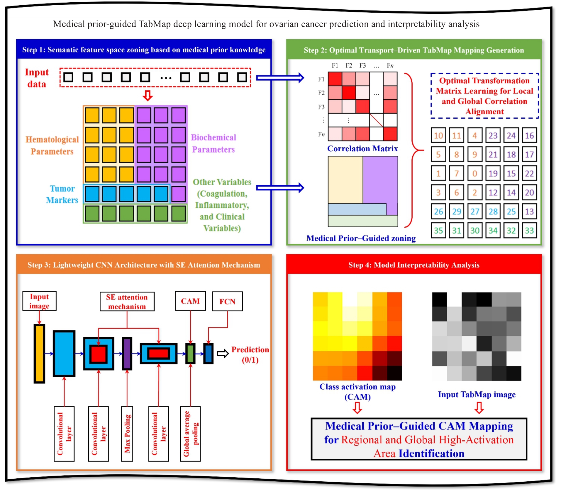
图1 医疗先验引导的TabMap深度学习模型用于卵巢癌预测及可解释性流程图
Fig.1 Medical prior-guided TabMap deep learning model for ovarian cancer prediction and interpretability.
| Model | Acc | Precision | Recall | F1-Score | BA |
|---|---|---|---|---|---|
| SVM | 77.45 | 75.32 | 73.28 | 0.7428 | 74.30 |
| RF | 81.23 | 79.45 | 77.56 | 0.7849 | 78.51 |
| LR | 76.89 | 74.67 | 72.34 | 0.7348 | 73.51 |
| XGBoost | 83.56 | 81.23 | 79.45 | 0.8033 | 80.34 |
| MLP | 78.22 | 78.34 | 76.45 | 0.7738 | 77.40 |
| CNN | 88.67 | 86.23 | 86.89 | 0.8655 | 85.06 |
| ResNet18 | 89.54 | 88.12 | 88.23 | 0.8817 | 86.18 |
| Ours | 91.82 | 89.96 | 89.45 | 0.8970 | 88.51 |
表2 不同模型在测试集上的性能对比
Tab.2 Performance comparison of different models on the test set
| Model | Acc | Precision | Recall | F1-Score | BA |
|---|---|---|---|---|---|
| SVM | 77.45 | 75.32 | 73.28 | 0.7428 | 74.30 |
| RF | 81.23 | 79.45 | 77.56 | 0.7849 | 78.51 |
| LR | 76.89 | 74.67 | 72.34 | 0.7348 | 73.51 |
| XGBoost | 83.56 | 81.23 | 79.45 | 0.8033 | 80.34 |
| MLP | 78.22 | 78.34 | 76.45 | 0.7738 | 77.40 |
| CNN | 88.67 | 86.23 | 86.89 | 0.8655 | 85.06 |
| ResNet18 | 89.54 | 88.12 | 88.23 | 0.8817 | 86.18 |
| Ours | 91.82 | 89.96 | 89.45 | 0.8970 | 88.51 |
| Model | AUC | Z-statistic | P |
|---|---|---|---|
| SVM | 0.8156 | 4.89 | <0.001 |
| RF | 0.8489 | 3.96 | <0.001 |
| LR | 0.8078 | 5.12 | <0.001 |
| XGBoost | 0.8723 | 3.45 | <0.001 |
| MLP | 0.8245 | 4.67 | <0.001 |
| CNN | 0.9089 | 2.15 | 0.0312 |
| ResNet18 | 0.9167 | 2.03 | 0.0421 |
| Ours | 0.9385 | - | - |
表3 不同模型与本文方法统计显著性检验结果对比
Tab.3 Comparison of statistical significance test results of different models with the proposed method
| Model | AUC | Z-statistic | P |
|---|---|---|---|
| SVM | 0.8156 | 4.89 | <0.001 |
| RF | 0.8489 | 3.96 | <0.001 |
| LR | 0.8078 | 5.12 | <0.001 |
| XGBoost | 0.8723 | 3.45 | <0.001 |
| MLP | 0.8245 | 4.67 | <0.001 |
| CNN | 0.9089 | 2.15 | 0.0312 |
| ResNet18 | 0.9167 | 2.03 | 0.0421 |
| Ours | 0.9385 | - | - |
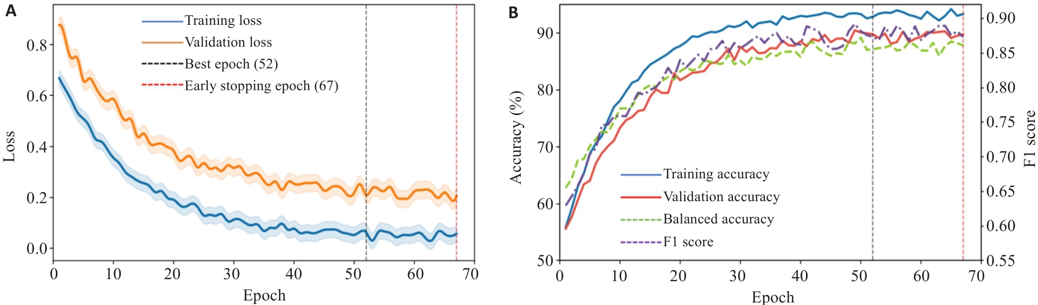
图2 模型训练过程中的收敛曲线
Fig.2 Convergence curves during the model training process. A: Changes in the loss function. B: Changes in accuracy rate.
| Imbalance handling strategy | Acc | Precision | Recall | F1-Score | BA |
|---|---|---|---|---|---|
| Baseline (No imbalance handling) | 90.83 | 86.30 | 82.05 | 0.8412 | 83.64 |
| Weighted loss only | 91.04 | 86.74 | 85.90 | 0.8632 | 85.72 |
| Weighted random sampling only | 90.62 | 86.84 | 87.18 | 0.8701 | 86.57 |
| Weighted loss+weighted sampling (Proposed Method) | 91.82 | 89.96 | 89.45 | 0.8970 | 88.51 |
表4 样本不平衡策略对比分析
Tab.4 Comparison and analysis of sample imbalance strategies
| Imbalance handling strategy | Acc | Precision | Recall | F1-Score | BA |
|---|---|---|---|---|---|
| Baseline (No imbalance handling) | 90.83 | 86.30 | 82.05 | 0.8412 | 83.64 |
| Weighted loss only | 91.04 | 86.74 | 85.90 | 0.8632 | 85.72 |
| Weighted random sampling only | 90.62 | 86.84 | 87.18 | 0.8701 | 86.57 |
| Weighted loss+weighted sampling (Proposed Method) | 91.82 | 89.96 | 89.45 | 0.8970 | 88.51 |
| Feature mapping strategy | Acc | BA | Performance gain over TabMap |
|---|---|---|---|
| No prior partition+optimal transport (TabMap) | 88.67 | 85.06 | - |
| Random partition+optimal transport | 87.95 | 84.28 | -0.72% / -0.78% |
| Prior-guided partitio +optimal transport | 91.82 | 88.51 | +3.15% / +3.45% |
表5 医疗先验引导策略消融实验结果对比
Tab.5 Comparison of experimental results of medical prior-guided strategy ablation
| Feature mapping strategy | Acc | BA | Performance gain over TabMap |
|---|---|---|---|
| No prior partition+optimal transport (TabMap) | 88.67 | 85.06 | - |
| Random partition+optimal transport | 87.95 | 84.28 | -0.72% / -0.78% |
| Prior-guided partitio +optimal transport | 91.82 | 88.51 | +3.15% / +3.45% |
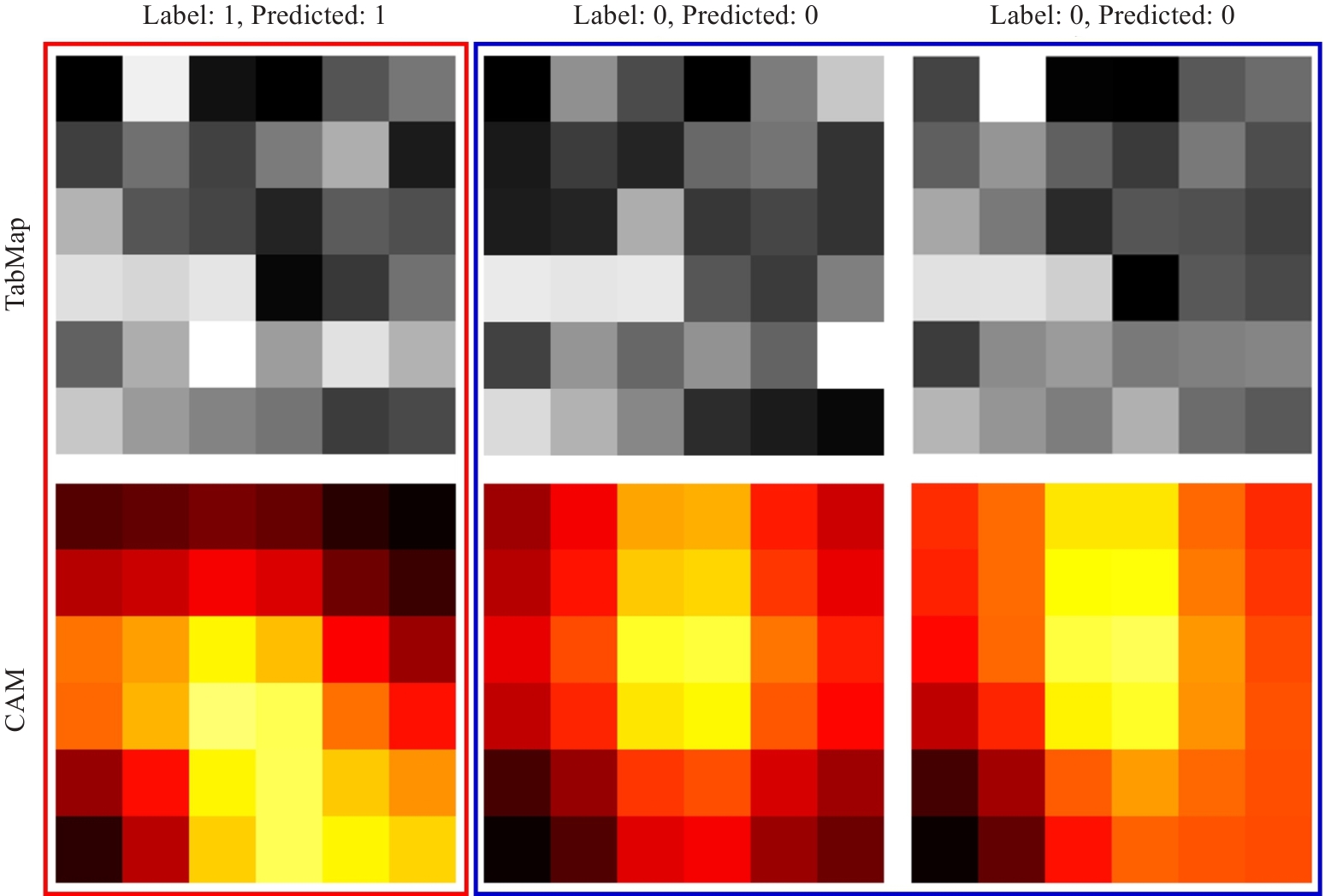
图3 3个测试样本的类激活映射可视化
Fig.3 Visualization of class activation maps for 3 test samples.
| Rank | Feature name | Activation strength | Assigned region | Clinical significance |
|---|---|---|---|---|
| 1 | Carbohydrate antigen 125 (CA125) | 0.7118 | Tumor Marker Region | The most important biomarker for ovarian cancer, with high sensitivity |
| 2 | Human epididymis Protein 4 (HE4) | 0.7040 | Tumor Marker Region | A specific biomarker for epithelial ovarian cancer |
| 3 | D-dimer | 0.7001 | Other (Coagulation, Inflammatory, and Clinical Variables) Region | Reflects a hypercoagulable state and is associated with tumor-related thrombotic risk |
| 4 | Fibrinogen (Fbg) | 0.6714 | Other (Coagulation, Inflammatory, and Clinical Variables) Region | An acute-phase reactant associated with tumor burden |
| 5 | C-reactive protein (CRP) | 0.6712 | Other (Coagulation, Inflammatory, and Clinical Variables) Region | An inflammatory marker reflecting the tumor microenvironment |
| 6 | Carbohydrate antigen 153 (CA153) | 0.6706 | Tumor Marker Region | An auxiliary biomarker for tumor differential diagnosis |
| 7 | Albumin (ALB) | 0.6685 | Biochemical Parameter Region | Reflects nutritional status and systemic inflammatory level |
| 8 | Age | 0.6682 | Other (Coagulation, Inflammatory, and Clinical Variables) Region | An important epidemiological risk factor for ovarian cancer |
| 9 | Carbohydrate antigen 724 (CA724) | 0.6676 | Tumor Marker Region | An auxiliary tumor biomarker |
| 10 | Alpha-fetoprotein (AFP) | 0.6571 | Tumor Marker Region | Used for differentiating germ cell tumors |
表6 基于类激活映射的关键特征重要性排名
Tab.6 Key feature importance ranking based on class activation mapping
| Rank | Feature name | Activation strength | Assigned region | Clinical significance |
|---|---|---|---|---|
| 1 | Carbohydrate antigen 125 (CA125) | 0.7118 | Tumor Marker Region | The most important biomarker for ovarian cancer, with high sensitivity |
| 2 | Human epididymis Protein 4 (HE4) | 0.7040 | Tumor Marker Region | A specific biomarker for epithelial ovarian cancer |
| 3 | D-dimer | 0.7001 | Other (Coagulation, Inflammatory, and Clinical Variables) Region | Reflects a hypercoagulable state and is associated with tumor-related thrombotic risk |
| 4 | Fibrinogen (Fbg) | 0.6714 | Other (Coagulation, Inflammatory, and Clinical Variables) Region | An acute-phase reactant associated with tumor burden |
| 5 | C-reactive protein (CRP) | 0.6712 | Other (Coagulation, Inflammatory, and Clinical Variables) Region | An inflammatory marker reflecting the tumor microenvironment |
| 6 | Carbohydrate antigen 153 (CA153) | 0.6706 | Tumor Marker Region | An auxiliary biomarker for tumor differential diagnosis |
| 7 | Albumin (ALB) | 0.6685 | Biochemical Parameter Region | Reflects nutritional status and systemic inflammatory level |
| 8 | Age | 0.6682 | Other (Coagulation, Inflammatory, and Clinical Variables) Region | An important epidemiological risk factor for ovarian cancer |
| 9 | Carbohydrate antigen 724 (CA724) | 0.6676 | Tumor Marker Region | An auxiliary tumor biomarker |
| 10 | Alpha-fetoprotein (AFP) | 0.6571 | Tumor Marker Region | Used for differentiating germ cell tumors |
| [1] | Han BF, Zheng RS, Zeng HM, et al. Cancer incidence and mortality in China, 2022[J]. J Natl Cancer Cent, 2024, 4(1): 47-53. doi:10.1016/j.jncc.2024.01.006 |
| [2] | Siegel RL, Giaquinto AN, Jemal A. Cancer statistics, 2024[J]. CA A Cancer J Clin, 2024, 74(1): 12-49. doi:10.3322/caac.21820 |
| [3] | Sundar S, Neal RD, Kehoe S. Diagnosis of ovarian cancer[J]. Bmj, 2015: h4443. doi:10.1136/bmj.h4443 |
| [4] | Dexter JM, Brubaker LW, Bitler BG, et al. Ovarian cancer think tank: an overview of the current status of ovarian cancer screening and recommendations for future directions[J]. Gynecol Oncol Rep, 2024, 53: 101376. doi:10.1016/j.gore.2024.101376 |
| [5] | Lawson-Michod KA, Watt MH, Grieshober L, et al. Pathways to ovarian cancer diagnosis: a qualitative study[J]. BMC Womens Health, 2022, 22(1): 430. doi:10.1186/s12905-022-02016-1 |
| [6] | Mataraso SJ, Espinosa CA, Seong D, et al. A machine learning approach to leveraging electronic health records for enhanced omics analysis[J]. Nat Mach Intell, 2025, 7(2): 293-306. doi:10.1038/s42256-024-00974-9 |
| [7] | Krones F, Marikkar U, Parsons G, et al. Review of multimodal machine learning approaches in healthcare[J]. Inf Fusion, 2025, 114: 102690. doi:10.1016/j.inffus.2024.102690 |
| [8] | He X, Bai XH, Chen H, et al. Machine learning models in evaluating the malignancy risk of ovarian tumors: a comparative study[J]. J Ovarian Res, 2024, 17(1): 219. doi:10.1186/s13048-024-01544-8 |
| [9] | Guido R, Ferrisi S, Lofaro D, et al. An overview on the advancements of support vector machine models in healthcare applications: a review[J]. Information, 2024, 15(4): 235. doi:10.3390/info15040235 |
| [10] | Barreñada L, Dhiman P, Timmerman D, et al. Understanding overfitting in random forest for probability estimation: a visualization and simulation study[J]. Diagn Progn Res, 2024, 8(1): 14. doi:10.1186/s41512-024-00177-1 |
| [11] | Madakkatel I, Lumsden AL, Mulugeta A, et al. Large-scale analysis to identify risk factors for ovarian cancer[J]. Int J Gynecol Cancer, 2025, 35(8): 101844. doi:10.1136/ijgc-2024-005424 |
| [12] | Zhu YT, Brettin T, Xia FF, et al. Converting tabular data into images for deep learning with convolutional neural networks[J]. Sci Rep, 2021, 11(1): 11325. doi:10.1038/s41598-021-90923-y |
| [13] | Sharma A, Vans E, Shigemizu D, et al. DeepInsight: a methodology to transform a non-image data to an image for convolution neural network architecture[J]. Sci Rep, 2019, 9(1): 11399. doi:10.1038/s41598-019-47765-6 |
| [14] | Bazgir O, Zhang RB, Dhruba SR, et al. Representation of features as images with neighborhood dependencies for compatibility with convolutional neural networks[J]. Nat Commun, 2020, 11(1): 4391. doi:10.1038/s41467-020-18197-y |
| [15] | Yan R, Islam MT, Xing L. Interpretable discovery of patterns in tabular data via spatially semantic topographic maps[J]. Nat Biomed Eng, 2025, 9(4): 471-82. doi:10.1038/s41551-024-01268-6 |
| [16] | Zhang TS, Pang AB, Lyu JG, et al. Application of nonlinear models combined with conventional laboratory indicators for the diagnosis and differential diagnosis of ovarian cancer[J]. J Clin Med, 2023, 12(3): 844. doi:10.3390/jcm12030844 |
| [17] | 叶应妩. 全国临床检验操作规程[M]. 3版. 南京: 东南大学出版社, 2006. |
| [18] | Burtis CA, Ashwood ER, Bruns DE. Tietz textbook of clinical chemistry and molecular diagnostics[M]. Elsevier, 2012. doi:10.1016/b978-1-4160-6164-9.00139-6 |
| [19] | Peyré G, Cuturi M, Solomon JM. Gromov-Wasserstein averaging of kernel and distance matrices[C]//International Conference on Machine Learning., 2016. |
| [20] | Xiong LL, Yi C, Xiong QL, et al. SEA-NET: medical image segmentation network based on spiral squeeze-and-excitation and attention modules[J]. BMC Med Imaging, 2024, 24(1): 17. doi:10.1186/s12880-024-01194-8 |
| [21] | Song ZY, Shi ZL, Yan XM, et al. An improved weighted cross-entropy-based convolutional neural network for auxiliary diagnosis of pneumonia[J]. Electronics, 2024, 13(15): 2929. doi:10.3390/electronics13152929 |
| [22] | Tharwat A. Classification assessment methods[J]. Appl Comput Inform, 2021, 17(1): 168-92. doi:10.1016/j.aci.2018.08.003 |
| [23] | Chen TQ, Guestrin C. XGBoost: a scalable tree boosting system[C]//Proceedings of the 22nd ACM SIGKDD International Conference on Knowledge Discovery and Data Mining. San Francisco California USA. ACM, 2016: 785-794. doi:10.1145/2939672.2939785 |
| [24] | Abu-Doush I, Ahmed B, Awadallah MA, et al. Enhancing multilayer perceptron neural network using archive-based Harris Hawks optimizer to predict gold prices[J]. J King Saud Univ Comput Inf Sci, 2023, 35(5): 101557. doi:10.1016/j.jksuci.2023.101557 |
| [25] | Krichen M. Convolutional neural networks: a survey[J]. Computers, 2023, 12(8): 151. doi:10.3390/computers12080151 |
| [26] | He KM, Zhang XY, Ren SQ, et al. Deep residual learning for image recognition[C]//2016 IEEE Conference on Computer Vision and Pattern Recognition (CVPR). June 27-30, 2016, Las Vegas, NV, USA. IEEE, 2016: 770-8. doi:10.1109/cvpr.2016.90 |
| [27] | Momenimovahed Z, Mazidimoradi A, Allahqoli L, et al. The role of CA-125 in the management of ovarian cancer: a systematic review[J]. Cancer Rep, 2025, 8(3): e70142. doi:10.1002/cnr2.70142 |
| [28] | Farzaneh F, Salimnezhad M, Hosseini MS, et al. D-dimer, fibrinogen and tumor marker levels in patients with benign and malignant ovarian tumors[J]. Asian Pac J Cancer Prev, 2023, 24(12): 4263-8. doi:10.31557/apjcp.2023.24.12.4263 |
| [29] | Zhang LY, Guo LL, Wang HY, et al. Knowledge graph and bibliometric analysis of inflammatory indicators in ovarian cancer[J]. Front Oncol, 2025, 15: 1533537. doi:10.3389/fonc.2025.1533537 |
| [1] | 张明旭, 吴迪恒, 林嘉泽, 吕祎君, 周凌宏, 甄鑫, 周浩, 秦耿耿. 基于Transformer的影像与检验数据多模态预测模型在医院获得性感染的应用研究[J]. 南方医科大学学报, 2026, 46(6): 1423-1433. |
| [2] | 许家宁, 毛钇骁, 张煜. 基于分层注意力驱动多实例学习的肾透明细胞癌数字病理分级分期[J]. 南方医科大学学报, 2026, 46(5): 1175-1184. |
| [3] | 王童, 段曹辉, 娄昕, 冯前进. 基于血管增强一致性保持扩散模型的3T到7T SWI生成方法[J]. 南方医科大学学报, 2026, 46(5): 1185-1192. |
| [4] | 陈博湧, 汪新怡, 赵新新, 宋婷, 李永宝. 基于Swin-ResViT网络的低质量动态cine-MR至高质量定位MR图像实时生成研究[J]. 南方医科大学学报, 2026, 46(4): 929-938. |
| [5] | 谭顺谦, 卓俐, 曾敏, 黄方俊, 朱君, 蔡光瑶, 甄鑫. 基于实验室指标的Transformer模型可高效鉴别卵巢癌[J]. 南方医科大学学报, 2026, 46(4): 939-945. |
| [6] | 卢学麒, 陈华元, 吴秋岑, 温耀棋, 刘国光, 陈超敏. 一种基于深度特征融合的可解释性12导联心电图自动诊断模型研究[J]. 南方医科大学学报, 2026, 46(1): 208-218. |
| [7] | 吴秋岑, 卢学麒, 温耀棋, 洪永, 吴煜良, 陈超敏. II导联心电图中心肌梗死检测与定位:基于多尺度残差模块融合改进通道注意力模型[J]. 南方医科大学学报, 2025, 45(8): 1777-1790. |
| [8] | 郑子瑜, 杨夏颖, 吴圣杰, 张诗婕, 吕国荣, 柳培忠, 王珺, 何韶铮. 多特征融合的产时超声胎方位识别模型[J]. 南方医科大学学报, 2025, 45(7): 1563-1570. |
| [9] | 渠梦, 傅蓉. ResLSTM-TemporalSE:多导联心电信号的自动分类[J]. 南方医科大学学报, 2025, 45(12): 2708-2717. |
| [10] | 谢辉荣, 胡潮滨, 梁国华, 韩红喆, 黄牧, 冯前进. 大学生心理压力智能评估:基于融合文本与影像的多模态模型的设计及验证[J]. 南方医科大学学报, 2025, 45(11): 2504-2510. |
| [11] | 贺亚迪, 周炫汝, 金锦辉, 宋婷. 基于PE-CycleGAN网络的鼻咽癌自适应放疗CBCT-sCT生成研究[J]. 南方医科大学学报, 2025, 45(1): 179-186. |
| [12] | 方威扬, 肖慧, 王爽, 林晓明, 陈超敏. 基于MRI影像和临床参数特征融合的深度学习模型预测术前肝细胞癌的细胞角蛋白19状态[J]. 南方医科大学学报, 2024, 44(9): 1738-1751. |
| [13] | 欧嘉志, 詹长安, 杨丰. 一维卷积神经网络的自编码癫痫发作异常检测模型[J]. 南方医科大学学报, 2024, 44(9): 1796-1804. |
| [14] | 汪辰, 蒙铭强, 李明强, 王永波, 曾栋, 边兆英, 马建华. 基于双域Transformer耦合特征学习的CT截断数据重建模型[J]. 南方医科大学学报, 2024, 44(5): 950-959. |
| [15] | 龙楷兴, 翁丹仪, 耿 舰, 路艳蒙, 周志涛, 曹 蕾. 基于多模态多示例学习的免疫介导性肾小球疾病自动分类方法[J]. 南方医科大学学报, 2024, 44(3): 585-593. |
| 阅读次数 | ||||||
|
全文 |
|
|||||
|
摘要 |
|
|||||
