
南方医科大学学报 ›› 2026, Vol. 46 ›› Issue (9): 2159-2172.doi: 10.12122/j.issn.1673-4254.2026.09.15
• • 上一篇
赵保凤1,2( ), 谭雪莹2, 梁雪雯2,3, 潘玉垚2,3, 原江水2(), 宋卫青2()
收稿日期:2026-01-20
出版日期:2026-09-20
发布日期:2026-09-30
通讯作者:
原江水,宋卫青
E-mail:zbf12340316@163.com;yjs19790104@163.com;songweiqing68@163.com
作者简介:赵保凤,在读硕士研究生,E-mail:zbf12340316@163.com
基金资助:
Baofeng ZHAO1,2(), Xueying TAN2, Xuewen LIANG2,3, Yuyao PAN2,3, Jiangshui YUAN2(), Weiqing SONG2()
Received:2026-01-20
Online:2026-09-20
Published:2026-09-30
Contact:
Jiangshui YUAN, Weiqing SONG
E-mail:zbf12340316@163.com;yjs19790104@163.com;songweiqing68@163.com
Supported by:摘要:
目的 探讨MIS18BP1通过PI3K/AKT信号通路调控肾癌细胞上皮-间充质转化(EMT)、增殖和迁移的作用及分子机制。 方法 基于TCGA和GEO数据库分析MIS18BP1在肾透明细胞癌(KIRC)中的表达特征,并通过免疫组化验证在肾癌及癌旁组织中的表达差异,进一步探讨其诊断价值及与临床病理参数、免疫浸润的相关性(P<0.05);采用加权基因共表达网络分析(WGCNA)筛选MIS18BP1相关基因模块并进行功能富集分析;通过慢病毒转染技术敲低肾癌细胞系786-O和ACHN中MIS18BP1的表达,采用CCK-8、克隆形成、Transwell与划痕愈合实验检测细胞增殖与迁移能力;Western blotting检测EMT标志蛋白(E-cadherin、N-cadherin、Vimentin)及PI3K/AKT通路关键蛋白(PI3K、p-AKT、AKT)的表达变化,进一步采用740Y-P(PI3K激活剂)处理进行回复实验。 结果 MIS18BP1在KIRC组织中高表达,与正常肾上皮细胞HK-2相比,MIS18BP1在786-O和ACHN中均升高,其表达水平与肿瘤TNM分期、病理分级呈正相关,且对KIRC具有良好的诊断效能(AUC=0.693~0.885);WGCNA与富集分析显示MIS18BP1相关基因富集于PI3K/AKT信号通路;细胞实验证实,敲低MIS18BP1可抑制肾癌细胞的增殖、迁移与克隆形成能力(P<0.05),同时下调N-cadherin、Vimentin表达,上调E-cadherin表达,并降低PI3K与AKT的磷酸化水平(P<0.05)。740Y-P处理可部分逆转敲低MIS18BP1所致的增殖和迁移抑制效应。 结论 MIS18BP1在肾癌中高表达且与不良临床特征相关,其可能通过激活PI3K/AKT信号通路促进EMT进程,从而增强肾癌细胞的增殖与迁移能力。
赵保凤, 谭雪莹, 梁雪雯, 潘玉垚, 原江水, 宋卫青. MIS18BP1经PI3K/AKT通路调控肾癌细胞上皮-间充质转化、增殖和迁移[J]. 南方医科大学学报, 2026, 46(9): 2159-2172.
Baofeng ZHAO, Xueying TAN, Xuewen LIANG, Yuyao PAN, Jiangshui YUAN, Weiqing SONG. MIS18BP1 regulates epithelial-mesenchymal transition, proliferation, and migration of renal cancer cells via the PI3K/AKT pathway[J]. Journal of Southern Medical University, 2026, 46(9): 2159-2172.
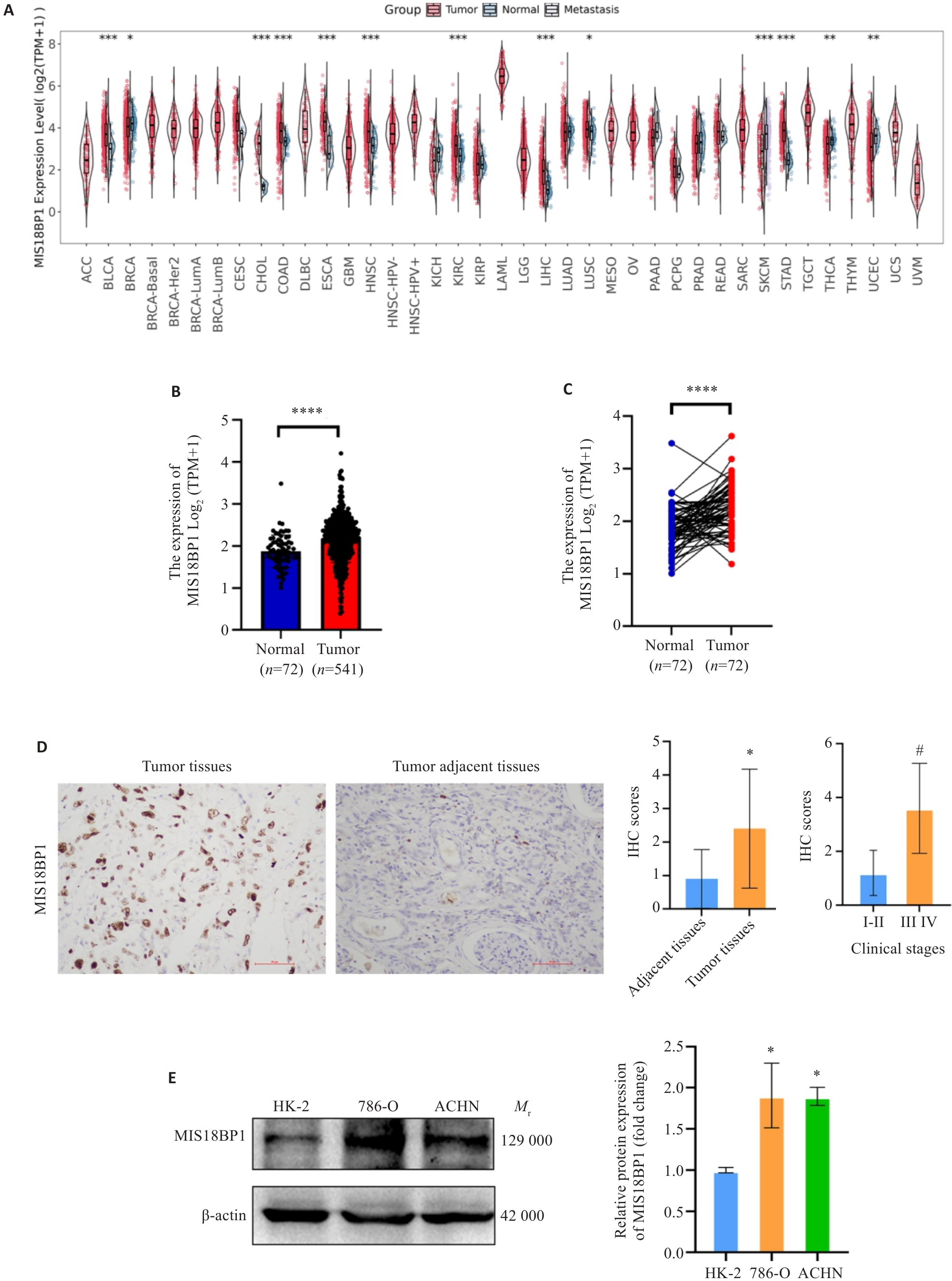
图1 MIS18BP1在泛癌及肾透明细胞癌中的表达特征
Fig.1 Expression patterns of MIS18BP1 in pan-cancer and renal clear cell carcinoma. A: Differential expression of MIS18BP1 in pan-cancer. *P<0.05, **P<0.01, ***P<0.001 vs Normal. B: Differential expression of MIS18BP1 in KIRC. ****P<0.0001. C: Paired difference analysis. ****P<0.0001. D: Expression of MIS18BP1 protein in renal clear cell carcinoma tissue and adjacent non-cancerous tissue (Original magnification: ×200). *P<0.05 vs adjacent tissues, #P<0.05 vs clinical stage I-II. E: Expression of MIS18BP1 protein in normal renal epithelial cells and renal cancer cell lines. *P<0.05 vs HK-2.
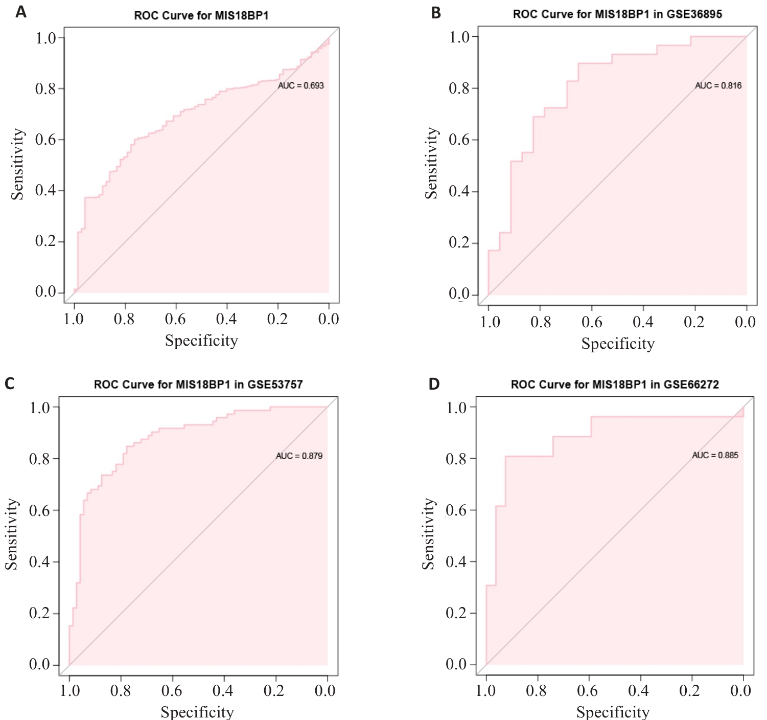
图2 MIS18BP1对KIRC的诊断价值
Fig.2 Diagnostic value of MIS18BP1 for KIRC. A: MIS18BP1 ROC curve for predicting overall survival in TCGA. B: ROC curve of MIS18BP1 for predicting overall survival in GSE36895. C: ROC curve of MIS18BP1 for predicting overall survival in GSE53757. D: ROC curve of MIS18BP1 for predicting overall survival in GSE66272.
| Dataset | AUC | Best cutoff | Sensitivity(%) | Specificity(%) | Youden index |
|---|---|---|---|---|---|
| TCGA | 0.693 | 2.097 | 60.07 | 76.39 | 0.3646 |
| GSE36895 | 0.816 | 5.017 | 89.66 | 65.22 | 0.5488 |
| GSE53757 | 0.879 | 6.759 | 84.72 | 77.78 | 0.6250 |
| GSE66272 | 0.885 | 0.0806 | 80.77 | 92.59 | 0.7336 |
表1 MIS18BP1在TCGA和GEO数据集中对KIRC的诊断效能
Tab.1 Diagnostic performance of MIS18BP1 for KIRC in TCGA and GEO datasets
| Dataset | AUC | Best cutoff | Sensitivity(%) | Specificity(%) | Youden index |
|---|---|---|---|---|---|
| TCGA | 0.693 | 2.097 | 60.07 | 76.39 | 0.3646 |
| GSE36895 | 0.816 | 5.017 | 89.66 | 65.22 | 0.5488 |
| GSE53757 | 0.879 | 6.759 | 84.72 | 77.78 | 0.6250 |
| GSE66272 | 0.885 | 0.0806 | 80.77 | 92.59 | 0.7336 |
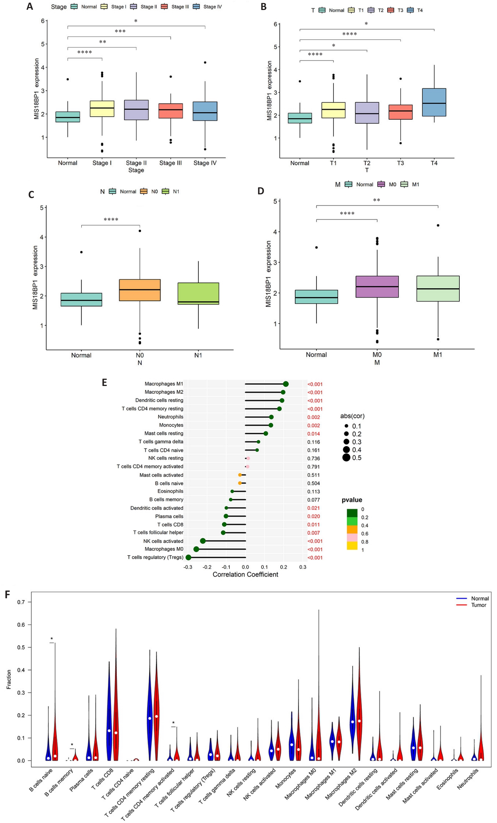
图3 MIS18BP1表达与KIRC临床病理特征及免疫浸润的关系
Fig.3 Correlation of MIS18BP1 expression level with clinical pathological features and immune infiltration in KIRC. A: Relationship between MIS18BP1 expression and T stage. B: Relationship between MIS18BP1 expression and N stage. C: Relationship between MIS18BP1 expression and M stage. D: Relationship between MIS18BP1 expression and grade. *P<0.05, **P<0.01, ***P<0.001, ****P<0.0001. E: Relationship between MIS18BP1 expression and immune cells. F: Differential expression of MIS18BP1 in immune cells. *P<0.05.
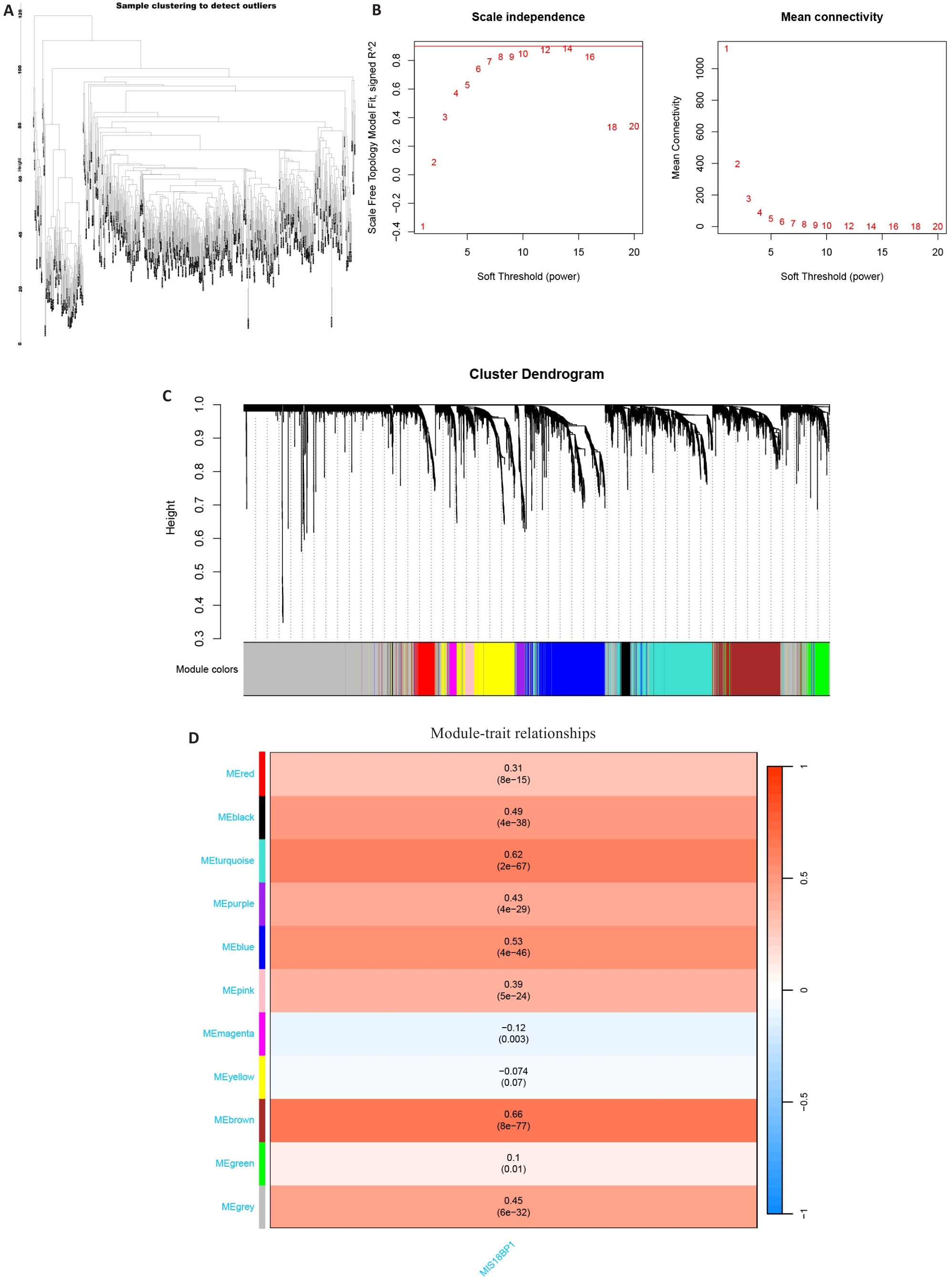
图4 WGCNA识别MIS18BP1相关基因模块
Fig.4 Weighted gene co-expression network analysis (WGCNA) for identifying MIS18BP1-related gene modules. A: Sample clustering dendrogram. B: Determination of the optimal soft threshold (left: scale independence assessment; right: average connectivity analysis), with the red reference line indicating R²=0.9. C: Gene clustering dendrogram (branches represent individual genes, and the colored blocks at the bottom indicate co-expression modules). D: Module-trait association heatmap (colored blocks represent co-expression modules, red and blue indicate positive and negative correlations with correlation coefficients and P-value annotations).
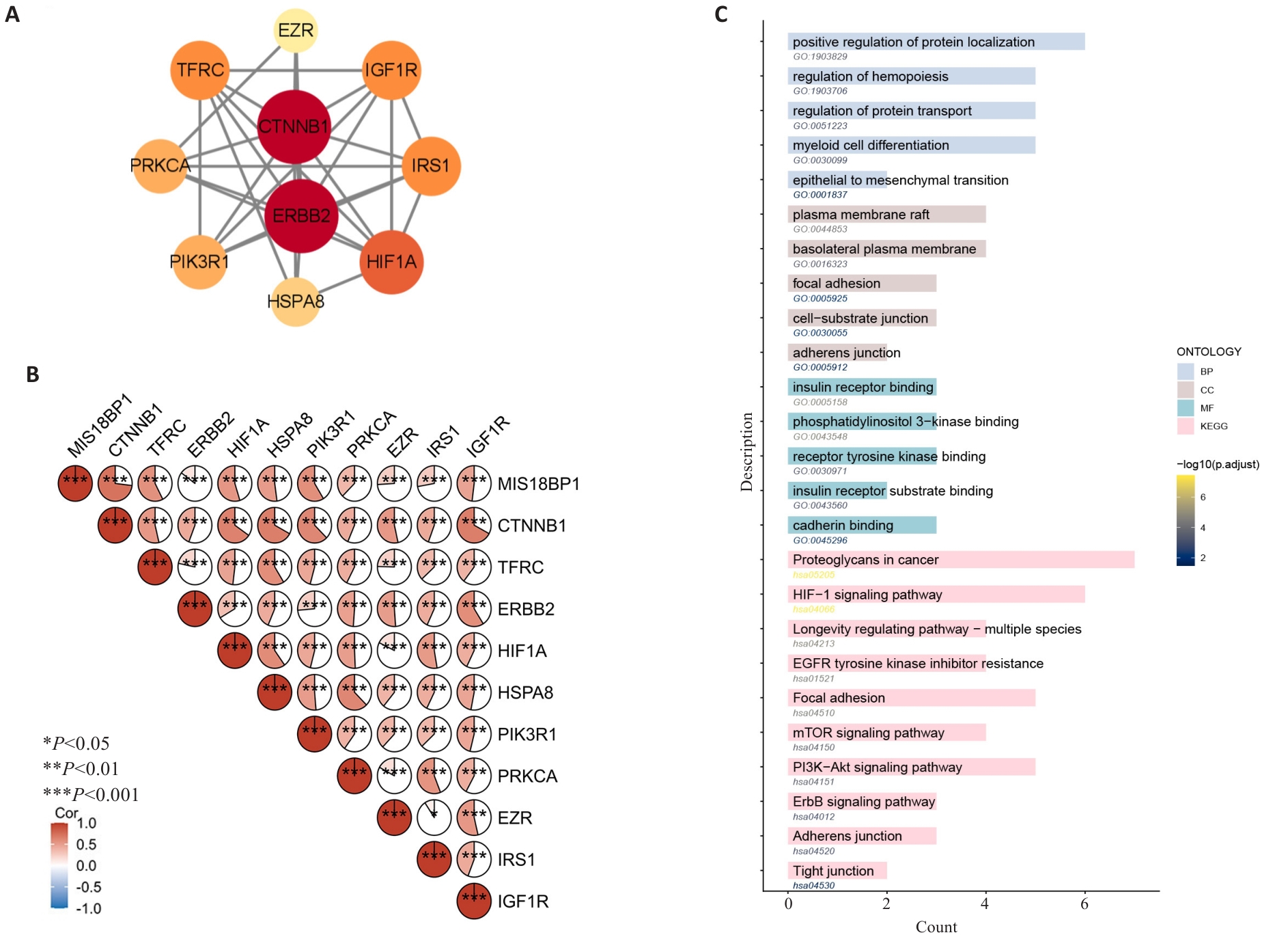
图5 MIS18BP1相关基因的筛选及其功能通路富集分析
Fig.5 Screening of MIS18BP1-related genes and their GO/KEGG analysis. A: Screening key genes related to MIS18BP1 based on the PPI network. Node size is proportional to degree centrality, and node color intensity is proportional to betweenness centrality (dark red indicates high betweenness). B: Correlation analysis between key genes and MIS18BP1 expression. *P<0.05, ***P<0.001. C: GO and KEGG enrichment analysis of key genes.
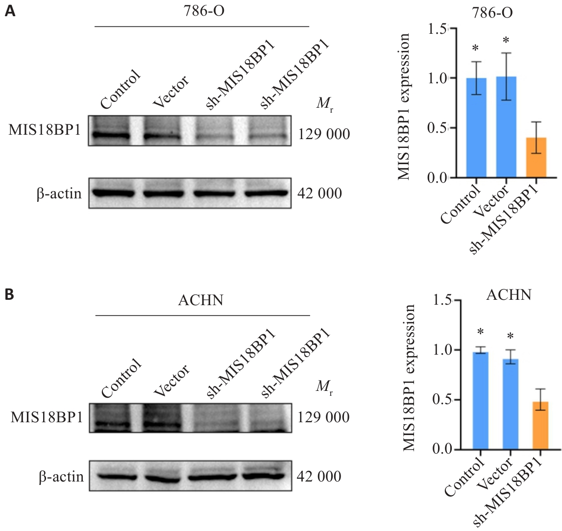
图6 敲低肾癌细胞中的MIS18BP1
Fig.6 Knockdown of MIS18BP1 in renal cancer cells. A: Western blotting for detection of MIS18BP1 protein expression in 786-O cell line. B: Western blotting for detection of MIS18BP1 protein expression in ACHN cell line. *P<0.05 vs sh-MIS18BP1.
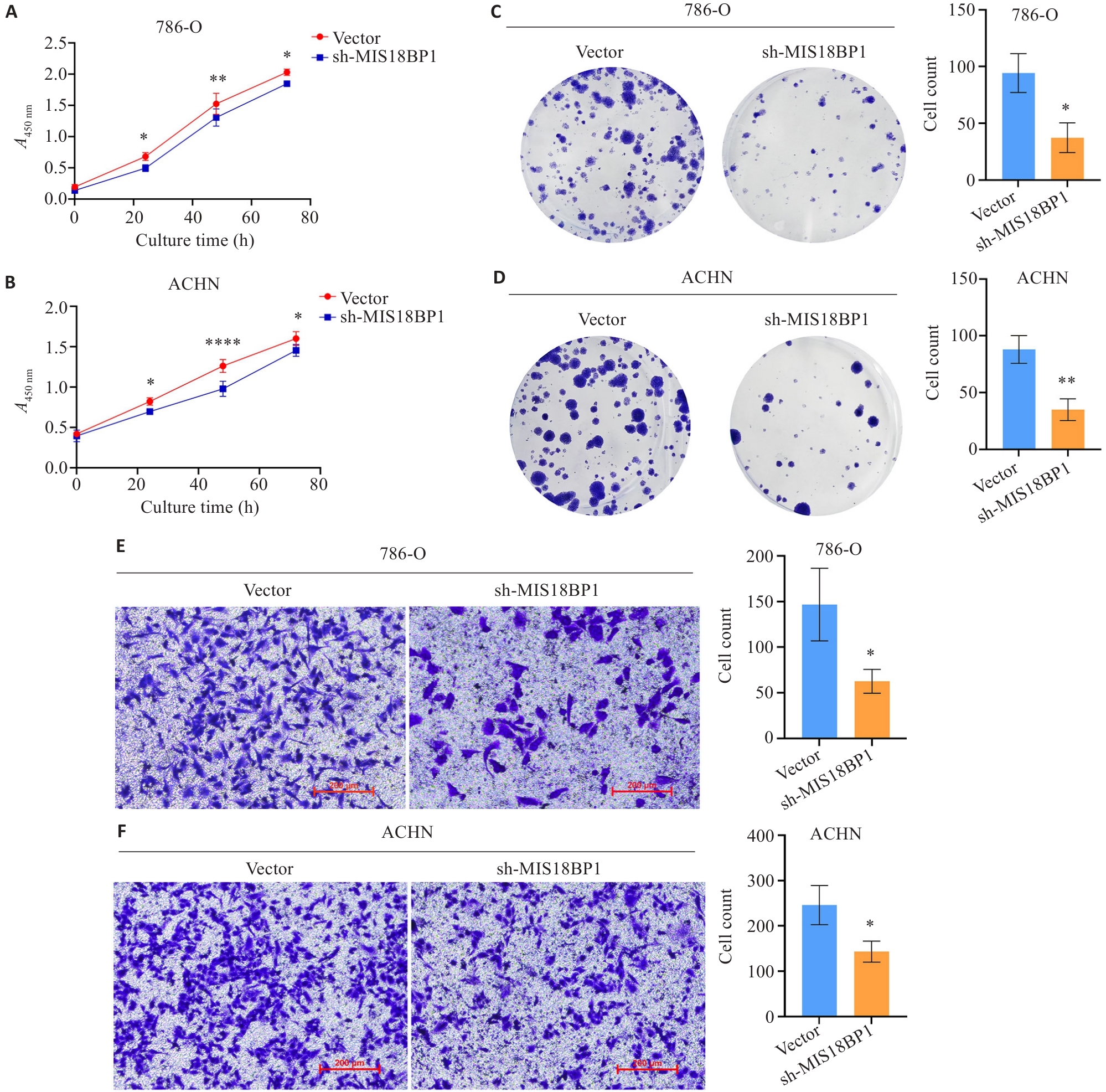
图7 敲低MIS18BP1蛋白抑制肾癌细胞的增殖能力
Fig.7 Knocking down MIS18BP1 inhibits proliferation of renal cancer cells. A, C, E: Knockdown of MIS18BP1 significantly inhibits proliferation of 786-O cells (Scale bar=200 μm). B, D, F: Knockdown of MIS18BP1 significantly inhibits proliferation of ACHN cells (Scale bar=200 μm). *P<0.05, **P<0.01, ****P<0.0001 vs Vector.
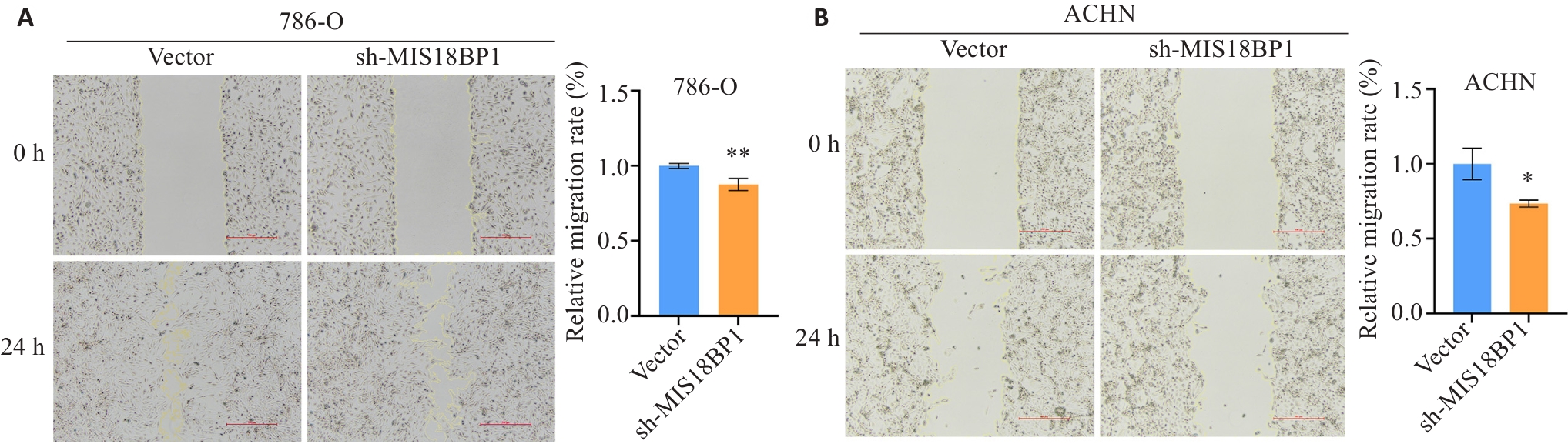
图8 敲低MIS18BP1蛋白抑制肾癌细胞的迁移能力
Fig.8 Knocking down MIS18BP1 inhibits migration ability of renal cancer cells. A: Knocking down MIS18BP1 in inhibits migration ability of 786-O cells (Scale bar=500 μm). B: Knocking down MIS18BP1 inhibits migration ability of ACHN cells (Scale bar=500 μm). *P<0.05, **P<0.01 vs Vector.
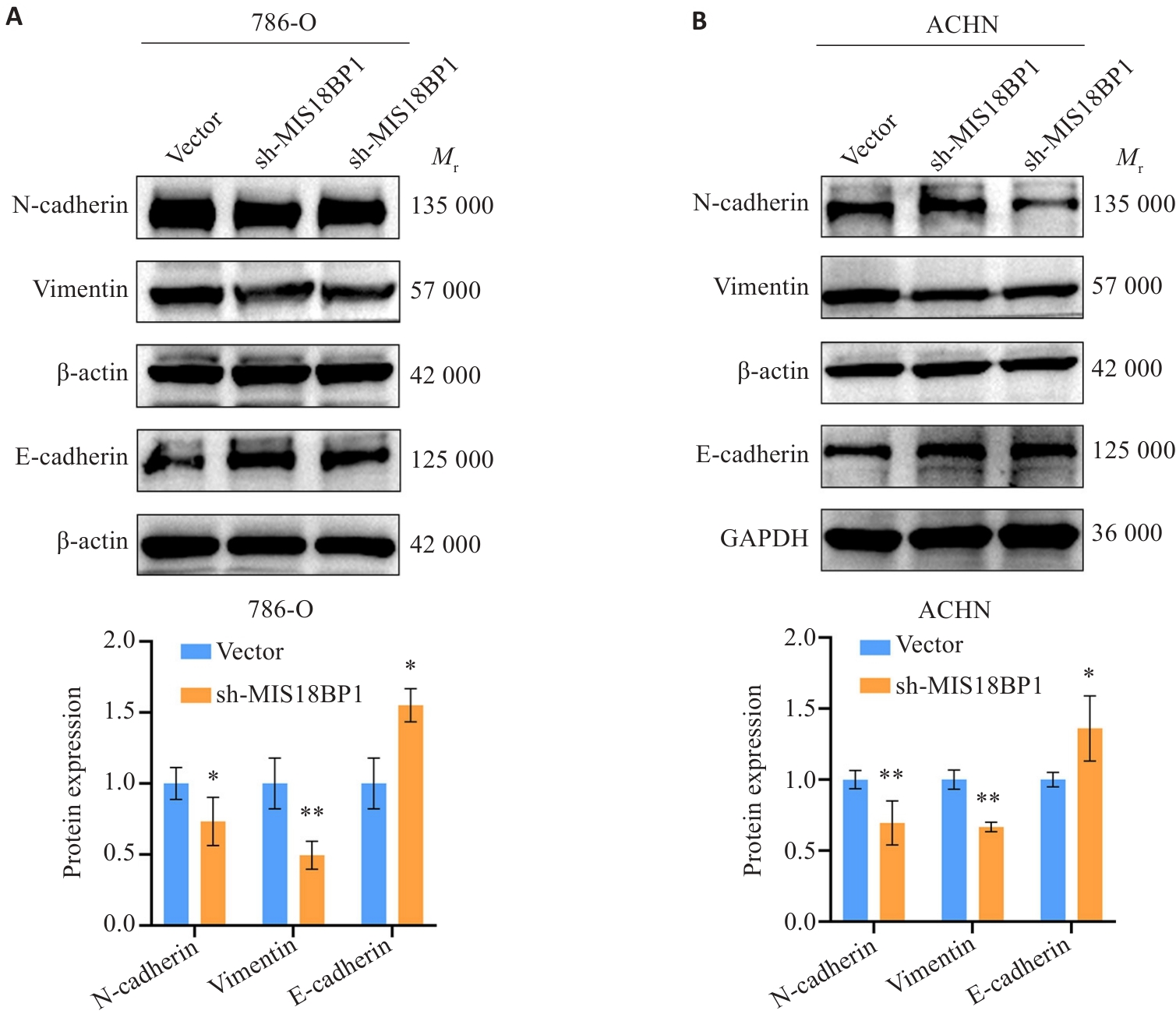
图9 MIS18BP1对肾癌细胞EMT相关蛋白表达的影响
Fig.9 Effect of MIS18BP1 on expression of EMT-related proteins in renal cancer cells. A: MIS18BP1 regulates the expression of EMT-related proteins in 786-O cells. B: MIS18BP1 regulates the expression of EMT-related proteins in ACHN cells. *P<0.05, **P<0.01 vs Vector.
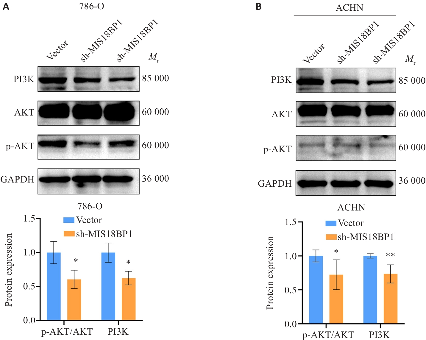
图10 MIS18BP1下调对PI3K/AKT信号通路的影响
Fig.10 Effect of MIS18BP1 knockdown on the PI3K/AKT signaling pathway. A: MIS18BP1 regulates the expression of key proteins in the PI3K/AKT signaling pathway in 786-O cells. B: MIS18BP1 regulates the expression of key proteins in the PI3K/AKT signaling pathway in ACHN cells. *P<0.05, **P<0.01 vs Vector.
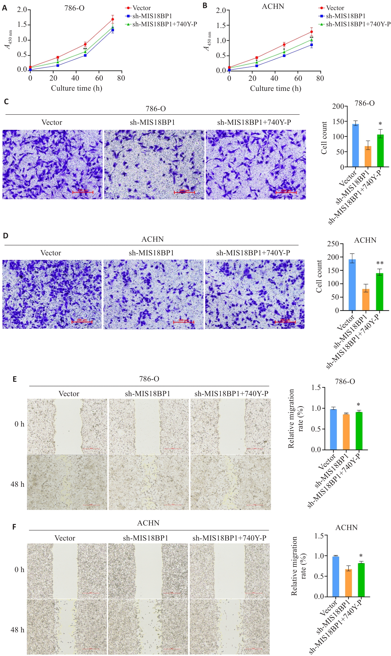
图11 敲低MIS18BP1抑制786-O和ACHN细胞增殖及迁移,该作用可被740Y-P部分逆转
Fig.11 Knockdown of MIS18BP1 inhibits proliferation and migration of 786-O and ACHN cells, which is partially reversed by 740Y P. A, B: CCK-8 assay for assessing proliferation activity of 786-O (A) and ACHN (B) cells. C, D: Wound healing assay for assessing migration ability of 786-O (C) and ACHN (D) cells (Scale bar=200 μm). E, F: Transwell assay for detecting 786-O (E) and ACHN (F) cells passing through the membrane (Scale bar=500 μm). *P<0.05, **P<0.01 vs sh-MIS18BP1 (C-F).
| [1] | Siegel RL, Giaquinto AN, Jemal A. Cancer statistics, 2024[J]. CA A Cancer J Clin, 2024, 74(1): 12-49. doi:10.3322/caac.21820 |
| [2] | Moch H, Amin MB, Berney DM, et al. The 2022 World Health Organization classification of tumours of the urinary system and male genital organs: part A: renal, penile, and testicular tumours[J]. Eur Urol, 2022, 82(5): 458-68. doi:10.1016/j.eururo.2022.06.016 |
| [3] | Hsieh JJ, Purdue MP, Signoretti S, et al. Renal cell carcinoma[J]. Nat Rev Dis Primers, 2017, 3: 17009. doi:10.1016/j.celrep.2017.02.074 |
| [4] | Ljungberg B, Albiges L, Bedke J, et al. EAU Guidelines on Renal Cell Carcinoma: The 2023 Update[J]. Eur Urol, 2023, 119-53. |
| [5] | Thamkachy R, Medina-Pritchard B, Park SH, et al. Structural basis for Mis18 complex assembly and its implications for centromere maintenance[J]. EMBO Rep, 2024, 25(8): 3348-72. doi:10.1038/s44319-024-00183-w |
| [6] | Fujita Y, Hayashi T, Kiyomitsu T, et al. Priming of centromere for CENP-a recruitment by human hMis18α, hMis18β, and M18BP1[J]. Dev Cell, 2007, 12(1): 17-30. doi:10.1016/j.devcel.2006.11.002 |
| [7] | McKinley KL, Cheeseman IM. The molecular basis for centromere identity and function[J]. Nat Rev Mol Cell Biol, 2016, 17(1): 16-29. doi:10.1038/nrm.2015.5 |
| [8] | Kixmoeller K, Allu PK, Black BE. The centromere comes into focus: from CENP-a nucleosomes to kinetochore connections with the spindle[J]. Open Biol, 2020, 10(6): 200051. doi:10.1098/rsob.200051 |
| [9] | Stellfox ME, Nardi IK, Knippler CM, et al. Differential binding partners of the Mis18α/β YIPPEE domains regulate Mis18 complex recruitment to centromeres[J]. Cell Rep, 2016, 15(10): 2127-35. doi:10.1016/j.celrep.2016.05.004 |
| [10] | Ohzeki JI, Shono N, Otake K, et al. KAT7/HBO1/MYST2 regulates CENP-a chromatin assembly by antagonizing Suv39h1-mediated centromere inactivation[J]. Dev Cell, 2016, 37(5): 413-27. doi:10.1016/j.devcel.2016.05.006 |
| [11] | Sun SY, Hu XT, Yu XF, et al. Nuclear translocation of ATG5 induces DNA mismatch repair deficiency (MMR-D)/microsatellite instability (MSI) via interacting with Mis18α in colorectal cancer[J]. Br J Pharmacol, 2021, 178(11): 2351-69. doi:10.1111/bph.15422 |
| [12] | Cao WJ, Tan XY, Li XZ, et al. MIS18BP1 promotes bladder cancer cell proliferation and growth via inactivating P53 signaling pathway[J]. Med Oncol, 2025, 42(5): 156. doi:10.1007/s12032-025-02704-6 |
| [13] | Lamouille S, Xu J, Derynck R. Molecular mechanisms of epithelial–mesenchymal transition[J]. Nat Rev Mol Cell Biol, 2014, 15(3): 178-96. doi:10.1038/nrm3758 |
| [14] | Thiery JP, Acloque H, Huang RYJ, et al. Epithelial-mesenchymal transitions in development and disease[J]. Cell, 2009, 139(5): 871-90. doi:10.1016/j.cell.2009.11.007 |
| [15] | Hemmings BA, Restuccia DF. PI3K-PKB/Akt pathway[J]. Cold Spring Harb Perspect Biol, 2012, 4(9): a011189. doi:10.1101/cshperspect.a011189 |
| [16] | Larue L, Bellacosa A. Epithelial–mesenchymal transition in development and cancer: role of phosphatidylinositol 3' kinase/AKT pathways[J]. Oncogene, 2005, 24(50): 7443-54. doi:10.1038/sj.onc.1209091 |
| [17] | Li TW, Fan JY, Wang BB, et al. TIMER: a web server for comprehensive analysis of tumor-infiltrating immune cells[J]. Cancer Res, 2017, 77(21): e108-10. doi:10.1158/0008-5472.can-17-0307 |
| [18] | Ahmed MM, Tazyeen S, Ali R, et al. Network centrality approaches used to uncover and classify most influential nodes with their related miRNAs in cardiovascular diseases[J]. Gene Rep, 2022, 27: 101555. doi:10.1016/j.genrep.2022.101555 |
| [19] | Muetze T, Goenawan IH, Wiencko HL, et al. Contextual Hub Analysis Tool (CHAT): a Cytoscape app for identifying contextually relevant hubs in biological networks[J]. F1000Research, 2016, 5: 1745. doi:10.12688/f1000research.9118.1 |
| [20] | Bray F, Laversanne M, Sung H, et al. Global cancer statistics 2022: GLOBOCAN estimates of incidence and mortality worldwide for 36 cancers in 185 countries[J]. CA A Cancer J Clin, 2024, 74(3): 229-63. doi:10.3322/caac.21834 |
| [21] | 张永贞, 杨国庆, 张思维, 等. 中国2009年肾及泌尿系统其他癌发病和死亡分析[J]. 中国肿瘤, 2013, 22(5): 333-7. |
| [22] | Chen YW, Wang LK, Panian J, et al. Treatment landscape of renal cell carcinoma[J]. Curr Treat Options Oncol, 2023, 24(12): 1889-916. doi:10.1007/s11864-023-01161-5 |
| [23] | Rini BI, Plimack ER, Stus V, et al. Pembrolizumab plus axitinib versus sunitinib for advanced renal-cell carcinoma[J]. N Engl J Med, 2019, 380(12): 1116-27. doi:10.1056/nejmoa1816714 |
| [24] | Pal SK, Albiges L, Tomczak P, et al. Atezolizumab plus cabozantinib versus cabozantinib monotherapy for patients with renal cell carcinoma after progression with previous immune checkpoint inhibitor treatment (CONTACT-03): a multicentre, randomised, open-label, phase 3 trial[J]. Lancet, 2023, 402(10397): 185-95. doi:10.1016/s0140-6736(23)00922-4 |
| [25] | McKay RR, McGregor BA, Xie WL, et al. Optimized management of nivolumab and ipilimumab in advanced renal cell carcinoma: a response-based phase II study (OMNIVORE)[J]. J Clin Oncol, 2020, 38(36): 4240-8. doi:10.1200/jco.20.02295 |
| [26] | Moree B, Meyer CB, Fuller CJ, et al. CENP-C recruits M18BP1 to centromeres to promote CENP-a chromatin assembly[J]. J Cell Biol, 2011, 194(6): 855-71. doi:10.1083/jcb.201106079 |
| [27] | Stellfox ME, Bailey AO, Foltz DR. Putting CENP-a in its place[J]. Cell Mol Life Sci, 2013, 70(3): 387-406. doi:10.1007/s00018-012-1048-8 |
| [28] | Musacchio A, Desai A. A molecular view of kinetochore assembly and function[J]. Biology, 2017, 6(1): 5. doi:10.3390/biology6010005 |
| [29] | Tao LS, Xie W, Du Y, et al. Activation of the PI3K/AKT pathway by EDIL3 induces EMT and promotes tumor progression in clear cell renal cell carcinoma[J]. Mol Genet Genom, 2026, 301(1): 47. doi:10.1007/s00438-026-02369-4 |
| [30] | Zhao HN, Wang EM. COL1A1 drives tumor progression in kidney renal clear cell carcinoma by regulating EMT through the PI3K/Akt pathway[J]. Cancer Cell Int, 2025, 25(1): 314. doi:10.1186/s12935-025-03956-y |
| [31] | Wang SJ, Xie DQ, Yue HZ, et al. Phospholipase C beta 2 as a key regulator of tumor progression and epithelial-mesenchymal transition via PI3K/AKT signaling in renal cell carcinoma[J]. Biomedicines, 2025, 13(2): 304. doi:10.3390/biomedicines13020304 |
| [32] | Zhan SZ, Bai XJ, Zhao YQ, et al. TGFBI promotes proliferation and epithelial-mesenchymal transition in renal cell carcinoma through PI3K/AKT/mTOR/HIF-1α pathway[J]. Cancer Cell Int, 2024, 24(1): 265. doi:10.1186/s12935-024-03454-7 |
| [33] | Zhou BP, Deng J, Xia WY, et al. Dual regulation of Snail by GSK-3β-mediated phosphorylation in control of epithelial–mesenchymal transition[J]. Nat Cell Biol, 2004, 6(10): 931-40. doi:10.1038/ncb1173 |
| [34] | Villagrasa P, Díaz VM, Viñas-Castells R, et al. Akt2 interacts with Snail1 in the E-cadherin promoter[J]. Oncogene, 2012, 31(36): 4022-33. doi:10.1038/onc.2011.562 |
| [35] | Yan D, Avtanski D, Saxena NK, et al. Leptin-induced epithelial-mesenchymal transition in breast cancer cells requires β‑catenin activation via Akt/GSK3- and MTA1/Wnt1 protein-dependent pathways[J]. J Biol Chem, 2012, 287(11): 8598-612. doi:10.1074/jbc.m111.322800 |
| [36] | Lamouille S, Derynck R. Cell size and invasion in TGF-beta-induced epithelial to mesenchymal transition is regulated by activation of the mTOR pathway[J]. J Cell Biol, 2007, 178(3): 437-51. doi:10.1083/jcb.200611146 |
| [37] | Yin SS, Liu L, Brobbey C, et al. PRMT5-mediated arginine methylation activates AKT kinase to govern tumorigenesis[J]. Nat Commun, 2021, 12: 3444. doi:10.1038/s41467-021-23833-2 |
| [1] | 但昱彤, 张庭辅, 刘红梅, 张鹏. 甲基结合蛋白1对人脐静脉内皮细胞迁移、增殖以及血管形成的影响[J]. 南方医科大学学报, 2026, 46(7): 1585-1592. |
| [2] | 许家宁, 毛钇骁, 张煜. 基于分层注意力驱动多实例学习的肾透明细胞癌数字病理分级分期[J]. 南方医科大学学报, 2026, 46(5): 1175-1184. |
| [3] | 范东伟, 李煊赫, 姚廷敬, 金从稳, 王兴亮, 黄诚. 毛兰素通过阻断Wnt/β-catenin信号抑制乳腺癌细胞增殖和迁移[J]. 南方医科大学学报, 2026, 46(4): 838-847. |
| [4] | 杨佳瑶, 何玉莲, 郭延垒, 尚芳红, 花雷, 阳勇, 张小梅, 魏江平. 党参-茯苓配伍调控ERα/PI3K/Akt信号通路改善单侧颈总动脉永久结扎痴呆大鼠的认知障碍[J]. 南方医科大学学报, 2026, 46(2): 247-258. |
| [5] | 邱佳惠, 陈萌, 满如, 陈鑫, 邱东瑞, 常启同, 马洪玉. 化瘀通便汤通过抑制PI3K/Akt信号通路促进慢传输型便秘大鼠肠道Cajal间质细胞的胞葬作用[J]. 南方医科大学学报, 2026, 46(2): 293-300. |
| [6] | 赵铖, 李稳, 郑宝寿, 王光明, 肖芝松, 李云鹏. lncRNA SNHG12与ELAVL1相互作用激活PI3K/AKT信号通路促进前列腺癌细胞多西他赛的耐药机制[J]. 南方医科大学学报, 2026, 46(1): 183-190. |
| [7] | 杨子为, 吕畅, 董柱, 计书磊, 毕生辉, 张雪花, 王晓武. 金樱子通过调控Src-AKT1轴抑制肺动脉高压平滑肌增殖[J]. 南方医科大学学报, 2025, 45(9): 1889-1902. |
| [8] | 饶璐, 丁家和, 魏江平, 阳勇, 张小梅, 王计瑞. 槐花通过抑制PI3K/AKT通路减轻炎症反应治疗银屑病[J]. 南方医科大学学报, 2025, 45(9): 1989-1996. |
| [9] | 赵华轩, 张桂潮, 刘家荣, 莫富添, 李韬恩, 类成勇, 吕世栋. 肾透明细胞癌患者的免疫细胞浸润特征与临床病理参数具有相关性[J]. 南方医科大学学报, 2025, 45(6): 1280-1288. |
| [10] | 徐皓男, 张放, 黄钰莹, 姚其盛, 管悦琴, 陈浩. 百蕊草通过调节肠道菌群和调控EGFR/PI3K/Akt信号通路改善小鼠抗生素相关性腹泻[J]. 南方医科大学学报, 2025, 45(2): 285-295. |
| [11] | 高俊杰, 叶开, 吴竞. 槲皮素通过调控TP53基因抑制肾透明细胞癌的增殖和迁移[J]. 南方医科大学学报, 2025, 45(2): 313-321. |
| [12] | 裴月娇, 刘慧敏, 昕宇, 刘波. miR-124通过调控PI3K/AKT信号通路改善睡眠剥夺大鼠认知功能[J]. 南方医科大学学报, 2025, 45(2): 340-346. |
| [13] | 刘柳青, 王坤, 王雪晴, 杜冰心. 枸杞多糖通过下调miR-23a减轻顺铂诱导的颗粒细胞损伤[J]. 南方医科大学学报, 2025, 45(11): 2340-2349. |
| [14] | 张可妮, 乔通, 尹林, 黄菊, 耿志军, 左芦根, 胡建国, 李静. 球松素靶向肠上皮细胞PI3K/AKT/CCL2轴抑制巨噬细胞肠道浸润缓解葡聚糖硫酸钠诱导的小鼠结肠炎[J]. 南方医科大学学报, 2025, 45(10): 2199-2209. |
| [15] | 陈晓睿, 魏青政, 张宗亮, 原江水, 宋卫青. 过表达带电多泡体蛋白2B基因抑制肾透明细胞癌细胞的增殖[J]. 南方医科大学学报, 2025, 45(1): 126-136. |
| 阅读次数 | ||||||
|
全文 |
|
|||||
|
摘要 |
|
|||||
