
南方医科大学学报 ›› 2026, Vol. 46 ›› Issue (7): 1533-1542.doi: 10.12122/j.issn.1673-4254.2026.07.07
• • 上一篇
王琳钰1,2( ), 王晓燕1,2,4, 朱季军1,2, 蒋波1,2, 徐洁1,2,4, 高彩月2,3,4, 李冬冬2,3,4, 王玉2,3,4, 李晓敏1,2,4()
收稿日期:2026-01-26
出版日期:2026-07-20
发布日期:2026-07-20
通讯作者:
李晓敏
E-mail:1007080916@qq.com;lxm1980326@sina.com
作者简介:王琳钰,在读硕士研究生,E-mail: 1007080916@qq.com
基金资助:
Linyu WANG1,2(), Xiaoyan WANG1,2,4, Jijun ZHU1,2, Bo JIANG1,2, Jie XU1,2,4, Caiyue GAO2,3,4, Dongdong LI2,3,4, Yu WANG2,3,4, Xiaomin LI1,2,4()
Received:2026-01-26
Online:2026-07-20
Published:2026-07-20
Contact:
Xiaomin LI
E-mail:1007080916@qq.com;lxm1980326@sina.com
Supported by:摘要:
目的 探究丝切蛋白1(CFL1)及其磷酸化形式p-CFL1在结肠癌中的表达情况,分析其与临床病理特征及预后的关系。揭示CFL1/p-CFL1调控结肠癌细胞迁移的分子机制,重点阐明其核定位在此过程中所发挥的作用及潜在机制。 方法 利用TCGA数据库、结肠癌组织芯片免疫组化分析CFL1和p-CFL1表达及临床病理参数相关性;应用Kaplan-Meier生存分析探究CFL1和p-CFL1表达水平与结肠癌患者生存期的关系;应用免疫荧光明确CFL1和p-CFL1在细胞核和细胞质的定位;构建野生型(CFL1-WT)与核定位信号突变型(CFL1-MUT)载体,在HCT116细胞中过表达,通过Transwell小室迁移实验检测细胞迁移能力;结合转录组测序与KEGG富集分析解析差异基因与信号通路。 结果 免疫组化结果显示,CFL1与p-CFL1在结肠癌组织中表达显著高于癌旁组织(P<0.0001),且二者表达呈正相关(r=0.4753,P<0.0001)。CFL1和p-CFL1高表达与淋巴结转移、临床分期晚及患者总生存期缩短相关(P<0.05)。免疫组化与免疫荧光显示CFL1/p-CFL1可定位于细胞质与细胞核。功能实验表明,过表达CFL1-WT与CFL1-MUT均能促进HCT116细胞迁移,且CFL1-WT作用更强。KEGG分析提示,CFL1-WT调控的基因显著富集于Ras、PI3K-Akt、细胞粘附分子等肿瘤转移相关通路。 结论 CFL1/p-CFL1在结肠癌中高表达,是预后不良的潜在生物标志物。CFL1可通过核定位进一步激活肿瘤转移相关信号通路,从而促进结肠癌迁移。
王琳钰, 王晓燕, 朱季军, 蒋波, 徐洁, 高彩月, 李冬冬, 王玉, 李晓敏. CFL1及p-CFL在结肠癌中高表达预示结肠癌患者预后不良并促进癌细胞迁移[J]. 南方医科大学学报, 2026, 46(7): 1533-1542.
Linyu WANG, Xiaoyan WANG, Jijun ZHU, Bo JIANG, Jie XU, Caiyue GAO, Dongdong LI, Yu WANG, Xiaomin LI. High expression of CFL1 and phospho-CFL1 predicts poor prognosis and promotes migration of colon cancer cells in vitro[J]. Journal of Southern Medical University, 2026, 46(7): 1533-1542.
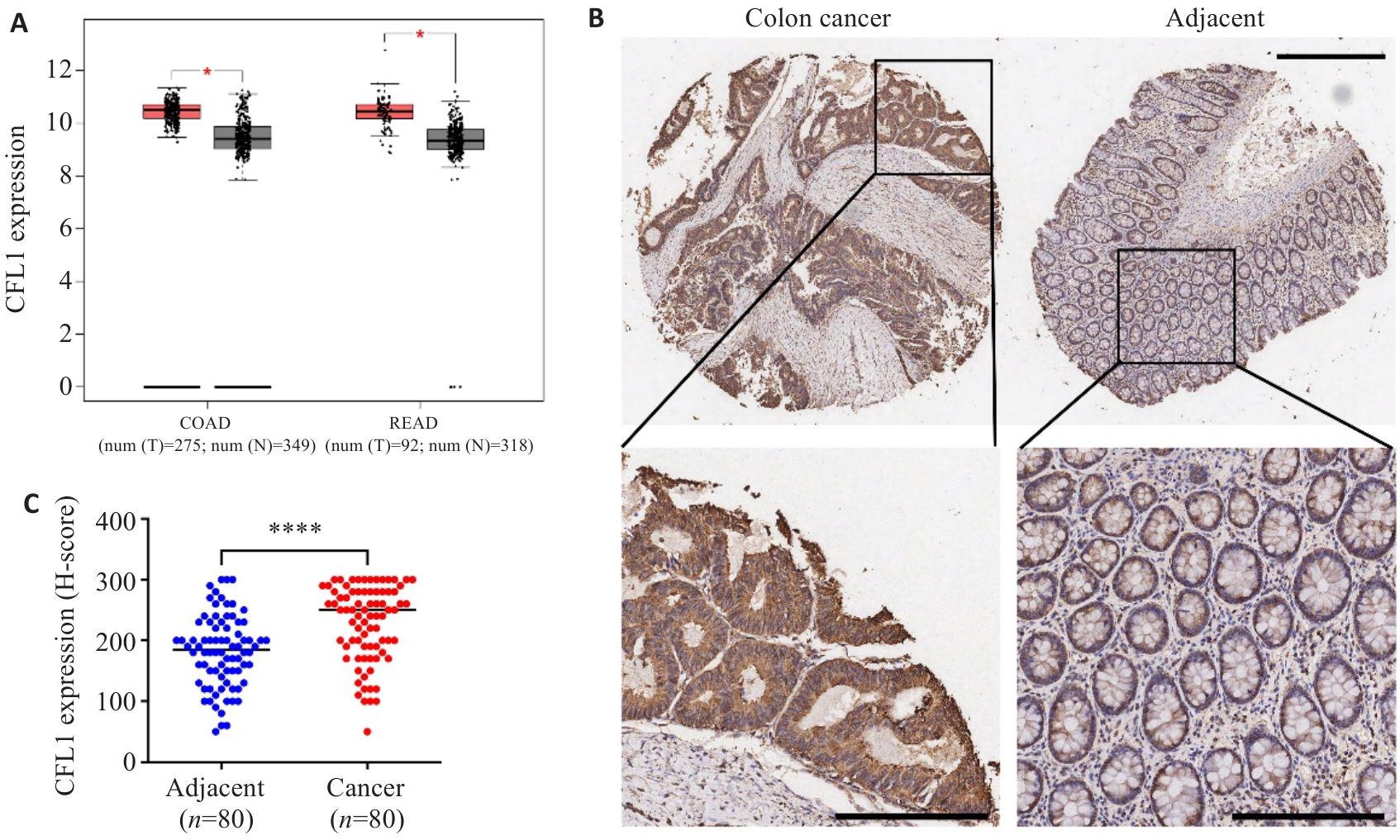
图1 结肠癌及癌旁组织CFL1的表达情况
Fig.1 CFL1 is highly expressed in colon cancer tissues. A: Expression levels of CFL1 in colon adenocarcinoma (COAD) and rectal adenocarcinoma (READ) from the TCGA database. B, C: Representative immunohistochemical staining and the corresponding H-score analysis of CFL1 in colon cancer tissues and paired adjacent normal tissues (n=80). Scale bar=500 µm (upper panel); Scale bar=200 µm (lower panel). *P<0.05, ****P<0.0001.
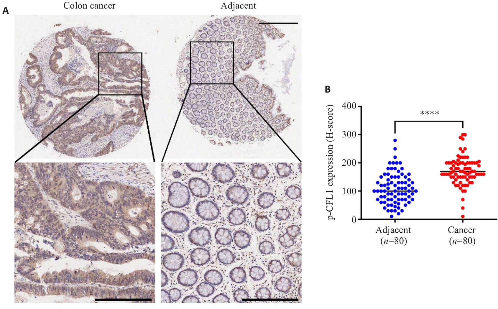
图2 结肠癌及癌旁组织p-CFL1的表达情况
Fig.2 p-CFL1 is highly expressed in colon cancer tissues. A, B: Representative immunohistochemical staining and the corresponding H-score analysis of p-CFL1 in colon cancer tissues and paired adjacent normal tissues (n=80). Scale bar=500 µm (upper panel); Scale bar=200 µm (lower panel). ****P<0.0001.
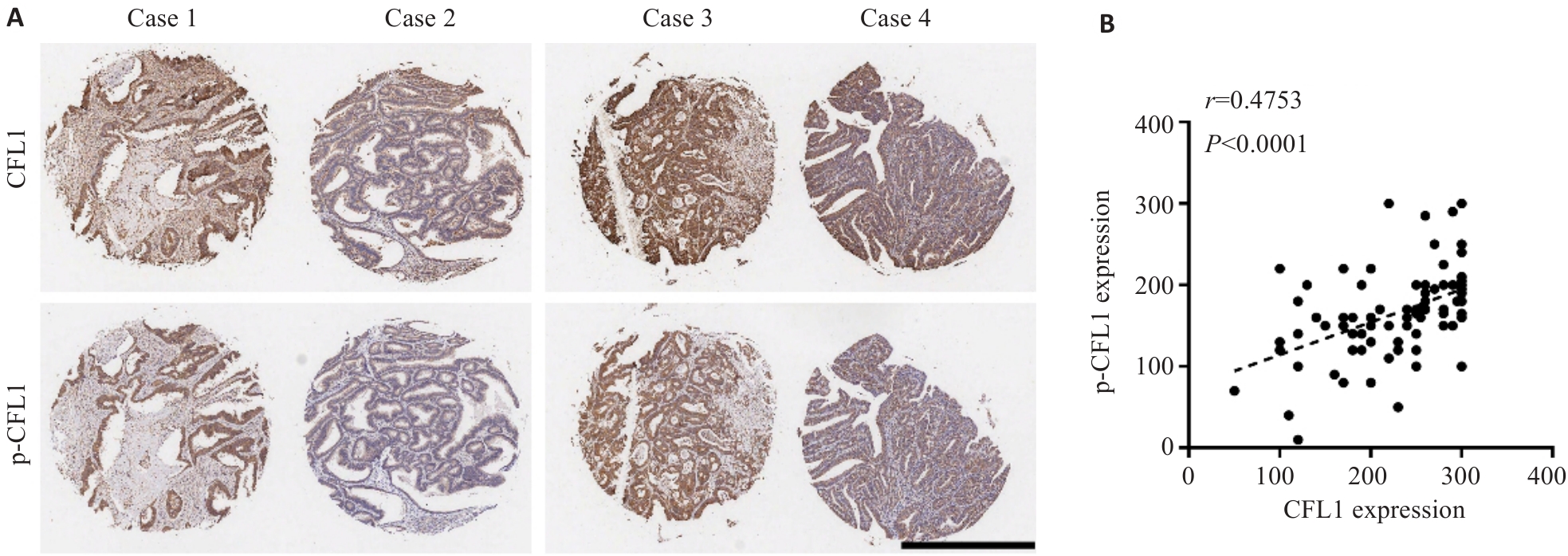
图3 CFL1和p-CFL1在结肠癌组织中的表达呈正相关
Fig.3 Positive correlation between expressions of CFL1 and p-CFL1 in colon cancer tissues. A: Representative images showing consistent expression trends of CFL1 and p-CFL1 in the same colon cancer tissue samples (Scale bar=100 μm). B: Pearson correlation analysis between CFL1 and p-CFL1 expression levels in colon cancer tissues (n=96).
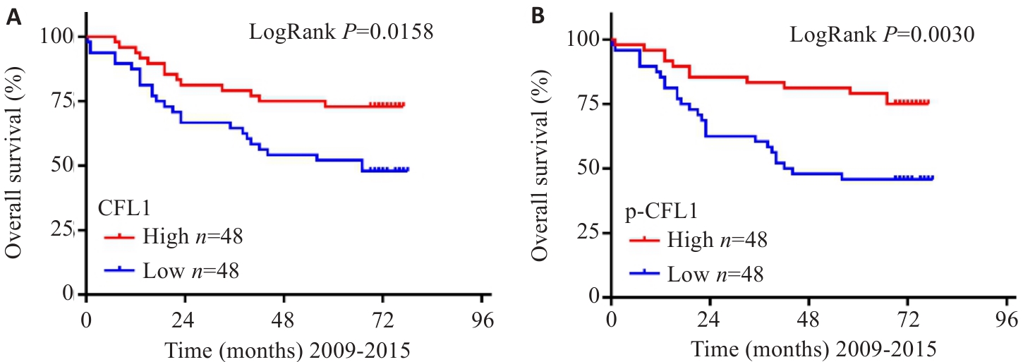
图4 CFL1和p-CFL1高表达对结肠癌患者术后生存情况的影响
Fig.4 Impact of high expression of CFL1 and p-CFL1 on overall survival of patients with colon cancer. A: Kaplan-Meier survival curves comparing overall survival between patients with high and low expression of CFL1 (P=0.0158). B: Kaplan-Meier survival curves comparing overall survival between patients with high and low expression of p-CFL1 (P=0.0030).
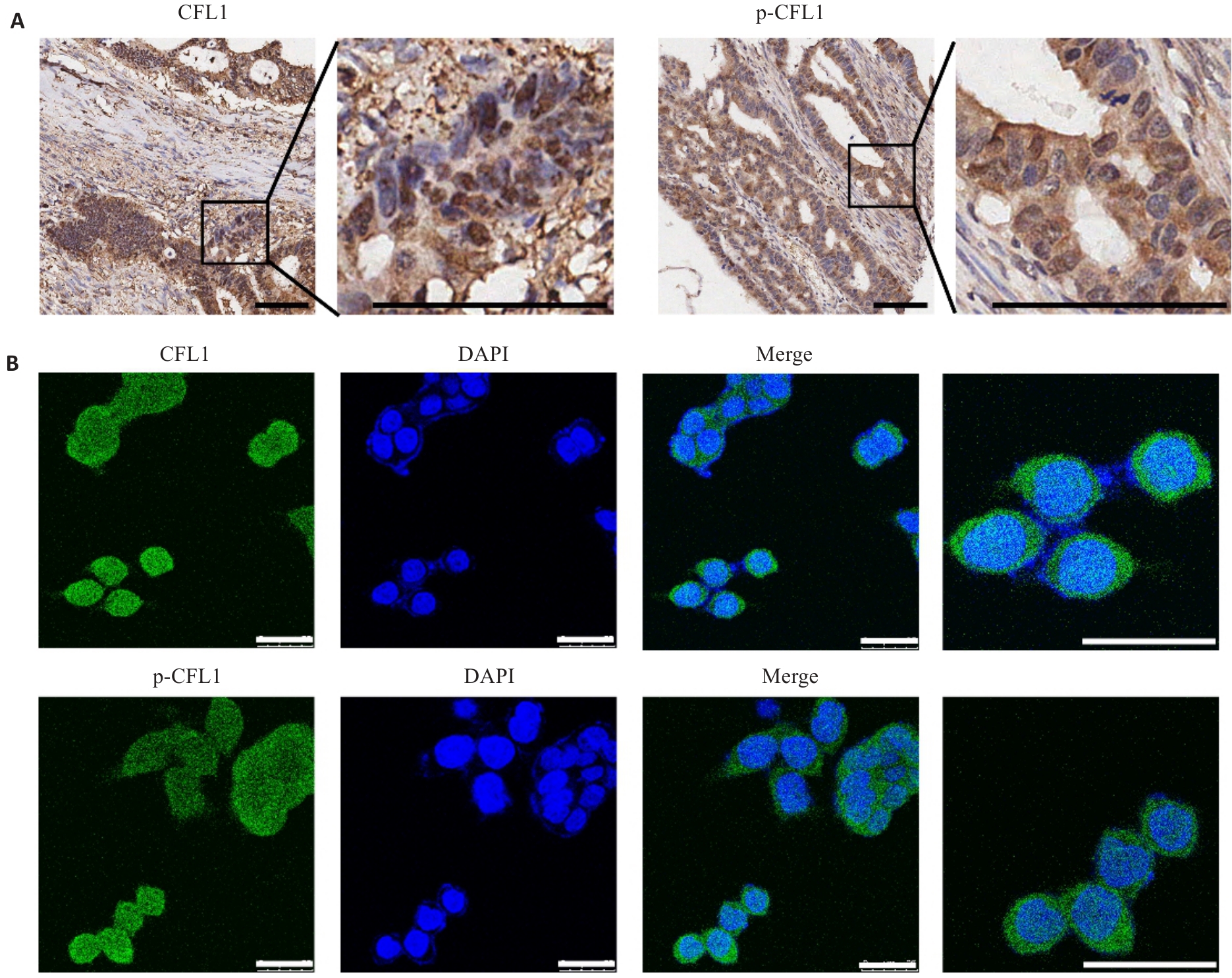
图5 CFL1和p-CFL1在部分结肠癌的细胞核中有阳性表达
Fig.5 Nuclear localization of CFL1 and p-CFL1 in a subset of colon cancer cases. A: Representative immunohistochemical images showing nuclear and cytoplasmic staining of CFL1 and p-CFL1 in colon cancer tissues (Scale bar=100 μm). B: Immunofluorescence analysis confirms the localization of CFL1 and p-CFL1 in both the nucleus and cytoplasm of HCT116 cells (Scale bar=25 μm).
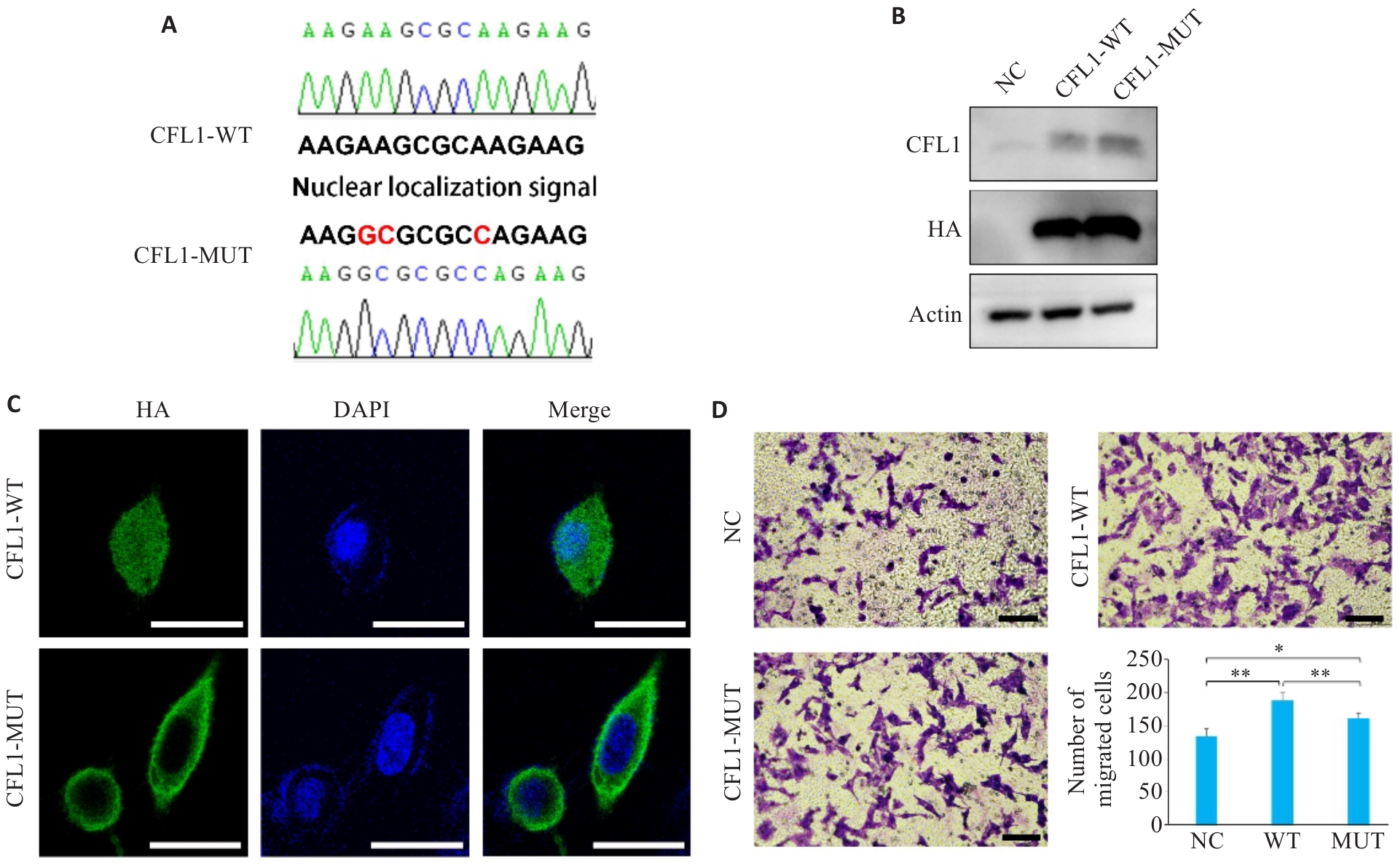
图6 过表达野生型和NLS突变型CFL1均促进结肠癌细胞迁移
Fig.6 Overexpression of wild-type and NLS-mutant CFL1 promotes migration of colon cancer cells. A: Sanger sequencing verification showing the presence of the NLS in the CFL1-WT vector and its mutation in the CFL1-MUT vector. B: Western blotting confirms overexpression of CFL1 in HCT116 cells transfected with CFL1-WT and CFL1-MUT vectors. C: Immunofluorescence staining showing that wild-type CFL1 overexpression leads to both nuclear and cytoplasmic distribution, while the NLS-mutant confines CFL1 to the cytoplasm (Scale bar=20 μm). D: Transwell migration assays demonstrating that both CFL1-WT and CFL1-MUT enhancemigration of colon cancer cells (Scale bar=100 μm; n=5). *P<0.05, **P<0.01.
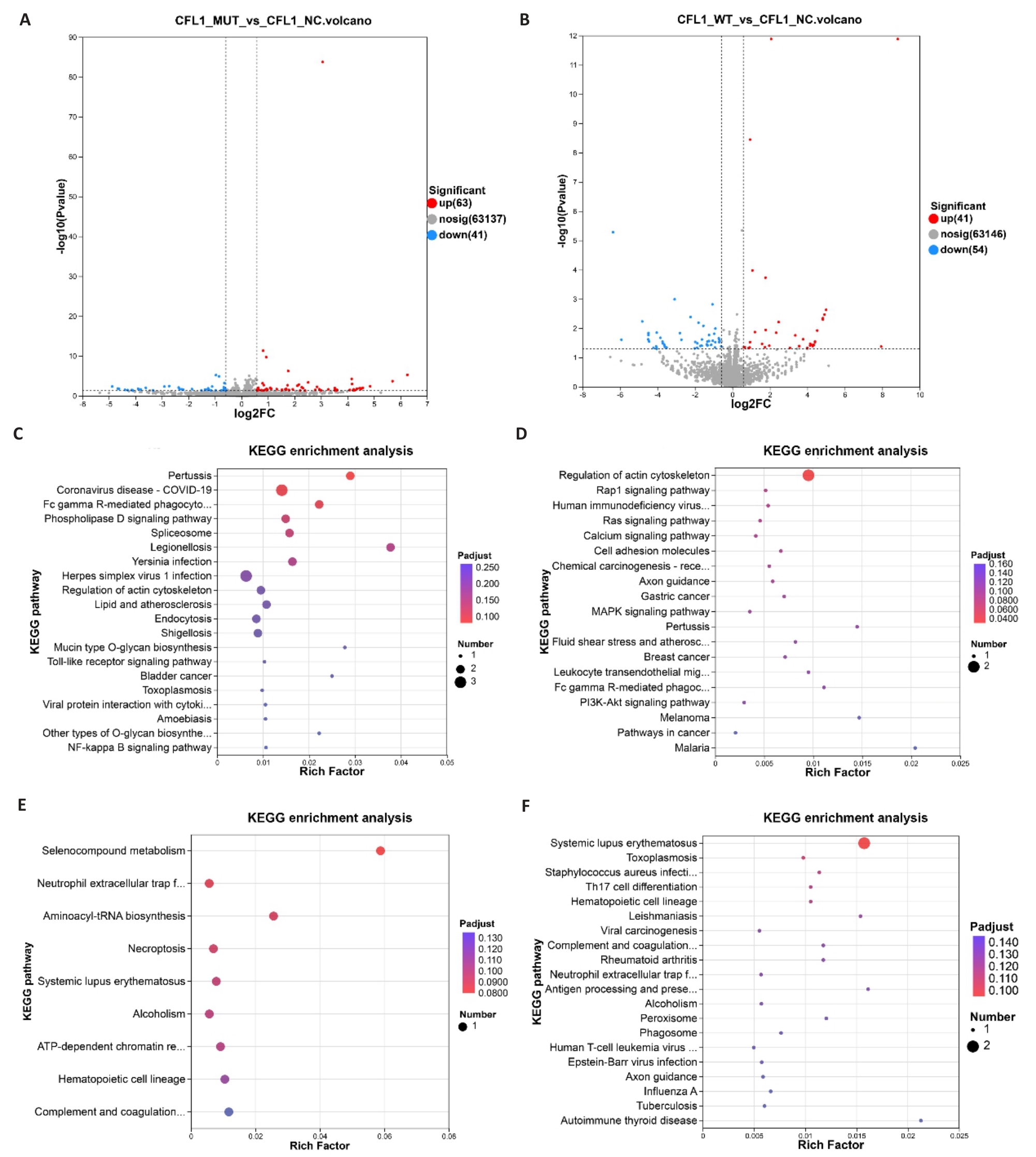
图7 野生型和NLS突变型CFL1的KEGG富集分析
Fig.7 KEGG enrichment analysis of wild-type and NLS-mutant CFL1. A, B: Volcano plots displaying the numbers of differentially expressed genes (DEGs) identified by RNA-seq in HCT116 cells transfected with CFL1-MUT, CFL1-WT, or control vectors. C, D: Bubble plots of KEGG pathway enrichment analysis for up-regulated DEGs (MUT vs NC: 63, WT vs NC: 41) in the CFL1-MUT and CFL1-WT groups, respectively. E, F: Bubble plots of KEGG pathway enrichment analysis for down-regulated DEGs (MUT vs NC: 41, WT vs NC: 54) in the CFL1-MUT and CFL1-WT groups, respectively.
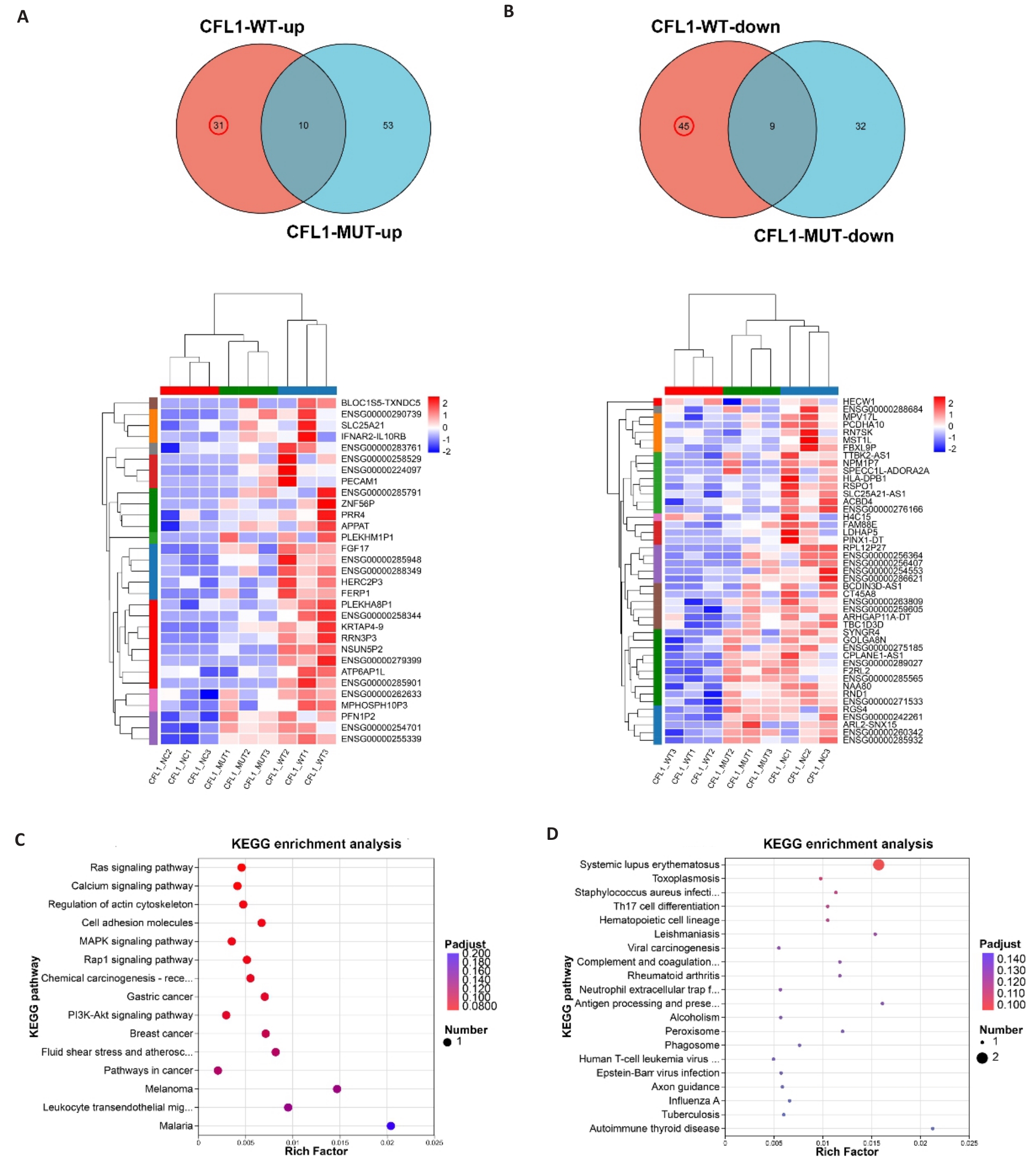
图8 核内定位的CFL1的KEGG富集分析
Fig.8 KEGG enrichment analysis of nuclear-localized CFL1. A, B: Venn diagram and heatmaps showing the 31 up-regulated and 45 down-regulated differentially expressed genes in CFL1-WT group compared to the control. C, D: KEGG enrichment analysis of the 31 up-regulated and 45 down-regulated DEGs.
| [1] | Sung H, Ferlay J, Siegel RL, et al. Global cancer statistics 2020: GLOBOCAN estimates of incidence and mortality worldwide for 36 cancers in 185 countries[J]. CA Cancer J Clin, 2021, 71(3): 209-49. doi:10.3322/caac.21660 |
| [2] | Zheng RS, Chen R, Han BF, et al. Cancer incidence and mortality in China, 2022[J]. Zhonghua Zhong Liu Za Zhi, 2024, 46(3): 221-31. |
| [3] | Mizuno K. Signaling mechanisms and functional roles of cofilin phosphorylation and dephosphorylation[J]. Cell Signal, 2013, 25(2): 457-69. doi:10.1016/j.cellsig.2012.11.001 |
| [4] | Daryabari SS, Fathi M, Mahdavi M, et al. Overexpression of CFL1 in gastric cancer and the effects of its silencing by siRNA with a nanoparticle delivery system in the gastric cancer cell line[J]. J Cell Physiol, 2020, 235(10): 6660-72. doi:10.1002/jcp.29562 |
| [5] | Castro MAA, Dal-Pizzol F, Zdanov S, et al. CFL1 expression levels as a prognostic and drug resistance marker in nonsmall cell lung cancer[J]. Cancer, 2010, 116(15): 3645-55. doi:10.1002/cncr.25125 |
| [6] | Branco MA, Kunst BM, Neto BS, et al. Progress and challenges in the use of Cofilin-1 as a prognostic and predictive biomarker for cancer management[J]. Gene, 2025, 968: 149746. doi:10.1016/j.gene.2025.149746 |
| [7] | Werle SD, Schwab JD, Tatura M, et al. Unraveling the molecular tumor-promoting regulation of cofilin-1 in pancreatic cancer[J]. Cancers, 2021, 13(4): 725. doi:10.3390/cancers13040725 |
| [8] | Chen LK, Cai JL, Huang YS, et al. Identification of cofilin-1 as a novel mediator for the metastatic potentials and chemoresistance of the prostate cancer cells[J]. Eur J Pharmacol, 2020, 880: 173100. doi:10.1016/j.ejphar.2020.173100 |
| [9] | Howard J, Goh CY, Gorzel KW, et al. The potential role of cofilin-1 in promoting triple negative breast cancer (TNBC) metastasis via the extracellular vesicles (EVs)[J]. Transl Oncol, 2022, 15(1): 101247. doi:10.1016/j.tranon.2021.101247 |
| [10] | Wang LX, Xiong L, Wu ZC, et al. Expression of UGP2 and CFL1 expression levels in benign and malignant pancreatic lesions and their clinicopathological significance[J]. World J Surg Oncol, 2018, 16(1): 11. doi:10.1186/s12957-018-1316-7 |
| [11] | Lu LI, Fu NI, Luo XU, et al. Overexpression of cofilin 1 in prostate cancer and the corresponding clinical implications[J]. Oncol Lett, 2015, 9(6): 2757-61. doi:10.3892/ol.2015.3133 |
| [12] | Yao BW, Li YZ, Chen TX, et al. Hypoxia-induced cofilin 1 promotes hepatocellular carcinoma progression by regulating the PLD1/AKT pathway[J]. Clin Transl Med, 2021, 11(3): e366. doi:10.1002/ctm2.366 |
| [13] | Mousavi S, Safaralizadeh R, Hosseinpour-Feizi M, et al. Study of cofilin 1 gene expression in colorectal cancer[J]. J Gastrointest Oncol, 2018, 9(5): 791-6. doi:10.21037/jgo.2018.05.17 |
| [14] | Kang X, Zhao CF, Liu YP, et al. The phosphorylation level of Cofilin-1 is related to the pathological subtypes of gastric cancer[J]. Medicine, 2022, 101(43): e31309. doi:10.1097/md.0000000000031309 |
| [15] | Qin YJ, Li WX, Long YL, et al. Relationship between p-cofilin and cisplatin resistance in patients with ovarian cancer and the role of p-cofilin in prognosis[J]. Cancer Biomark, 2019, 24(4): 469-75. doi:10.3233/cbm-182209 |
| [16] | Luo YY, Lu B, Hu YQ, et al. Triptonoterpene promotes the mitochondrial translocation of cofilin-1 to induce apoptosis in gastric cancer cells by regulating actin dynamics[J]. Biochem Pharmacol, 2025, 241: 117173. doi:10.1016/j.bcp.2025.117173 |
| [17] | Wang WS, Zhong HJ, Xiao DW, et al. The expression of CFL1 and N-WASP in esophageal squamous cell carcinoma and its correlation with clinicopathological features[J]. Dis Esophagus, 2010, 23(6): 512-21. doi:10.1111/j.1442-2050.2009.01035.x |
| [18] | Li XM, Wang JJ, Zhang C, et al. Circular RNA circITGA7 inhibits colorectal cancer growth and metastasis by modulating the Ras pathway and upregulating transcription of its host gene ITGA7 [J]. J Pathol, 2018, 246(2): 166-79. doi:10.1002/path.5125 |
| [19] | Li XM, Chen T, Cai YJ, et al. CircSETD3 interrupts the bidirectional positive feedback of ErbB3 and Akt by sponging miR-4667-5p to inhibit colorectal cancer progression and cetuximab resistance[J]. Int J Biol Macromol, 2025, 318(Pt 4): 145352. doi:10.1016/j.ijbiomac.2025.145352 |
| [20] | Li XM, Wang JJ, Lin WH, et al. circEXOC6B interacting with RRAGB, an mTORC1 activator, inhibits the progression of colorectal cancer by antagonizing the HIF1A-RRAGB-mTORC1 positive feedback loop[J]. Mol Cancer, 2022, 21(1): 135. doi:10.1186/s12943-022-01600-1 |
| [21] | Sousa-Squiavinato ACM, Rocha MR, Barcellos-de-Souza P, et al. Cofilin-1 signaling mediates epithelial-mesenchymal transition by promoting actin cytoskeleton reorganization and cell-cell adhesion regulation in colorectal cancer cells[J]. Biochim Biophys Acta Mol Cell Res, 2019, 1866(3): 418-29. doi:10.1016/j.bbamcr.2018.10.003 |
| [22] | Hensley PJ, Zetter D, Horbinski CM, et al. Association of epithelial-mesenchymal transition and nuclear cofilin with advanced urothelial cancer[J]. Hum Pathol, 2016, 57: 68-77. doi:10.1016/j.humpath.2016.06.020 |
| [23] | Madak-Erdogan Z, Ventrella R, Petry L, et al. Novel roles for ERK5 and cofilin as critical mediators linking ERα-driven transcription, actin reorganization, and invasiveness in breast cancer[J]. Mol Cancer Res, 2014, 12(5): 714-27. doi:10.1158/1541-7786.mcr-13-0588 |
| [24] | Domingues C, Geraldo AM, Anjo SI, et al. Cofilin-1 is a mechanosensitive regulator of transcription[J]. Front Cell Dev Biol, 2020, 8: 678. doi:10.3389/fcell.2020.00678 |
| [25] | Liu Z, Liu J, Dong XC, et al. Tn antigen promotes human colorectal cancer metastasis via H-Ras mediated epithelial-mesenchymal transition activation[J]. J Cell Mol Med, 2019, 23(3): 2083-92. doi:10.1111/jcmm.14117 |
| [26] | Abd Rahman NI, Tham CL, Abd Hamid R. Inhibition of angiogenesis and metastasis in colorectal cancer cell lines through KRAS-associated signaling pathways by 2-methoxy-6-undecyl-1, 4-benzoquinone[J]. Chem Biol Interact, 2024, 399: 111151. doi:10.1016/j.cbi.2024.111151 |
| [27] | Dai SP, Zhuang HW, Li ZZ, et al. miR-122/NEGR1 axis contributes colorectal cancer liver metastasis by PI3K/AKT pathway and macrophage modulation[J]. J Transl Med, 2024, 22(1): 1060. doi:10.1186/s12967-024-05901-5 |
| [28] | Shin AE, Sugiura K, Kariuki SW, et al. LIN28B-mediated PI3K/AKT pathway activation promotes metastasis in colorectal cancer models[J]. J Clin Invest, 2025, 135(8): e186035. doi:10.1172/jci186035 |
| [29] | Sun XJ, Zhang JL, Dong BQ, et al. Targeting SLITRK4 restrains proliferation and liver metastasis in colorectal cancer via regulating PI3K/AKT/NFκB pathway and tumor-associated macrophage[J]. Adv Sci, 2025, 12(1): 2400367. doi:10.1002/advs.202400367 |
| [30] | Paschos KA, Canovas D, Bird NC. The role of cell adhesion molecules in the progression of colorectal cancer and the development of liver metastasis[J]. Cell Signal, 2009, 21(5): 665-74. doi:10.1016/j.cellsig.2009.01.006 |
| [31] | Arabzadeh A, Chan C, Nouvion AL, et al. Host-related carcinoembryonic antigen cell adhesion molecule 1 promotes metastasis of colorectal cancer[J]. Oncogene, 2013, 32(7): 849-60. doi:10.1038/onc.2012.112 |
| [1] | 丁宁, 彭天书, 莫黎, 胡响当, 杨宗亮, 肖佑, 付肖冰, 赵建政, 李磊磊, 张英杰. 多组学整合揭示结直肠癌中的免疫-代谢异质性:针对ANGPTL4/FABP4/RBP7的预后模型及治疗策略[J]. 南方医科大学学报, 2026, 46(5): 977-993. |
| [2] | 宋淇乐, 苗益恺, 冯小桐, 王一凡, 刘伟, 魏琪, 于新汝, 陈文文, 付晓艳. 硒代胱氨酸通过诱导活性氧产生启动氧化应激损伤抑制结肠癌细胞生长[J]. 南方医科大学学报, 2026, 46(3): 532-540. |
| [3] | 梁笑笑, 栾慧林, 马鹏珍, 邓文珠, 金佳欣, 全晨阳, 陈婷婷, 杨友财, 谢颖桢. 基于生命八要素的心血管健康评分与缺血性脑卒中预后相关性及临床检验指标的中介效应:一项多中心前瞻性队列研究[J]. 南方医科大学学报, 2026, 46(2): 239-246. |
| [4] | 余晖豪, 邵玉, 程倩倩, 周新瑞, 耿海萍, 杨燕. 肝细胞癌中MPP6高表达预测患者不良预后并促进肿瘤恶性生物学行为[J]. 南方医科大学学报, 2026, 46(2): 394-402. |
| [5] | 赵新丽, 王豪杰, 宋银春, 袁帅, 张振, 周星琦, 李姗姗, 李娴, 李锋. ERI3在肝癌中高表达并与患者不良预后相关[J]. 南方医科大学学报, 2026, 46(1): 175-182. |
| [6] | 沙桐, 王文研, 宣佳斌, 吴洁, 石能贤, 何劲, 胡鸿彬, 张耀元. 基于Th1/Th2细胞因子检测的脓毒症免疫状态分型及预后分析:一项回顾性研究[J]. 南方医科大学学报, 2026, 46(1): 6-22. |
| [7] | 王莹, 李静, 王伊迪, 华明钰, 胡玮彬, 张晓智. 原发性肝癌患者的临床结局与治疗反应预测模型:基于失巢凋亡和免疫基因[J]. 南方医科大学学报, 2025, 45(9): 1967-1979. |
| [8] | 张瑜, 李海涛, 潘玉卿, 曹杰贤, 翟丽, 张曦. MZB1基因在泛癌中的表达及其与免疫浸润及预后的关系[J]. 南方医科大学学报, 2025, 45(9): 2006-2018. |
| [9] | 王子良, 陈孝华, 杨晶晶, 严晨, 张志郅, 黄炳轶, 赵萌, 刘嵩, 葛思堂, 左芦根, 陈德利. 高表达SURF4通过抑制紧密连接蛋白表达促进胃癌细胞的恶性生物学行为[J]. 南方医科大学学报, 2025, 45(8): 1732-1742. |
| [10] | 庞金龙, 赵新丽, 张振, 王豪杰, 周星琦, 杨玉梅, 李姗姗, 常小强, 李锋, 李娴. 皮肤黑色素瘤中MMRN2高表达促进肿瘤细胞的侵袭和迁移并与不良预后相关[J]. 南方医科大学学报, 2025, 45(7): 1479-1489. |
| [11] | 吴璇, 方家敏, 韩玮玮, 陈琳, 孙菁, 金齐力. 高表达PRELID1促进胃癌细胞上皮间质转化并与不良预后相关[J]. 南方医科大学学报, 2025, 45(7): 1535-1542. |
| [12] | 王康, 李海宾, 余靖, 孟源, 张虹丽. ELFN1高表达是结肠癌的预后生物标志物并促进结肠癌细胞的增殖转移[J]. 南方医科大学学报, 2025, 45(7): 1543-1553. |
| [13] | 张毅, 沈昱, 万志强, 陶嵩, 柳亚魁, 王栓虎. CDKN3高表达促进胃癌细胞的迁移和侵袭:基于调控p53/NF-κB信号通路和抑制胃癌细胞凋亡[J]. 南方医科大学学报, 2025, 45(4): 853-861. |
| [14] | 黄晴晴, 张文静, 张小凤, 王炼, 宋雪, 耿志军, 左芦根, 王月月, 李静, 胡建国. 高表达MYO1B促进胃癌细胞增殖、迁移和侵袭并与患者的不良预后有关[J]. 南方医科大学学报, 2025, 45(3): 622-631. |
| [15] | 李华莉, 宋婷, 刘嘉雯, 李永宝, 姜兆静, 窦文, 周凌宏. 预后导向的肺癌调强放疗计划优化新方法[J]. 南方医科大学学报, 2025, 45(3): 643-649. |
| 阅读次数 | ||||||
|
全文 |
|
|||||
|
摘要 |
|
|||||
