
南方医科大学学报 ›› 2026, Vol. 46 ›› Issue (6): 1203-1215.doi: 10.12122/j.issn.1673-4254.2026.06.01
• •
王炎炎1,2,3( ), 宋志会1,2(), 郭流漓1,2(), 肖扬4, 陈瑞5, 王怡1,2()
收稿日期:2025-04-09
接受日期:2026-04-30
出版日期:2026-06-20
发布日期:2026-06-24
通讯作者:
王怡
E-mail:wangyanyan@tjutcm.edu.cn;songzhihui22@126.com;912770041@qq.com;wangyi@tjutcm.edu.cn
作者简介:王炎炎,博士研究生,E-mail: wangyanyan@tjutcm.edu.cn
Yanyan WANG1,2,3(), Zhihui SONG1,2(), Liuli GUO1,2(), Yang XIAO4, Rui CHEN5, Yi WANG1,2()
Received:2025-04-09
Accepted:2026-04-30
Online:2026-06-20
Published:2026-06-24
Contact:
Yi WANG
E-mail:wangyanyan@tjutcm.edu.cn;songzhihui22@126.com;912770041@qq.com;wangyi@tjutcm.edu.cn
About author:First author contact:WANG Yanyan, SONG Zhihui, GUO Liuli contributed equally to this work.
Supported by:摘要:
目的 通过动物和细胞实验揭示通脉养心丸(TMYX)改善MIRI的作用及机制。 方法 在SD大鼠中建立MIRI模型,通过心脏超声心动图、心肌梗死面积、心肌病理、心肌酶及血清炎性细胞因子水平评估TMYX改善心肌损伤的作用。采用H9c2细胞建立体外缺氧/复氧(H/R)损伤模型。通过检测细胞活力、凋亡率、活性氧水平、细胞质和线粒体Ca²+浓度、线粒体膜电位、线粒体通透性转换孔(mPTP)开放状态及线粒体呼吸功能来评估TMYX对H9c2细胞的保护作用及其对线粒体功能的影响。最后,用ERK抑制剂和/或TMYX处理体外H/R处理的H9c2细胞,检测线粒体相关指标,以进一步验证TMYX通过调节ERK信号通路改善线粒体功能来减轻H9c2细胞损伤的机制。 结果 在体内,TMYX改善了MIRI大鼠的心功能、心肌损伤和心肌病理结构,降低了炎症因子水平、氧化应激水平和心肌梗死面积。在体外,TMYX提高了H/R损伤后H9c2细胞的活力,提高了细胞内ATP和ATP酶水平以及H/R损伤后的线粒体膜电位,降低了细胞凋亡率、ROS水平和mPTP开放程度。当加入ERK抑制剂时,TMYX改善线粒体功能的能力下降。 结论 TMYX通过激活ERK信号通路促进线粒体功能来缓解MIRI。
王炎炎, 宋志会, 郭流漓, 肖扬, 陈瑞, 王怡. 通脉养心丸通过激活ERK信号通路增强线粒体功能减轻大鼠心肌缺血再灌注损伤[J]. 南方医科大学学报, 2026, 46(6): 1203-1215.
Yanyan WANG, Zhihui SONG, Liuli GUO, Yang XIAO, Rui CHEN, Yi WANG. Tongmai Yangxin pills alleviate myocardial ischemia-reperfusion injury by enhancing mitochondrial function via activating the ERK signaling pathway[J]. Journal of Southern Medical University, 2026, 46(6): 1203-1215.
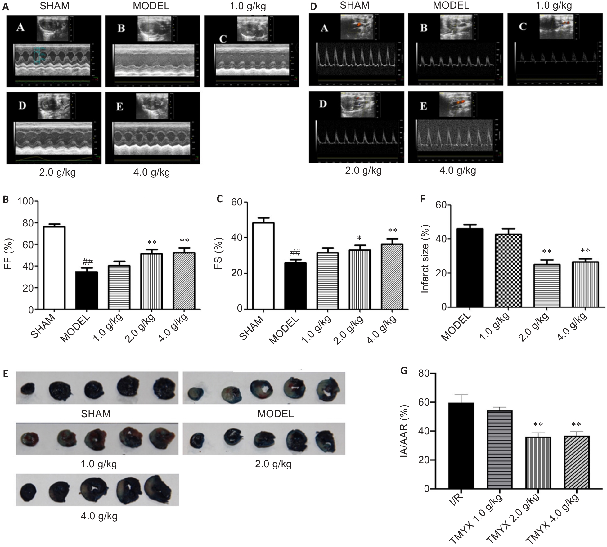
Fig.1 Effect of Tongmai Yangxin Pills (TMYX) on cardiac function in rats with myocardial ischemia-reperfusion injury (MIRI). A: Representative echocardiographic findings in each group. B: Comparison of left ventricular ejection fraction (EF) among the groups (n=12). C: Comparison of left ventricular short-axis shortening (FS) among the groups (n=12). D: Representative echocardiography for assessing cardiac blood flow in each group. E: Cardiac infarct size detected using Evans blue-TTC double staining in each group (n=6). F: Comparison of the mean cardiac infarct size among MIRI group and TMYX-treated groups (n=6). G: Comparison of infarct area normalized to area-at-risk (IA/AAR) among MIRI group and TMYX-treated groups (n=6). #P<0.05, ##P<0.01 vs SHAM group; *P<0.05, **P<0.01 vs MIRI model group.
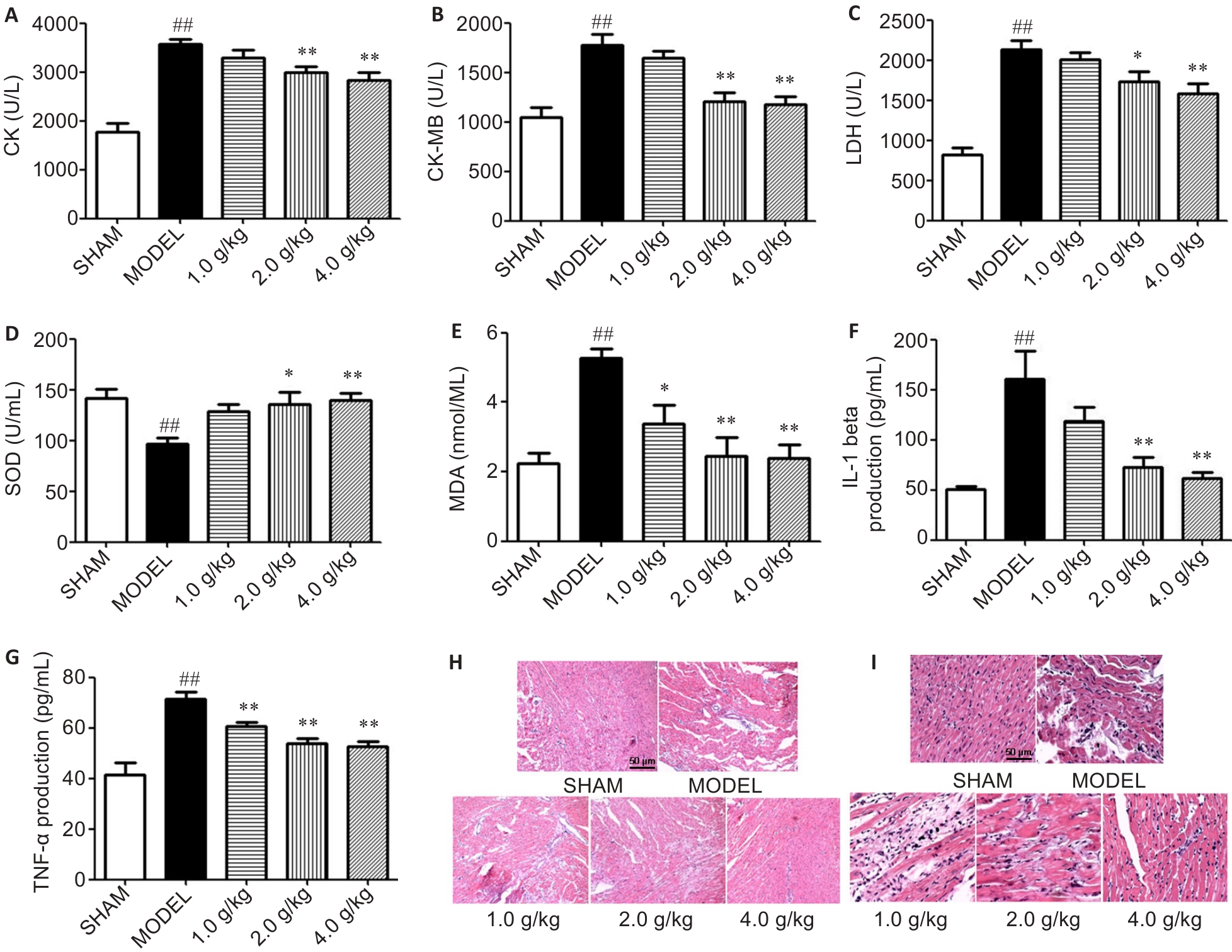
Fig.2 Effects of TMYX on myocardial injury in MIRI rats. A-G: Comparison of serum levels of creatine kinase (CK) (A; n=6), creatine kinase MB (CK-MB) (B; n=6), lactate dehydrogenase (LDH) (C; n=6), superoxide dismutase (SOD) (D; n=12), malondialdehyde (MDA) (E; n=12), tumor necrosis factor-α (TNF-α) (F; n=8), and interleukin-1β (IL-1β) (G; n=8) levels among the 5 groups of rats. H, I: HE staining of rat myocardial tissues in each group (H: original magnification:×100; I: ×200). ##P<0.01 vs sham group; *P<0.05, **P<0.01 vs model group.
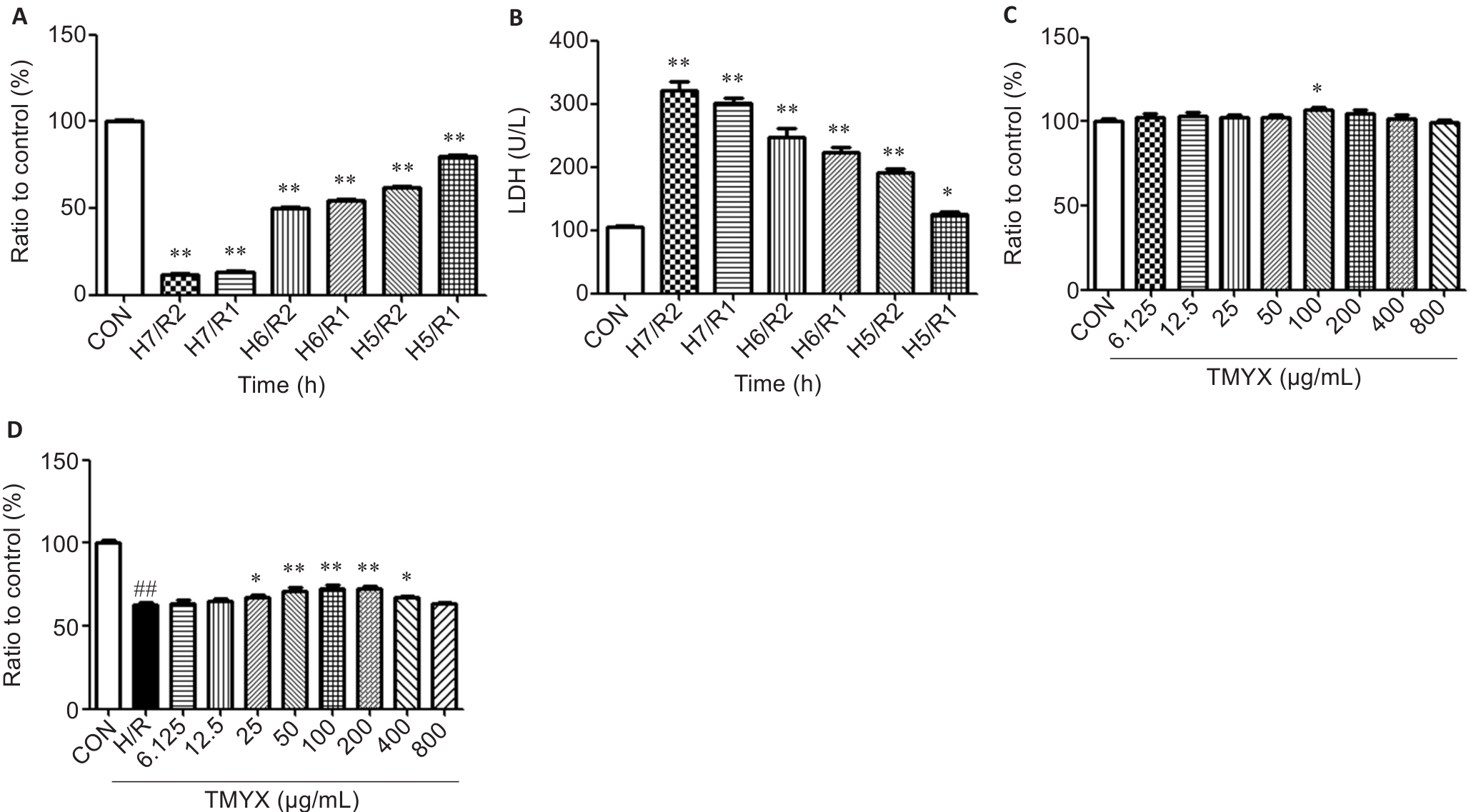
Fig.3 Hypoxia/reoxygenation (H/R) modeling and TMYX treatment conditions in H9c2 cells. A: H9c2 cell viability exposed to different hypoxia and reoxygenation times (n=30). B: LDH levels in H9c2 cells after exposure to different hypoxia and reoxygenation times (n=30). C: Effect of different concentrations of TMYX on H9c2 cell viability in normoxia (n=30). D: Effect of different concentrations of TMYX on H9c2 cell viability following hypoxia for 5 h and reoxygenation for 2 h (n=30). ##P<0.01 vs CON group; *P<0.05, **P<0.01 vs H/R group.
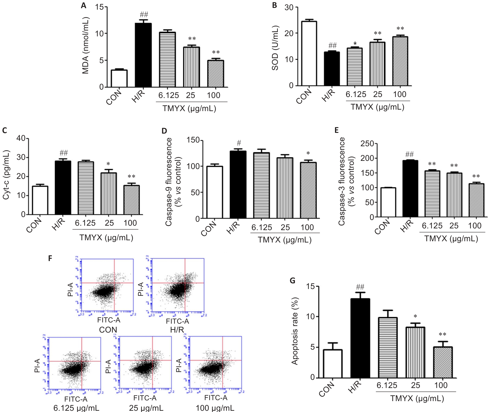
Fig.4 Effect of TMYX on H/R injury in H9c2 cells. A-E: Levels of MDA (A; n=30), SOD (B; n=30), Cyt-c (C; n=8), caspase-9 (D; n=9) and caspase-3 (E; n=9) in H9c2 cells with different treatments. F, G: Results of flow cytometry for analyzing apoptosis of H9c2 cells with different treatments (n=6). #P<0.05, ##P<0.01 vs CON group; *P<0.05, **P<0.01 vs H/R group.
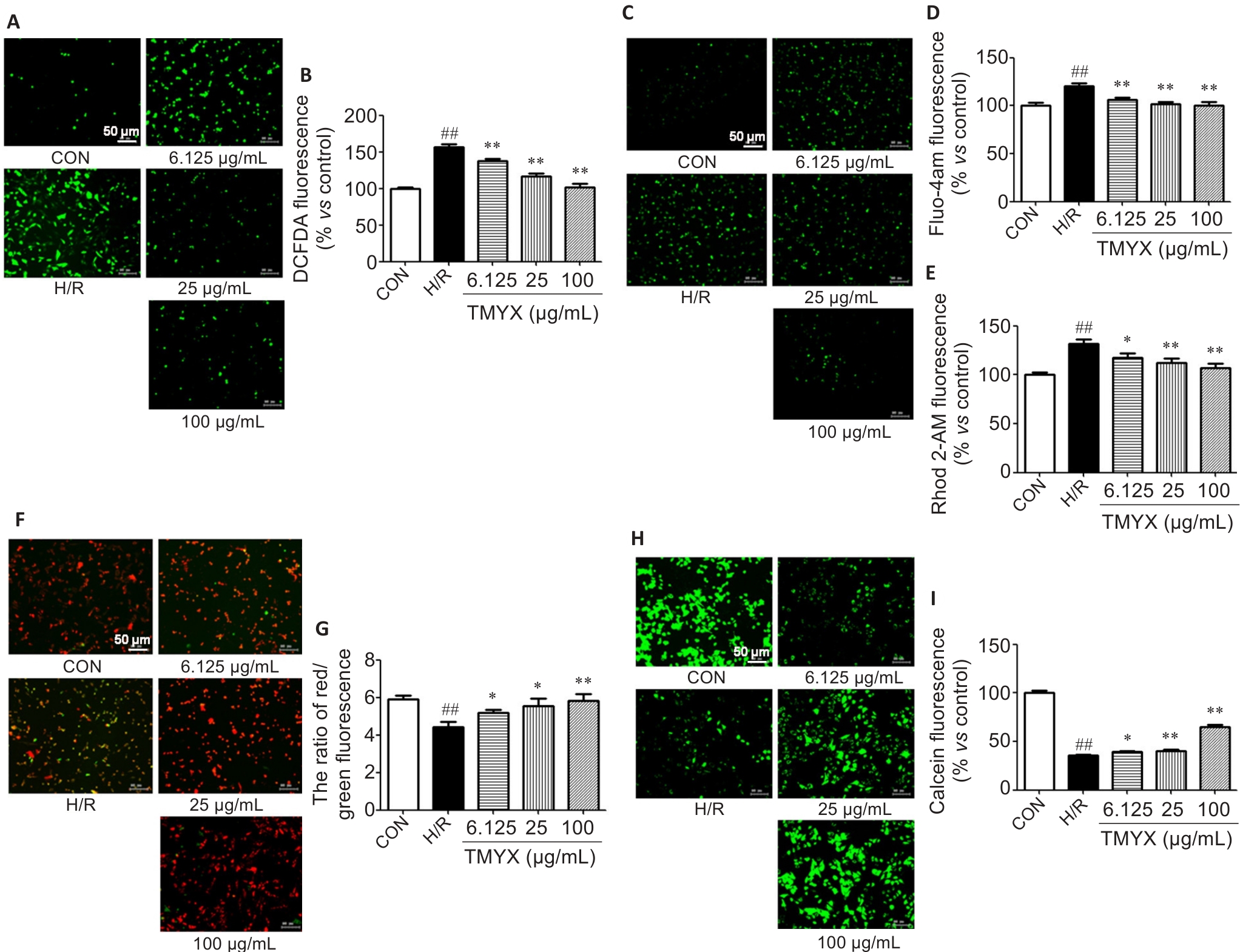
Fig.5 Effect of TMYX on mitochondrial function in H/R-exposed H9c2 cells. A, B: Fluorescence probe detection of reactive oxygen species (ROS) in H9c2 cells (×200). C, D: Detection of intracellular Ca2+ concentration using Fluo-4am in H9c2 cells in each group. E: Detection of mitochondrial Ca2+ concentration using Rhod 2-AM probe. F, G: Detection of mitochondrial membrane potential in H9c2 cells. H, I: Detection of mitochondrial permeability transition pore opening level in H9c2 cells. Data are presented as Mean±SD (n=30). ##P<0.01 vs CON group; *P<0.05, **P<0.01 vs H/R group.
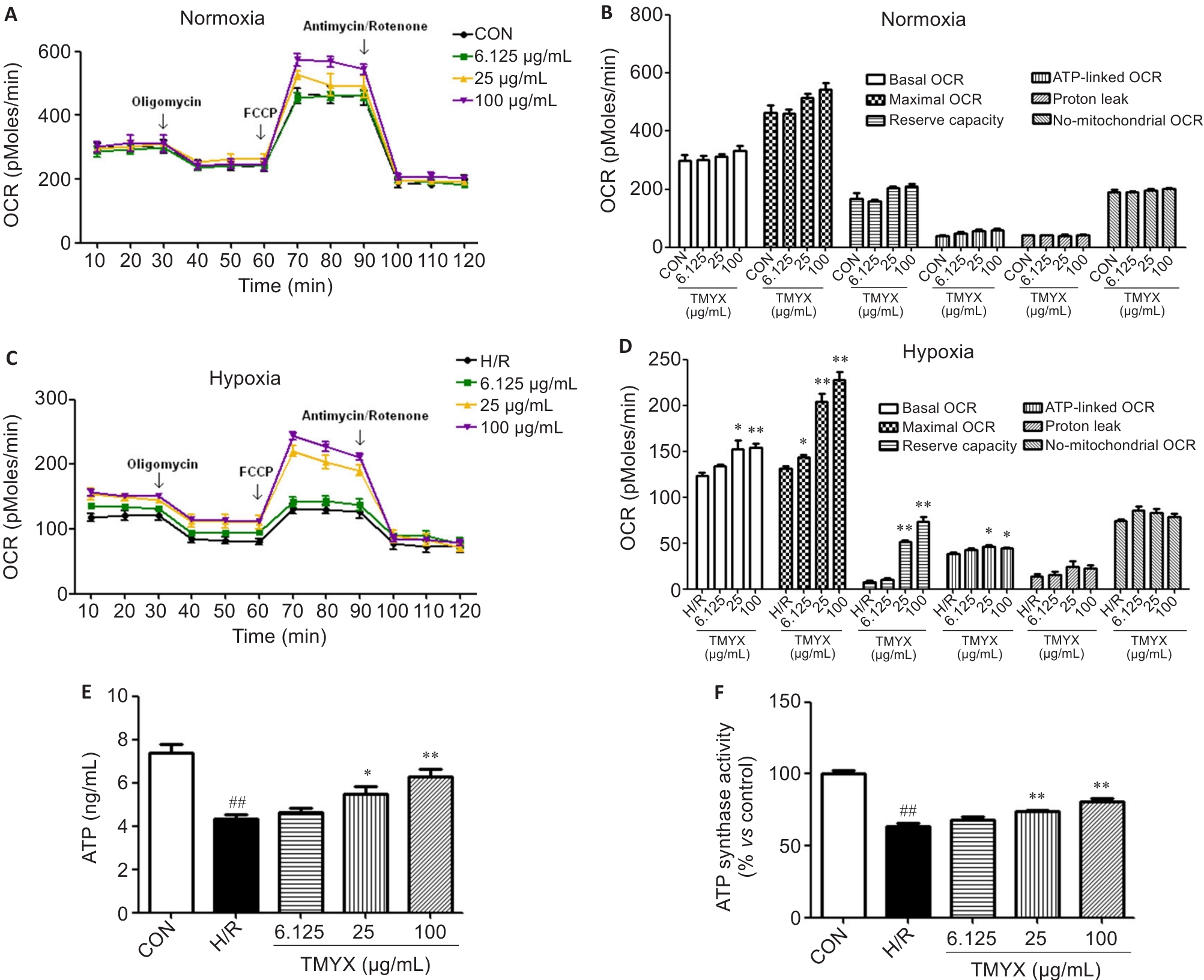
Fig.6 Effect of TMYX on mitochondrial energy metabolism in H/R-exposed H9c2 cells. A: Changes in mitochondrial respiratory oxygen consumption rate (OCR) in control and TMYX-treated H9c2 cells under normoxic conditions. B: Effects of TMYX on mitochondrial energy metabolism in H9c2 cells under normoxic conditions. C: Effect of TMYX on mitochondrial respiratory OCR in H/R-exposed H9c2 cells. D: Effects of TMYX mitochondrial energy metabolism in H/R-exposed H9c2 cells. E, F: Effect of TMYX on ATP and ATPase level in in H/R-exposed H9c2 cells. Data are presented as Mean±SD (n=30). ##P<0.01 vs CON group; *P<0.05, **P<0.01 vs H/R group.
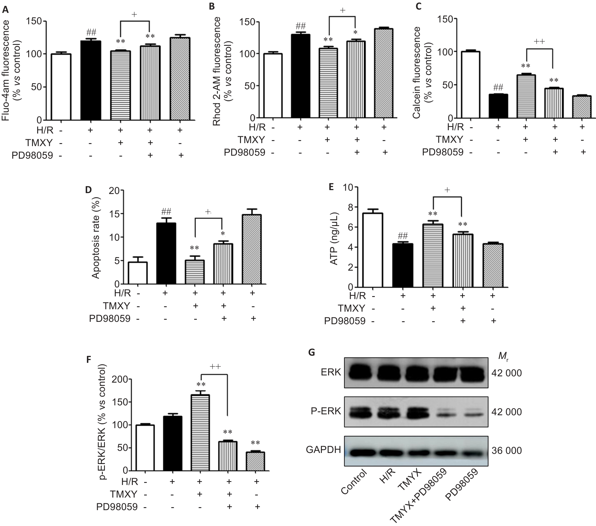
Fig.7 Effect of TMYX on ERK signaling pathway in H/R-exposed H9c2 cells. A: Intracellular cytoplasmic Ca2+ concentration in H9c2 cells with different treatments detected using Fluo-4am probe (n=30). B: Intramitochondrial Ca2+ concentration in H9c2 cells with different treatments detected using Rhod 2-AM probe (n=30). C: Degree of mPTP opening in H9c2 cells with different treatments (n=30). D: Apoptosis rates of the cells in different groups (n=30). E, F: ATP level (n=30) and p-ERK/ERK levels (n=6) in H9c2 cells with different treatments. G: Western blotting for detecting p-ERK and ERK protein expressions in H9c2 cells (n=6). Data are presented as Mean±SD. ##P<0.01 vs CON group; *P<0.05, **P<0.01 vs H/R group; +P<0.05, ++P<0.01 vs TMYX group.
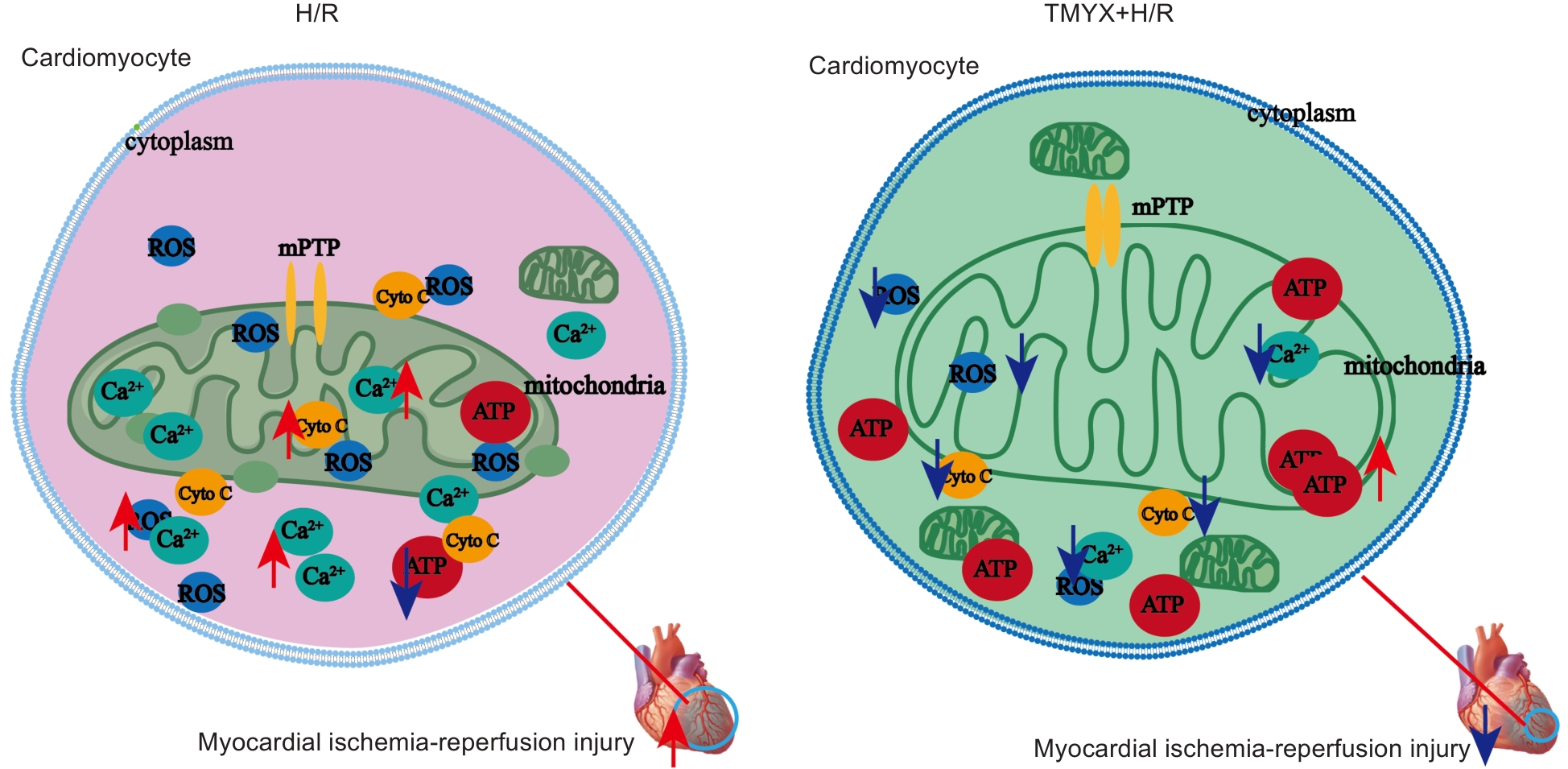
Fig.8 Diagram illustrating the therapeutic mechanism of TMYX against MIRI.
| [1] | Roth GA, Mensah GA, Johnson CO, et al. Global burden of cardiovascular diseases and risk factors, 1990-2019: update from the GBD 2019 study[J]. J Am Coll Cardiol, 2020, 76(25): 2982-3021. doi:10.1016/j.jacc.2020.11.010. |
| [2] | Cai W, Liu L, Shi X, et al. Alox15/15-HpETE aggravates myocardial ischemia-reperfusion injury by promoting cardiomyocyte ferroptosis [J]. Circulation, 2023, 147(19): 1444-60. doi:10.1161/circulationaha.122.060257 |
| [3] | Chen M, Zhong G, Liu M, et al. Integrating network analysis and experimental validation to reveal the mitophagy-associated mechanism of Yiqi Huoxue (YQHX) prescription in the treatment of myocardial ischemia/reperfusion injury[J]. Pharmacol Res, 2023, 189: 106682. doi:10.1016/j.phrs.2023.106682 |
| [4] | Zhang W, Chen R, Xu K, et al. Protective effect of Xinmai'an tablets via mediation of the AMPK/SIRT1/PGC-1alpha signaling pathway on myocardial ischemia-reperfusion injury in rats[J]. Phytomedicine, 2023, 120: 155034. doi:10.1016/j.phymed.2023.155034 |
| [5] | Ong SB, Samangouei P, Kalkhoran SB, et al. The mitochondrial permeability transition pore and its role in myocardial ischemia reperfusion injury[J]. J Mol Cell Cardiol, 2015, 78: 23-34. doi:10.1016/j.yjmcc.2014.11.005 |
| [6] | Piao L, Fang YH, Fisher M, et al. Dynamin-related protein 1 is a critical regulator of mitochondrial calcium homeostasis during myocardial ischemia/reperfusion injury[J]. FASEB J, 2024, 38(1): e23379. doi:10.1096/fj.202301040RR |
| [7] | Chouchani ET, Pell VR, Gaude E, et al. Ischaemic accumulation of succinate controls reperfusion injury through mitochondrial ROS[J]. Nature, 2014, 515(7527): 431-5. doi:10.1038/nature13909 |
| [8] | Zhang CX, Cheng Y, Liu DZ, et al. Mitochondria-targeted cyclosporin A delivery system to treat myocardial ischemia reperfusion injury of rats[J]. J Nanobiotechnology, 2019, 17(1): 18. doi:10.1186/s12951-019-0451-9 |
| [9] | Song Z, Yang Z, Tian L, et al. Targeting mitochondrial circadian rhythms: The potential intervention strategies of traditional Chinese medicine for myocardial ischaemia-reperfusion injury[J]. Biomed Pharmacother, 2023, 166: 115432. doi:10.1016/j.biopha.2023.115432 |
| [10] | Yu W, Xu M, Zhang T, et al. Mst1 promotes cardiac ischemia-reperfusion injury by inhibiting the ERK-CREB pathway and repressing FUNDC1-mediated mitophagy[J]. J Physiol Sci, 2019, 69(1): 113-27. doi:10.1007/S12576-018-0627-3 |
| [11] | Xie H, Zhang J, Zhu J, et al. Sevoflurane post-conditioning protects isolated rat hearts against ischemia-reperfusion injury via activation of the ERK1/2 pathway[J]. Acta Pharmacol Sin, 2014, 35(12): 1504-13. doi:10.1038/aps.2014.78 |
| [12] | Hernandez-Resendiz S, Zazueta C. PHO-ERK1/2 interaction with mitochondria regulates the permeability transition pore in cardioprotective signaling [J]. Life Sci, 2014, 108(1): 13-21. doi:10.1016/j.lfs.2014.04.037 |
| [13] | Yang Y, Li X, CHEN G, et al. Traditional Chinese medicine compound (Tongxinluo) and clinical outcomes of patients with acute myocardial infarction: The CTS-AMI randomized clinical trial[J]. JAMA, 2023, 330(16): 1534-45. doi:10.1001/jama.2023.19524 |
| [14] | Chen R, Chen T, Wang T, et al. Tongmai Yangxin pill reduces myocardial no-reflow by regulating apoptosis and activating PI3K/Akt/eNOS pathway[J]. J Ethnopharmacol, 2020, 261: 113069. doi:10.1016/j.jep.2020.113069 |
| [15] | Fan Y, Man S, Li H, et al. Analysis of bioactive components and pharmacokinetic study of herb-herb interactions in the traditional Chinese patent medicine Tongmai Yangxin Pill[J]. J Pharm Biomed Anal, 2016, 120: 364-73. doi:10.1016/j.jpba.2015.12.032 |
| [16] | Huang WW, Wang MY, Shi HM, et al. Comparative study of bioactive constituents in crude and processed Glycyrrhizae radix and their respective metabolic profiles in gastrointestinal tract in vitro by HPLC-DAD and HPLC-ESI/MS analyses[J]. Arch Pharm Res, 2012, 35(11): 1945-52. doi:10.1007/s12272-012-1111-x |
| [17] | Aiyasiding X, Liao HH, Feng H, et al. Liquiritin attenuates pathological cardiac hypertrophy by activating the PKA/LKB1/AMPK pathway[J]. Front Pharmacol, 2022, 13: 870699. doi:10.3389/fphar.2022.870699 |
| [18] | Cui Y, Yang Y, Tang X, et al. Cinnamic acid alleviates hypertensive left ventricular hypertrophy by antagonizing the vasopressor activity and the pro-cardiac hypertrophic signaling of angiotensin II[J]. Front Pharmacol, 2025, 16: 1555991. doi:10.3389/fphar.2025.1555991 |
| [19] | Bai Y, Tan D, Deng Q, et al. Cinnamic acid alleviates endothelial dysfunction and oxidative stress by targeting PPARdelta in obesity and diabetes[J]. Chin Med, 2025, 20(1): 13. doi:10.1186/s13020-025-01064-7 |
| [20] | Xu Z, Li M, Lyu D, et al. Cinnamaldehyde activates AMPK/PGC-1alpha pathway via targeting GRK2 to ameliorate heart failure[J]. Phytomedicine, 2024, 133: 155894. doi:10.1016/j.phymed.2024.155894 |
| [21] | Cui Y, Wang P, Li M, et al. Cinnamic acid mitigates left ventricular hypertrophy and heart failure in part through modulating FTO-dependent N(6)‑methyladenosine RNA modification in cardio-myocytes[J]. Biomed Pharmacother, 2023, 165: 115168. doi:10.1016/j.biopha.2023.115168 |
| [22] | Koczurkiewicz-Adamczyk P, Klas K, Gunia-Krzyzak A, et al. Cinnamic acid derivatives as cardioprotective agents against oxidative and structural damage induced by doxorubicin[J]. Int J Mol Sci, 2021, 22(12): 6217. doi:10.3390/ijms22126217 |
| [23] | Liu LJ, Zhu GH, Luo HY, et al. Tongmai Yangxin Pill combined with metoprolol or metoprolol alone for the treatment of symptomatic premature ventricular complex: a multicenter, randomized, parallel-controlled clinical study[J]. J Geriatr Cardiol, 2022, 19(4): 284-91. doi:10.11909/j.issn.1671-5411.2022.04.008 |
| [24] | Wang Y, Wang X, Wang J, et al. Tongmai Yangxin intervening in myocardial remodeling after PCI for coronary heart disease: study protocol for a double-blind, randomized controlled trial[J]. Trials, 2020, 21(1): 287. doi:10.1186/s13063-020-4208-4 |
| [25] | Chen R, Chen T, Wang T, et al. Tongmai Yangxin pill reduces myocardial No-reflow via endothelium-dependent NO-cGMP signaling by activation of the cAMP/PKA pathway[J]. J Ethnopharmacol, 2021, 267: 113462. doi:10.1016/j.jep.2020.113462 |
| [26] | Yang Z, Lin S, Liu Y, et al. Traditional Chinese medicine in coronary microvascular disease[J]. Front Pharmacol, 2022, 13: 929159. doi:10.3389/fphar.2022.929159 |
| [27] | Niccoli G, Montone RA, Ibanez B, et al. Optimized Treatment of ST-Elevation Myocardial Infarction[J]. Circ Res, 2019, 125(2): 245-58. doi:10.1161/CIRCRESAHA.119.315344 |
| [28] | Fan Y, Liu J, Miao J, et al. Anti-inflammatory activity of the Tongmai Yangxin pill in the treatment of coronary heart disease is associated with estrogen receptor and NF-kappaB signaling pathway [J]. J Ethnopharmacol, 2021, 276: 114106. doi:10.1016/j.jep.2021.114106 |
| [29] | Piantoni C, Paina M, Molla D, et al. Chinese natural compound decreases pacemaking of rabbit cardiac sinoatrial cells by targeting second messenger regulation of f-channels[J]. Elife, 2022, 11: e75119. doi:10.7554/eLife.75119 |
| [30] | Chen T, Zhang Y, Chen M, et al. Tongmai Yangxin pill alleviates myocardial no-reflow by activating GPER to regulate HIF-1alpha signaling and downstream potassium channels[J]. Pharm Biol, 2023, 61(1): 499-513. doi:10.1080/13880209.2023.2184481 |
| [31] | Ramachandra CJA, Hernandez-Resendiz S, Crespo-Avilan G E, et al. Mitochondria in acute myocardial infarction and cardioprotection [J]. EBioMedicine, 2020, 57: 102884. doi:10.1016/j.ebiom.2020.102884 |
| [32] | Dai DF, Rabinovitch PS, Ungvari Z. Mitochondria and cardiovascular aging[J]. Circ Res, 2012, 110(8): 1109-24. doi:10.1161/circresaha.111.246140 |
| [33] | Bugger H, Pfeil K. Mitochondrial ROS in myocardial ischemia reperfusion and remodeling[J]. Biochim Biophys Acta Mol Basis Dis, 2020, 1866(7): 165768. doi:10.1016/j.bbadis.2020.165768 |
| [34] | Gong Y, Lin J, Ma Z, et al. Mitochondria-associated membrane-modulated Ca2+ transfer: A potential treatment target in cardiac ischemia reperfusion injury and heart failure[J]. Life Sci, 2021, 278: 119511. doi:10.1016/j.lfs.2021.119511 |
| [35] | Halestrap AP, Clarke SJ, Javadov SA. Mitochondrial permeability transition pore opening during myocardial reperfusion-a target for cardioprotection[J]. Cardiovasc Res, 2004, 61(3): 372-85. doi:10.1016/s0008-6363(03)00533-9 |
| [36] | Zhang H, Yang N, He H, et al. The zinc transporter ZIP7 (Slc39a7) controls myocardial reperfusion injury by regulating mitophagy[J]. Basic Res Cardiol, 2021, 116(1): 54. doi:10.1007/s00395-021-00894-4 |
| [37] | Jonassen AK, Sack MN, Mjos OD, et al. Myocardial protection by insulin at reperfusion requires early administration and is mediated via Akt and p70s6 kinase cell-survival signaling[J]. Circ Res, 2001, 89(12): 1191-8. doi:10.1161/hh2401.101385 |
| [38] | Shalbueva N, Mareninova OA, Gerloff A, et al. Effects of oxidative alcohol metabolism on the mitochondrial permeability transition pore and necrosis in a mouse model of alcoholic pancreatitis[J]. Gastroenterology, 2013, 144(2): 437-46.e6. doi:10.1053/j.gastro.2012.10.037 |
| [39] | Robichaux DJ, Harata M, Murphy E, et al. Mitochondrial permeability transition pore-dependent necrosis[J]. J Mol Cell Cardiol, 2023, 174: 47-55. doi:10.1016/j.yjmcc.2022.11.003 |
| [40] | Dong L, Shen Z, Chi H, et al. Research progress of Chinese medicine in the treatment of myocardial ischemia-reperfusion injury[J]. Am J Chin Med, 2023, 51(1): 1-17. doi:10.1142/s0192415x23500015 |
| [41] | Wang F, Wang H, Liu X, et al. Neuregulin-1 alleviates oxidative stress and mitigate inflammation by suppressing NOX4 and NLRP3/caspase-1 in myocardial ischaemia-reperfusion injury[J]. J Cell Mol Med, 2021, 25(3): 1783-95. doi:10.1111/jcmm.16287 |
| [42] | Su RY, Geng XY, Yang Y, et al. Nesfatin-1 inhibits myocardial ischaemia/reperfusion injury through activating Akt/ERK pathway-dependent attenuation of endoplasmic reticulum stress[J]. J Cell Mol Med, 2021, 25(11): 5050-9. doi:10.1111/jcmm.16481 |
| [43] | Chen P, Lu Y, He B, et al. Rab32 promotes glioblastoma migration and invasion via regulation of ERK/Drp1-mediated mitochondrial fission[J]. Cell Death Dis, 2023, 14(3): 198. doi:10.1038/s41419-023-05721-3 |
| [44] | Hu C, Sun Y, Li W, et al. Hypoxia improves self-renew and migration of urine-derived stem cells by upregulating autophagy and mitochondrial function through ERK signal pathway[J]. Mitochondrion, 2023, 73: 1-9. doi:10.1016/j.mito.2023.09.001 |
| [1] | 雷艳萍, 宋嘉晟, 徐乐吾, 刘睿, 赵岳. 麦冬皂苷D通过激活β-catenin/FUNDC1/线粒体自噬轴减轻阿霉素诱导的小鼠心肌肥厚[J]. 南方医科大学学报, 2026, 46(4): 803-815. |
| [2] | 杨剑明, 杨龙, 洪铠文, 耿贝贝, 赵满, 王耀光, 夏婷, 董津睿. 腹腔注射薤白碳量子点可改善顺铂诱导的小鼠急性肾损伤并修复线粒体功能[J]. 南方医科大学学报, 2026, 46(3): 505-512. |
| [3] | 刘泽, 毛樟坤, 尤达, 王俊杰, 何咏梅, 余伊雯, 文志强, 方会龙, 何汶霞. 宽缨酮靶向抑制STAT3减轻线粒体功能障碍和炎症缓解急性肾损伤[J]. 南方医科大学学报, 2026, 46(3): 570-581. |
| [4] | 林心君, 何昱霖, 施红, 刘佳绣, 胡海霞. 石斛合剂通过调控Sirt3介导的线粒体自噬通路缓解大鼠糖尿病心肌病[J]. 南方医科大学学报, 2026, 46(1): 47-54. |
| [5] | 赵锦燕, 彭娇, 林明和, 朱晓勤, 黄彬, 林久茂. 清解扶正颗粒通过抑制线粒体依赖的凋亡、激活AMPK-PGC-1α通路缓解5-氟尿嘧啶引起的骨骼肌损伤[J]. 南方医科大学学报, 2026, 46(1): 94-103. |
| [6] | 闫爱丽, 罗梦瑶, 常晋瑞, 李新华, 朱娟霞. 橙皮素通过调控AMPK/NLRP3通路减轻阿霉素诱导的小鼠心肌毒性[J]. 南方医科大学学报, 2025, 45(9): 1850-1858. |
| [7] | 欧泽金, 李瀛, 陈诗, 王梓译, 何美仪, 陈志成, 唐侍豪, 孟晓静, 王致. 抑制铁死亡减轻敌草快引起的斑马鱼急性肾损伤的机制[J]. 南方医科大学学报, 2025, 45(8): 1743-1750. |
| [8] | 张璐, 丁焕章, 许浩燃, 陈珂, 许博文, 杨勤军, 吴迪, 童佳兵, 李泽庚. 参芪补中方通过激活AMPK/SIRT1/PGC-1α改善COPD肺脾气虚证大鼠线粒体功能障碍[J]. 南方医科大学学报, 2025, 45(5): 969-976. |
| [9] | 卞芬兰, 倪诗垚, 赵鹏, 戚毛男星, 唐碧, 王洪巨, 康品方, 刘进军. 积雪草苷通过抑制NLRP3炎症体介导的细胞焦亡减轻大鼠心肌缺血再灌注损伤[J]. 南方医科大学学报, 2025, 45(5): 977-985. |
| [10] | 刘露玉, 公茂伟, 廖国松, 赵维星, 傅强. 高血压通过UCP2下调介导的线粒体功能障碍加重大鼠术后学习记忆损伤[J]. 南方医科大学学报, 2025, 45(4): 725-735. |
| [11] | 廖茗, 钟文华, 张冉, 梁娟, 徐文陶睿, 万文珺, 吴超, 李曙. 源自蛇毒的蛋白C激活剂通过调控HIF-1α抑制BNIP3活性氧生成保护人脐静脉内皮细胞免受缺氧-复氧损伤[J]. 南方医科大学学报, 2025, 45(3): 614-621. |
| [12] | 丁珊珊, 廖颖, 白雪, 黄娇阳, 浅川哲也. 敲低Cav1基因可抑制小鼠肝细胞线粒体功能及关键基因mRNA的m6A修饰和表达[J]. 南方医科大学学报, 2025, 45(12): 2607-2615. |
| [13] | 秦萌, 孙思宇, 刘佳琪, 高玉娇, 汪昊, 王友坤, 孙奥, 严加纯, 汪金宝, 于影. 白藜芦醇改善PM2.5诱导的脑缺血再灌注损伤小鼠血脑屏障及维持线粒体动力学平衡[J]. 南方医科大学学报, 2025, 45(12): 2690-2698. |
| [14] | 陈一镠, 马民, 苏燃, 朱寅宾, 冯晴, 罗嘉丽, 冯伟峰, 颜显欣. 理冲消癥颗粒通过上调ANT3介导的线粒体凋亡增强小鼠卵巢癌移植瘤对顺铂的敏感性[J]. 南方医科大学学报, 2025, 45(11): 2309-2319. |
| [15] | 马丹丹, 程洁, 张虹, 刘广, 宋凯. 电针通过Bcl-2/Bax/caspase-3信号通路修复海马线粒体损伤改善创伤后应激障碍大鼠的焦虑症状[J]. 南方医科大学学报, 2025, 45(11): 2375-2384. |
| 阅读次数 | ||||||
|
全文 |
|
|||||
|
摘要 |
|
|||||
