Journal of Southern Medical University ›› 2025, Vol. 45 ›› Issue (6): 1212-1219.doi: 10.12122/j.issn.1673-4254.2025.06.10
Previous Articles Next Articles
Xiaoxiang ZHANG( ), Ying TIAN, Lilan FU, Yin ZHANG, Ye DONG, Fei XIE, Li CHEN, Yanchao HUANG, Hubing WU(), Jianer TAN()
Received:2025-01-06
Online:2025-06-20
Published:2025-06-27
Contact:
Hubing WU, Jianer TAN
E-mail:xxzhang23@163.com;wuhbym@163.com;Jianer.tan@foxmail.com
Supported by:Xiaoxiang ZHANG, Ying TIAN, Lilan FU, Yin ZHANG, Ye DONG, Fei XIE, Li CHEN, Yanchao HUANG, Hubing WU, Jianer TAN. 68Ga-DOTATATE and 18F-FDG PET/CT dual-modality imaging enhances precision of staging and treatment decision for gastroenteropancreatic neuroendocrine neoplasms[J]. Journal of Southern Medical University, 2025, 45(6): 1212-1219.
Add to citation manager EndNote|Ris|BibTeX
URL: https://www.j-smu.com/EN/10.12122/j.issn.1673-4254.2025.06.10

Fig.1 Classification of detection patterns of GEP-NEN by 18F-FDG and 68Ga-DOTATATE PET/CT.
| Characteristics | Value | Ratio (%) |
|---|---|---|
| Gender (n) | ||
| Male | 29 | 59.2 |
| Female | 20 | 40.8 |
| Age [year, M(P25,P75)] | 55 (45.5-60) | - |
| Diagnosis (n) | ||
| Newly diagnosed | 34 | 69.4 |
| Recurrence or metastasis after treatment | 15 | 30.6 |
| Primary tumor (n) | ||
| Gastric | 6 | 12.2 |
| Colorectal | 18 | 36.7 |
| Pancreatic | 25 | 51.0 |
| Histopathology (n) | ||
| G1 NET | 13 | 26.5 |
| G2 NET | 24 | 49.0 |
| G3 NET | 6 | 12.2 |
| NEC | 6 | 12.2 |
| Patient with metastasis (n) | ||
| Yes | 30 | 61.2 |
| No | 19 | 38.8 |
Tab.1 Characteristics of 49 patients with GEP-NEN
| Characteristics | Value | Ratio (%) |
|---|---|---|
| Gender (n) | ||
| Male | 29 | 59.2 |
| Female | 20 | 40.8 |
| Age [year, M(P25,P75)] | 55 (45.5-60) | - |
| Diagnosis (n) | ||
| Newly diagnosed | 34 | 69.4 |
| Recurrence or metastasis after treatment | 15 | 30.6 |
| Primary tumor (n) | ||
| Gastric | 6 | 12.2 |
| Colorectal | 18 | 36.7 |
| Pancreatic | 25 | 51.0 |
| Histopathology (n) | ||
| G1 NET | 13 | 26.5 |
| G2 NET | 24 | 49.0 |
| G3 NET | 6 | 12.2 |
| NEC | 6 | 12.2 |
| Patient with metastasis (n) | ||
| Yes | 30 | 61.2 |
| No | 19 | 38.8 |
| Lesion | Number of patients (n) | Number of lesions (n) | 18F-FDG positive lesion detection [n(%)] | 68Ga-DOTATATE positive lesion detection [n(%)] | P |
|---|---|---|---|---|---|
| Total | 49 | 539 | 347 (64.4) | 447 (82.9) | <0.001 |
| Primary/recurrent | 45 | 45 | 32 (71.1) | 42 (93.3) | 0.011 |
| Lymph nodes | 29 | 109 | 89 (81.7) | 107 (98.2) | <0.001 |
| Liver | 1 | 277 | 193 (69.7) | 220 (79.4) | 0.011 |
| Bone | 9 | 65 | 11 (16.9) | 62 (95.4) | <0.001 |
| Lung | 3 | 23 | 9 (39.1) | 2 (8.7) | 0.035 |
| Peritoneum | 2 | 6 | 6 (100) | 1 (16.7) | 0.015 |
| Adrenal | 2 | 3 | 3 (100) | 3 (100) | >0.999 |
| Brain | 1 | 3 | 2 (66.7) | 3 (100) | >0.999 |
| Kidney | 1 | 1 | 1 (100) | 1 (100) | >0.999 |
| Spleen | 1 | 2 | 0 (0.0) | 2 (100) | 0.333 |
| Pancreas | 1 | 1 | 0 (0.0) | 1 (100) | >0.999 |
| Ovary | 1 | 1 | 0 (0.0) | 1 (100) | >0.999 |
| Soft tissue | 2 | 3 | 1 (33.3) | 2 (66.7) | >0.999 |
Tab.2 Comparison of 18F-FDG and 68Ga-DOTATATE PET/CT for detection of primary/recurrent and metastatic lesion
| Lesion | Number of patients (n) | Number of lesions (n) | 18F-FDG positive lesion detection [n(%)] | 68Ga-DOTATATE positive lesion detection [n(%)] | P |
|---|---|---|---|---|---|
| Total | 49 | 539 | 347 (64.4) | 447 (82.9) | <0.001 |
| Primary/recurrent | 45 | 45 | 32 (71.1) | 42 (93.3) | 0.011 |
| Lymph nodes | 29 | 109 | 89 (81.7) | 107 (98.2) | <0.001 |
| Liver | 1 | 277 | 193 (69.7) | 220 (79.4) | 0.011 |
| Bone | 9 | 65 | 11 (16.9) | 62 (95.4) | <0.001 |
| Lung | 3 | 23 | 9 (39.1) | 2 (8.7) | 0.035 |
| Peritoneum | 2 | 6 | 6 (100) | 1 (16.7) | 0.015 |
| Adrenal | 2 | 3 | 3 (100) | 3 (100) | >0.999 |
| Brain | 1 | 3 | 2 (66.7) | 3 (100) | >0.999 |
| Kidney | 1 | 1 | 1 (100) | 1 (100) | >0.999 |
| Spleen | 1 | 2 | 0 (0.0) | 2 (100) | 0.333 |
| Pancreas | 1 | 1 | 0 (0.0) | 1 (100) | >0.999 |
| Ovary | 1 | 1 | 0 (0.0) | 1 (100) | >0.999 |
| Soft tissue | 2 | 3 | 1 (33.3) | 2 (66.7) | >0.999 |
| Grades | Patterns A | Patterns B | Patterns C | Patterns D |
|---|---|---|---|---|
| Total | 23 (46.9) | 19 (38.8) | 6 (12.2) | 1 (2.0) |
| G1 NET | 6 (12.2) | 7 (14.3) | 0 (0.0) | 0 (0.0) |
| G2 NET | 14 (28.6) | 8 (16.3) | 2 (4.1) | 0 (0.0) |
| G3 NET | 2 (4.1) | 1 (2.0) | 2 (4.1) | 1 (2.0) |
| NEC | 1 (2.0) | 3 (6.1) | 2 (4.1) | 0 (0.0) |
Tab.3 Comparison of 18F-FDG and 68Ga-DOTATATE PET/CT for detecting different grades of GEP-NEN [n(%)]
| Grades | Patterns A | Patterns B | Patterns C | Patterns D |
|---|---|---|---|---|
| Total | 23 (46.9) | 19 (38.8) | 6 (12.2) | 1 (2.0) |
| G1 NET | 6 (12.2) | 7 (14.3) | 0 (0.0) | 0 (0.0) |
| G2 NET | 14 (28.6) | 8 (16.3) | 2 (4.1) | 0 (0.0) |
| G3 NET | 2 (4.1) | 1 (2.0) | 2 (4.1) | 1 (2.0) |
| NEC | 1 (2.0) | 3 (6.1) | 2 (4.1) | 0 (0.0) |
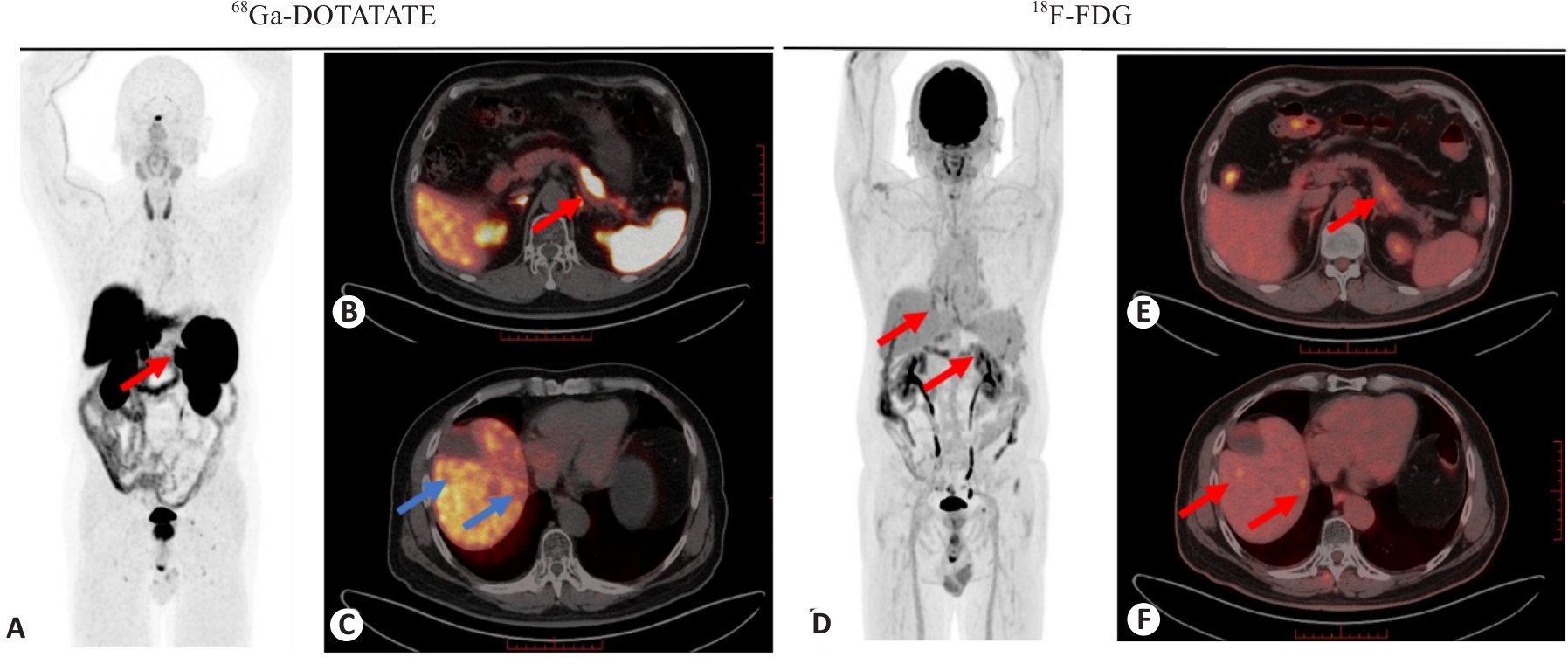
Fig.2 A G2 NET patient underwent dual tracer PET/CT scans, and the staging was changed due to additional detection of multiple liver metastases by 18F-FDG PET/CT imaging. The 66-year-old male patient was diagnosed with pancreatic neuroendocrine tumor (G2 NET) with multiple liver metastases. 68Ga-DOTATATE PET/CT showed a high uptake lesion in the tail of the pancreas (A, B, red arrows, SUVmax: 35.2), while 18F-FDG PET/CT showed a mild hypermetabolic lesion in the tail of the pancreas (D, E, red arrows, SUVmax: 4.2). 18F-FDG PET/CT displayed multiple hypermetabolic lesions in the liver (D, F, red arrows, SUVmax: 4.0 and 3.6, respectively), while 68Ga-DOTATATE PET/CT did not show abnormal uptake at the corresponding site (C, blue arrows).
| 1 | Pavel M, Öberg K, Falconi M, et al. Gastroenteropancreatic neuroendocrine neoplasms: ESMO Clinical Practice Guidelines for diagnosis, treatment and follow-up[J]. Ann Oncol, 2020, 31(7): 844-60. doi:10.1016/j.annonc.2020.03.304 |
| 2 | Oronsky B, Ma PC, Morgensztern D, et al. Nothing but NET: a review of neuroendocrine tumors and carcinomas[J]. Neoplasia, 2017, 19(12): 991-1002. doi:10.1016/j.neo.2017.09.002 |
| 3 | Christofer Juhlin C, Mete O, Baloch ZW. The 2022 WHO classification of thyroid tumors: novel concepts in nomenclature and grading[J]. Endocr Relat Cancer, 2022, 30(2): e220293. doi:10.1530/erc-22-0293 |
| 4 | Rindi G, Klimstra DS, Abedi-Ardekani B, et al. A common classification framework for neuroendocrine neoplasms: an International Agency for Research on Cancer (IARC) and World Health Organization (WHO) expert consensus proposal[J]. Mod Pathol, 2018, 31(12): 1770-86. doi:10.1038/s41379-018-0110-y |
| 5 | Zhang XB, Fan YB, Jing R, et al. Gastroenteropancreatic neuroendocrine neoplasms: current development, challenges, and clinical perspectives[J]. Mil Med Res, 2024, 11(1): 35. doi:10.1186/s40779-024-00535-6 |
| 6 | Zhang PP, Yu JY, Li J, et al. Clinical and prognostic value of PET/CT imaging with combination of 68Ga-DOTATATE and 18F-FDG in gastroenteropancreatic neuroendocrine neoplasms[J]. Contrast Media Mol Imaging, 2018, 2018: 2340389. doi:10.1155/2018/2340389 |
| 7 | 张青菊, 杨卫东, 王胜军, 等. Ga-DOTANOC PET/CT显像在胰腺神经内分泌肿瘤诊断及分期中的应用价值[J]. 中华核医学与分子影像杂志, 2019, 39(8): 453-7. doi:10.3760/cma.j.issn.2095-2848.2019.08.002 |
| 8 | Panzuto F, Ramage J, Pritchard DM, et al. European Neuroendocrine Tumor Society (ENETS) 2023 guidance paper for gastroduodenal neuroendocrine tumours (NETs) G1-G3[J]. J Neuroendocrinol, 2023, 35(8): e13306. doi:10.1111/jne.13306 |
| 9 | You H, Kandathil A, Beg M, et al. Ga-68 DOTATATE PET/CT and F-18 FDG PET/CT in the evaluation of low and intermediate versus high-grade neuroendocrine tumors[J]. Nucl Med Commun, 2020, 41(10): 1060-5. doi:10.1097/mnm.0000000000001255 |
| 10 | Chan DL, Pavlakis N, Schembri GP, et al. Dual somatostatin receptor/FDG PET/CT imaging in metastatic neuroendocrine tumours: proposal for a novel grading scheme with prognostic significance[J]. Theranostics, 2017, 7(5): 1149-58. doi:10.7150/thno.18068 |
| 11 | Naswa N, Sharma P, Gupta SK, et al. Dual tracer functional imaging of gastroenteropancreatic neuroendocrine tumors using 68Ga-DOTA-NOC PET-CT and 18F-FDG PET-CT: competitive or complimentary?[J]. Clin Nucl Med, 2014, 39(1): e27-34. doi:10.1097/rlu.0b013e31827a216b |
| 12 | Kaewput C, Vinjamuri S. Role of combined 68Ga DOTA-Peptides and 18F FDG PET/CT in the evaluation of gastroenteropancreatic neuroendocrine neoplasms[J]. Diagnostics, 2022, 12(2): 280. doi:10.3390/diagnostics12020280 |
| 13 | Carideo L, Prosperi D, Panzuto F, et al. Role of combined 68Ga Ga-DOTA-SST analogues and 18F FDG PET/CT in the management of GEP-NENs: a systematic review[J]. J Clin Med, 2019, 8(7): 1032. doi:10.3390/jcm8071032 |
| 14 | Partelli S, Rinzivillo M, Maurizi A, et al. The role of combined Ga-DOTANOC and 18FDG PET/CT in the management of patients with pancreatic neuroendocrine tumors[J]. Neuroendocrinology, 2014, 100(4): 293-9. doi:10.1159/000368609 |
| 15 | Has Simsek D, Kuyumcu S, Turkmen C, et al. Can complementary 68Ga-DOTATATE and 18F-FDG PET/CT establish the missing link between histopathology and therapeutic approach in gastro-enteropancreatic neuroendocrine tumors[J]? J Nucl Med, 2014, 55(11): 1811-7. doi:10.2967/jnumed.114.142224 |
| 16 | Chauhan A, Chan K, Halfdanarson TR, et al. Critical updates in neuroendocrine tumors: version 9 American joint committee on cancer staging system for gastroenteropancreatic neuroendocrine tumors[J]. CA Cancer J Clin, 2024, 74(4): 359-67. doi:10.3322/caac.21840 |
| 17 | Kryza D, Tadino V, Filannino MA, et al. Fully automated 18F fluorocholine synthesis in the TracerLab MX FDG Coincidence synthesizer[J]. Nucl Med Biol, 2008, 35(2): 255-60. doi:10.1016/j.nucmedbio.2007.11.008 |
| 18 | He Q, Zhang ZK, Zhang LQ, et al. Head-to-head comparison between 68Ga Ga-DOTA-NOC and 18F DOPA PET/CT in a diverse cohort of patients with pheochromocytomas and paragangliomas[J]. Eur J Nucl Med Mol Imaging, 2024, 51(7): 1989-2001. doi:10.1007/s00259-024-06622-z |
| 19 | Velikyan I. 68Ga-Based radiopharmaceuticals: production and application relationship[J]. Molecules, 2015, 20(7): 12913-43. doi:10.3390/molecules200712913 |
| 20 | Hope TA, Allen-Auerbach M, Bodei LS, et al. SNMMI procedure standard/EANM practice guideline for SSTR PET: imaging neuroendocrine tumors[J]. J Nucl Med, 2023, 64(2): 204-10. doi:10.2967/jnumed.122.264860 |
| 21 | 余浩军, 顾宇参, 杨 志, 等. 神经内分泌肿瘤68Ga-DOTATATE联合18F-FDG两日法全身PET/CT显像操作规范专家共识[EB/OL]. [2025-01-21]. . |
| 22 | Wang LJ, Tang GH, Hu KZ, et al. Comparison of 68Ga-FAPI and 18F-FDG PET/CT in the evaluation of advanced lung cancer[J]. Radiology, 2022, 303(1): 191-9. doi:10.1148/radiol.211424 |
| 23 | Zamora V, Cabanne A, Salanova R, et al. Immunohistochemical expression of somatostatin receptors in digestive endocrine tumours[J]. Dig Liver Dis, 2010, 42(3): 220-5. doi:10.1016/j.dld.2009.07.018 |
| 24 | Virgolini I, Ambrosini V, Bomanji JB, et al. Procedure guidelines for PET/CT tumour imaging with 68Ga-DOTA-conjugated peptides: 68Ga-DOTA-TOC, 68Ga-DOTA-NOC, 68Ga-DOTA-TATE[J]. Eur J Nucl Med Mol Imaging, 2010, 37(10): 2004-10. doi:10.1007/s00259-010-1512-3 |
| 25 | Hofman MS, Eddie Lau WF, Hicks RJ. Somatostatin receptor imaging with 68Ga DOTATATE PET/CT: clinical utility, normal patterns, pearls, and pitfalls in interpretation[J]. Radiographics, 2015, 35(2): 500-16. doi:10.1148/rg.352140164 |
| 26 | Putzer D, Gabriel M, Henninger B, et al. Bone metastases in patients with neuroendocrine tumor: 68Ga-DOTA-Tyr3-octreotide PET in comparison to CT and bone scintigraphy[J]. J Nucl Med, 2009, 50(8): 1214-21. doi:10.2967/jnumed.108.060236 |
| 27 | 唐文鑫, 王琦新, 杨松松, 等. 68Ga-DOTATATE PET/CT与18F-FDG PET/CT对分化良好和分化不良的胃肠胰神经内分泌肿瘤显像的对比研究[J]. 肿瘤影像学, 2022, 31(3): 230-5. doi:10.19732/j.cnki.2096-6210.2022.03.003 |
| 28 | Muffatti F, Partelli S, Cirocchi R, et al. Combined 68Ga-DOTA-peptides and 18F-FDG PET in the diagnostic work-up of neuroendocrine neoplasms (NEN)[J]. Clin Transl Imag, 2019, 7(3): 181-8. doi:10.1007/s40336-019-00328-1 |
| 29 | 赵 帅, 程 超, 左长京. Ga-SSA/F-FDG PET/CT联合显像在神经内分泌肿瘤诊治中的应用价值[J]. 中华核医学与分子影像杂志, 2020, 40(1): 47-51. doi:10.3760/cma.j.issn.2095-2848.2020.01.012 |
| 30 | 中国抗癌协会神经内分泌肿瘤专业委员会. 中国抗癌协会神经内分泌肿瘤诊治指南(2022年版)[J]. 中国癌症杂志, 2022, 32(6): 545-79. doi:10.19401/j.cnki.1007-3639.2022.06.010 |
| 31 | Yang ZH, Tang LH, Klimstra DS. Effect of tumor heterogeneity on the assessment of Ki67 labeling index in well-differentiated neuroendocrine tumors metastatic to the liver: implications for prognostic stratification[J]. Am J Surg Pathol, 2011, 35(6): 853-60. doi:10.1097/pas.0b013e31821a0696 |
| 32 | Kayani I, Bomanji JB, Groves A, et al. Functional imaging of neuroendocrine tumors with combined PET/CT using 68Ga-DOTATATE (DOTA-DPhe1, Tyr3-octreotate) and 18F-FDG[J]. Cancer, 2008, 112(11): 2447-55. doi:10.1002/cncr.23469 |
| 33 | Abgral R, Leboulleux S, Déandreis D, et al. Performance of 18Fluorodeoxyglucose-positron emission tomography and somatostatin receptor scintigraphy for high Ki67 (≥10%) well-differentiated endocrine carcinoma staging[J]. J Clin Endocrinol Metab, 2011, 96(3): 665-71. doi:10.1210/jc.2010-2022 |
| 34 | Centonze G, Maisonneuve P, Simbolo M, et al. Lung carcinoid tumours: histology and Ki-67, the eternal rivalry[J]. Histopathology, 2023, 82(2): 324-39. doi:10.1111/his.14819 |
| 35 | Reubi JC, Waser B. Concomitant expression of several peptide receptors in neuroendocrine tumours: molecular basis for in vivo multireceptor tumour targeting[J]. Eur J Nucl Med Mol Imaging, 2003, 30(5): 781-93. doi:10.1007/s00259-003-1184-3 |
| 36 | Vesterinen T, Leijon H, Mustonen H, et al. Somatostatin receptor expression is associated with metastasis and patient outcome in pulmonary carcinoid tumors[J]. J Clin Endocrinol Metab, 2019, 104(6): 2083-93. doi:10.1210/jc.2018-01931 |
| 37 | Ivanidze J, Roytman M, Sasson A, et al. Molecular imaging and therapy of somatostatin receptor positive tumors[J]. Clin Imaging, 2019, 56: 146-54. doi:10.1016/j.clinimag.2019.04.006 |
| 38 | Hu XW, Li DD, Wang R, et al. Comparison of the application of 18F-FDG and 68Ga-DOTATATE PET/CT in neuroendocrine tumors: a retrospective study[J]. Medicine, 2023, 102(19): e33726. doi:10.1097/md.0000000000033726 |
| 39 | 杜长治, 谢 卿, 翟士桢, 等. Ga-DOTATATE与F-FDG PET/CT显像探测神经内分泌肿瘤骨转移的对比研究[J]. 中华核医学与分子影像杂志, 2021, 41(9): 520-4. |
| 40 | Shen ZH, Zhang XJ, Li QX, et al. Comparison of 18F-FDG PET/CT and 18F-DOTATATE PET/CT in the diagnosis of multiple metastases in rectal neuroendocrine neoplasms[J]. Radiol Case Rep, 2024, 19(9): 3757-62. doi:10.1016/j.radcr.2024.03.051 |
| 41 | 王 玲, 胡桂兰, 乔 真, 等. 神经内分泌肿瘤转移灶PET/CT生长抑素受体显像特点分析[J]. 中华核医学与分子影像杂志, 2017, 37(3): 132-6. doi:10.3760/cma.j.issn.2095-2848.2017.03.002 |
| 42 | Shastry M, Kayani I, Wild D, et al. Distribution pattern of 68Ga-DOTATATE in disease-free patients[J]. Nucl Med Commun, 2010, 31(12): 1025-32. doi:10.1097/mnm.0b013e32833f635e |
| 43 | Yi CA, Shin KM, Lee KS, et al. Non-small cell lung cancer staging: efficacy comparison of integrated PET/CT versus 3.0-T whole-body MR imaging[J]. Radiology, 2008, 248(2): 632-42. doi:10.1148/radiol.2482071822 |
| 44 | Zhou Y, Li L, Wang H, et al. Heterogeneous uptake of 68Ga-DOTATATE and 18F-FDG in initial diagnosed neuroendocrine tumors patients: which patients are suitable for dual-tracer PET imaging[J]? Clin Nucl Med, 2024, 49(6): 516-20. doi:10.1097/rlu.0000000000005231 |
| 45 | Adams S, Baum R, Rink T, et al. Limited value of fluorine-18 fluorodeoxyglucose positron emission tomography for the imaging of neuroendocrine tumours[J]. Eur J Nucl Med, 1998, 25(1): 79-83. doi:10.1007/s002590050197 |
| 46 | Fang JM, Li J, Shi JQ. An update on the diagnosis of gastroenteropancreatic neuroendocrine neoplasms[J]. World J Gastroenterol, 2022, 28(10): 1009-23. doi:10.3748/wjg.v28.i10.1009 |
| 47 | Skoura E, Michopoulou S, Mohmaduvesh M, et al. The impact of 68Ga-DOTATATE PET/CT imaging on management of patients with neuroendocrine tumors: experience from a national referral center in the United Kingdom[J]. J Nucl Med, 2016, 57(1): 34-40. doi:10.2967/jnumed.115.166017 |
| 48 | Tierney JF, Kosche C, Schadde E, et al. 68Gallium-DOTATATE positron emission tomography-computed tomography (PET CT) changes management in a majority of patients with neuroendocrine tumors[J]. Surgery, 2019, 165(1): 178-85. doi:10.1016/j.surg.2018.03.030 |
| 49 | Ghobrial SN, Menda Y, Zamba GK, et al. Prospective analysis of the impact of 68Ga-DOTATOC positron emission tomography-computerized axial tomography on management of pancreatic and small bowel neuroendocrine tumors[J]. Pancreas, 2020, 49(8): 1033-6. doi:10.1097/mpa.0000000000001625 |
| [1] | Xiaoyu ZHANG, Hao WANG, Dong ZENG, Zhaoying BIAN. A low-dose CT image restoration method based on central guidance and alternating optimization [J]. Journal of Southern Medical University, 2025, 45(4): 844-852. |
| [2] | Zhixiong ZENG, Yongbo WANG, Zongyue LIN, Zhaoying BIAN, Jianhua MA. A segmented backprojection tensor degradation feature encoding model for motion artifacts correction in dental cone beam computed tomography [J]. Journal of Southern Medical University, 2025, 45(2): 422-436. |
| [3] | Shizhou TANG, Ruolan SU, Shuting LI, Zhenzhen LAI, Jinhong HUANG, Shanzhou NIU. A low-dose CT reconstruction method using sub-pixel anisotropic diffusion [J]. Journal of Southern Medical University, 2025, 45(1): 162-169. |
| [4] | Shengwang PENG, Yongbo WANG, Zhaoying BIAN, Jianhua MA, Jing HUANG. A dual-domain cone beam computed tomography reconstruction framework with improved differentiable domain transform for cone-angle artifact correction [J]. Journal of Southern Medical University, 2024, 44(6): 1188-1197. |
| [5] | Zongyue LIN, Yongbo WANG, Zhaoying BIAN, Jianhua MA. A deep blur learning-based motion artifact reduction algorithm for dental cone-beam computed tomography images [J]. Journal of Southern Medical University, 2024, 44(6): 1198-1208. |
| [6] | CHEN Shixuan, ZENG Dong, BIAN Zhaoying, MA Jianhua. A low-dose CT reconstruction algorithm across different scanners based on federated feature learning [J]. Journal of Southern Medical University, 2024, 44(2): 333-343. |
| [7] | ZHONG Weixiong, LIANG Fangrong, YANG Ruimeng, ZHEN Xin. Prediction of microvascular invasion in hepatocellular carcinoma based on multi-phase dynamic enhanced CT radiomics feature and multi-classifier hierarchical fusion model [J]. Journal of Southern Medical University, 2024, 44(2): 260-269. |
| [8] | Caolin LIU, Qingqing ZOU, Menghong WANG, Qinmei YANG, Liwen SONG, Zixiao LU, Qianjin FENG, Yinghua ZHAO. Identification of osteoid and chondroid matrix mineralization in primary bone tumors using a deep learning fusion model based on CT and clinical features: a multi-center retrospective study [J]. Journal of Southern Medical University, 2024, 44(12): 2412-2420. |
| [9] | Jingyi LIAO, Shengwang PENG, Yongbo WANG, Zhaoying BIAN. A dual-domain cone beam computed tomography sparse-view reconstruction method based on generative projection interpolation [J]. Journal of Southern Medical University, 2024, 44(10): 2044-2054. |
| [10] | ZHOU Hao, ZENG Dong, BIAN Zhaoying, MA Jianhua. A semi-supervised network-based tissue-aware contrast enhancement method for CT images [J]. Journal of Southern Medical University, 2023, 43(6): 985-993. |
| [11] | WANG Weiran, SUN Zeyu, XIN Ran, DING Yipu, LIU Zinuan, WANG Xi, WANG Jing, SHAN Dongkai, LIU Changfu. Calcification distributional density of the aortic-valvular complex is an independent risk factor for conduction block following self-expanding transcatheter aortic valve replacement [J]. Journal of Southern Medical University, 2023, 43(11): 1901-1908. |
| [12] | HE Fawei, WANG Yongbo, TAO Xi, ZHU Manman, HONG Zixuan, BIAN Zhaoying, MA Jianhua. Low-dose helical CT projection data restoration using noise estimation [J]. Journal of Southern Medical University, 2022, 42(6): 849-859. |
| [13] | LIU Changfu, SUN Zeyu, WANG Jing, WANG Minhan, XIN Ran, DING Yipu, WANG Xi, MU Yang, CHEN Tao, JIANG Bo, WANG Lin, ZHANG Ming, SHAN Dongkai, CHEN Yundai. Anythink for CT-based aorta root measurements before transcatheter aortic valve replacement: measurement consistency with 3mensio and impact on short-term prognosis [J]. Journal of Southern Medical University, 2022, 42(11): 1646-1654. |
| [14] |
.
Four-dimensional cone-beam CT reconstruction based on motion-compensated robust principal component analysis
[J]. Journal of Southern Medical University, 2021, 41(2): 243-249.
|
| [15] | WANG Haoru, CHEN Xin, LIU Huan, YU Chunlin, HE Ling. Computed tomography-based radiomics for differential of retroperitoneal neuroblastoma and ganglioneuroblastoma in children [J]. Journal of Southern Medical University, 2021, 41(10): 1569-1576. |
| Viewed | ||||||
|
Full text |
|
|||||
|
Abstract |
|
|||||