南方医科大学学报 ›› 2025, Vol. 45 ›› Issue (6): 1336-1342.doi: 10.12122/j.issn.1673-4254.2025.06.23
董振翔1,2( ), 郭义昊1,2, 刘蔷1,2, 张益哲1,2, 丘倩怡3, 张晓东3, 冯衍秋1,2(
 )
)
收稿日期:2025-03-13
出版日期:2025-06-20
发布日期:2025-06-27
通讯作者:
冯衍秋
E-mail:3464737526@qq.com;foree@163.com
作者简介:董振翔,在读硕士研究生,E-mail: 3464737526@qq.com
基金资助:
Zhenxiang DONG1,2( ), Yihao GUO1,2, Qiang LIU1,2, Yizhe ZHANG1,2, Qianyi QIU3, Xiaodong ZHANG3, Yanqiu FENG1,2(
 )
)
Received:2025-03-13
Online:2025-06-20
Published:2025-06-27
Contact:
Yanqiu FENG
E-mail:3464737526@qq.com;foree@163.com
Supported by:摘要:
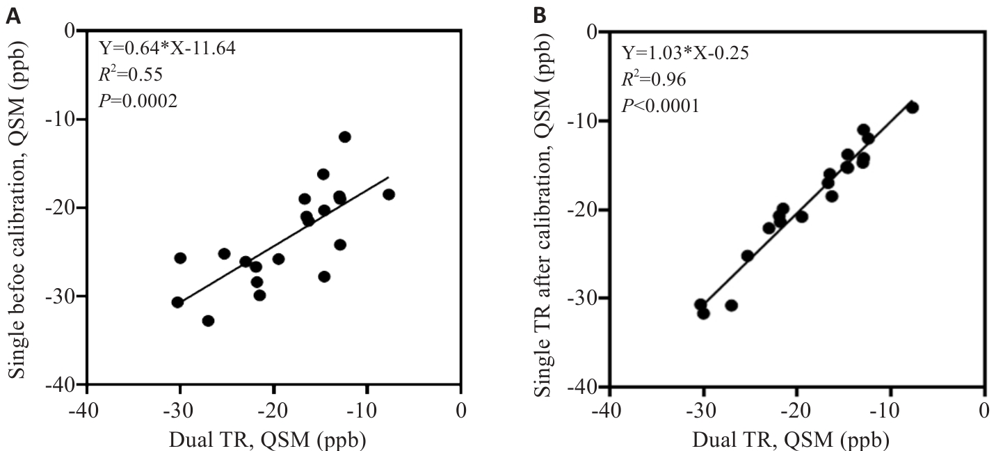
目的 提出一种利用双极读出梯度的单重复时间(TR)腰椎定量磁化率成像方法,并比较单TR与双TR方法在不同腰椎骨密度受试者的定量磁化率结果。 方法 提出平移校正方法解决了正负梯度读出图像之间沿频率编码方向空间错位问题,提出相位校正方法消除正负梯度读出图像之间的相位差。使用单TR和双TR方法采集了6名正常人,2名骨质减少患者以及2名骨质疏松患者的腰椎L1-L5的数据,重建出磁化率图后,将校正前后单TR与双TR方法的定量结果进行比较。 结果 单TR方法校正前与双TR方法所得腰椎磁化率值的线性回归结果为:Y=0.64*X-11.61。单TR方法校正后与双TR方法腰椎磁化率值的线性回归结果为:Y=1.03*X+0.25。说明校正后的单TR方法与双TR方法的结果高度一致,并且校正后的单TR方法能够有效地区分正常、骨量减少及骨质疏松患者。 结论 经过校正的单TR双极读出梯度方法能够生成无伪影的腰椎定量磁化率分布图,并且将数据采集时间缩短了50%。
董振翔, 郭义昊, 刘蔷, 张益哲, 丘倩怡, 张晓东, 冯衍秋. 基于双极读出梯度的单重复时间腰椎定量磁化率成像[J]. 南方医科大学学报, 2025, 45(6): 1336-1342.
Zhenxiang DONG, Yihao GUO, Qiang LIU, Yizhe ZHANG, Qianyi QIU, Xiaodong ZHANG, Yanqiu FENG. A single repetition time quantitative magnetic susceptibility imaging method for the lumbar spine using bipolar readout gradient[J]. Journal of Southern Medical University, 2025, 45(6): 1336-1342.
| MR scan sequence | Dual TR gradient echo sequence | Single TR gradient echo sequence |
|---|---|---|
| Field of view | 256×184×152 mm3 | 256×184×152 mm3 |
| Voxel size | 2 mm isotropic | 2 mm isotropic |
| Flip angle | 15° | 15° |
| Echo time | TE1=1.54 ms, TE2=2.3 ms, | TE1=1.2 ms, NTE =6 |
| Bandwidth | 996 hz/pixel | 996 hz/pixel |
| TR | 25 ms | 25 ms |
| Scan time | 9 min10 s | 4 min35 s |
| SENSE | SENSE=1 | SENSE=1 |
表1 双TR和单TR梯度回波序列的MR扫描参数
Tab.1 MR scan parameters of dual TR and single TR gradient echo sequences
| MR scan sequence | Dual TR gradient echo sequence | Single TR gradient echo sequence |
|---|---|---|
| Field of view | 256×184×152 mm3 | 256×184×152 mm3 |
| Voxel size | 2 mm isotropic | 2 mm isotropic |
| Flip angle | 15° | 15° |
| Echo time | TE1=1.54 ms, TE2=2.3 ms, | TE1=1.2 ms, NTE =6 |
| Bandwidth | 996 hz/pixel | 996 hz/pixel |
| TR | 25 ms | 25 ms |
| Scan time | 9 min10 s | 4 min35 s |
| SENSE | SENSE=1 | SENSE=1 |

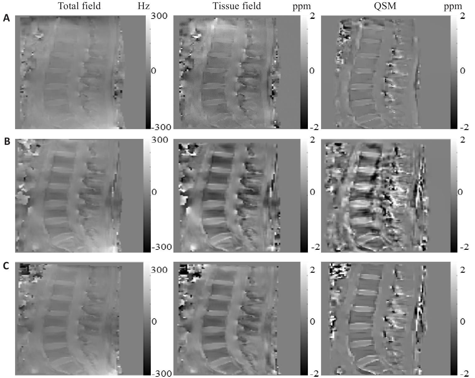
图4 正常志愿者的总场图,组织场图以及QSM结果
Fig.4 Total field map, tissue field map, and QSM results of the lumbar spine of normal volunteers.A: Dual TR method fitting results. B: Single TR method fitting results before calibration. C: Single TR method fitting results after calibration.

图5 单TR方法与双TR方法的QSM量化结果比较
Fig.5 Comparison of quantitative results between dual TR lumbar spine QSM and single TR. A: Comparison of dual TR and single TR before calibration. B: Comparison of dual TR and single TR after calibration.

图6 单TR方法在正常志愿者, 骨量减少以及骨质疏松患者的QSM结果
Fig.6 QSM results of the single TR method in normal individuals and in patients with osteopenia and osteoporosis. ***P<0.01.
| 1 | Wang Y, Liu T. Quantitative susceptibility mapping (QSM): decoding MRI data for a tissue magnetic biomarker[J]. Magn Reson Med, 2015, 73(1): 82-101. doi:10.1002/mrm.25358 |
| 2 | Wisnieff C, Ramanan S, Olesik J, et al. Quantitative susceptibility mapping (QSM) of white matter multiple sclerosis lesions: Interpreting positive susceptibility and the presence of iron[J]. Magn Reson Med, 2015, 74(2): 564-70. doi:10.1002/mrm.25420 |
| 3 | Ravanfar P, Loi SM, Syeda WT, et al. Systematic review: quantitative susceptibility mapping (QSM) of brain iron profile in neurodegenerative diseases[J]. Front Neurosci, 2021, 15: 618435. doi:10.3389/fnins.2021.618435 |
| 4 | Yang J, Lv M, Han L, et al. Evaluation of brain iron deposition in different cerebral arteries of acute ischaemic stroke patients using quantitative susceptibility mapping[J]. Clin Radiol, 2024, 79(4): e592-8. doi:10.1016/j.crad.2024.01.007 |
| 5 | Guo W, Zhang D, Sun J, et al. Quantitative susceptibility mapping of subcortical iron deposition in Parkinson disease and multiple system atrophy: clinical correlations and diagnostic implications[J]. Quant Imaging Med Surg, 2024, 14(7): 4464-74. doi:10.21037/qims-24-168 |
| 6 | Carpenter KLH, Li W, Wei HJ, et al. Magnetic susceptibility of brain iron is associated with childhood spatial IQ[J]. NeuroImage, 2016, 132: 167-74. doi:10.1016/j.neuroimage.2016.02.028 |
| 7 | Li X, Allen RP, Earley CJ, et al. Brain iron deficiency in idiopathic restless legs syndrome measured by quantitative magnetic susceptibility at 7 tesla[J]. Sleep Med, 2016, 22: 75-82. doi:10.1016/j.sleep.2016.05.001 |
| 8 | Kataike VM, Desmond PM, Steward C, et al. Iron changes within infarct tissue in ischemic stroke patients after successful reperfusion quantified using QSM[J]. Neuroradiology, 2024, 66(12): 2233-42. doi:10.1007/s00234-024-03444-6 |
| 9 | Sharma SD, Hernando D, Horng DE, et al. Quantitative susceptibility mapping in the abdomen as an imaging biomarker of hepatic iron overload[J]. Magn Reson Med, 2015, 74(3): 673-83. doi:10.1002/mrm.25448 |
| 10 | Lin HM, Wei HJ, He NY, et al. Quantitative susceptibility mapping in combination with water-fat separation for simultaneous liver iron and fat fraction quantification[J]. Eur Radiol, 2018, 28(8): 3494-504. doi:10.1007/s00330-017-5263-4 |
| 11 | Jafari R, Sheth S, Spincemaille P, et al. Rapid automated liver quantitative susceptibility mapping[J]. J Magn Reson Imaging, 2019, 50(3): 725-32. doi:10.1002/jmri.26632 |
| 12 | Buelo CJ, Velikina J, Mao L, et al. Multicenter, multivendor validation of liver quantitative susceptibility mapping in patients with iron overload at 1.5 T and 3 T[J]. Magn Reson Med, 2025, 93(1): 330-40. doi:10.1002/mrm.30251 |
| 13 | Jung M, Bresson X, Chan TF, et al. Nonlocal Mumford-Shah regularizers for color image restoration[J]. IEEE Trans Image Process, 2011, 20(6): 1583-98. doi:10.1109/tip.2010.2092433 |
| 14 | Lebenatus A, Kuster J, Straub S, et al. In-vitro detection of intramammary-like macrocalcifications using susceptibility-weighted MR imaging techniques at 1.5T[J]. Magn Reson Med Sci, 2024: mp. 2024-75. doi:10.2463/mrms.mp.2024-0075 |
| 15 | Boehm C, Sollmann N, Meineke J, et al. Preconditioned water-fat total field inversion: Application to spine quantitative susceptibility mapping[J]. Magn Reson Med, 2022, 87(1): 417-30. doi:10.1002/mrm.28903 |
| 16 | Diefenbach MN, Meineke J, Ruschke S, et al. On the sensitivity of quantitative susceptibility mapping for measuring trabecular bone density[J]. Magn Reson Med, 2019, 81(3): 1739-54. doi:10.1002/mrm.27531 |
| 17 | Guo Y, Chen Y, Zhang X, et al. Magnetic susceptibility and fat content in the lumbar spine of postmenopausal women with varying bone mineral density[J]. J Magn Reson Imaging, 2019, 49(4): 1020-8. doi:10.1002/jmri.26279 |
| 18 | Guo Y, Liu Z, Wen Y, et al. Quantitative susceptibility mapping of the spine using in-phase echoes to initialize inhomogeneous field and R2* for the nonconvex optimization problem of fat-water separation[J]. NMR Biomed, 2019, 32(11): e4156. doi:10.1002/nbm.4156 |
| 19 | Jerban S, Ma Y, Jang H, et al. MRI-based bone biomarkers; proceedings of the Seminars in musculoskeletal. Radiology, F, 2024 [C]. doi:10.1055/s-0040-1710355 |
| 20 | Li N, Li XM, Xu L, et al. Comparison of QCT and DXA: osteoporosis detection rates in postmenopausal women[J]. Int J Endocrinol, 2013, 2013: 895474. doi:10.1155/2013/895474 |
| 21 | Wáng YXJ, Yu W, Leung JCS, et al. More evidence to support a lower quantitative computed tomography (QCT) lumbar spine bone mineral density (BMD) cutpoint value for classifying osteoporosis among older East Asian women than for Caucasians[J]. Quant Imaging Med Surg, 2024, 14(5): 3239-47. doi:10.21037/qims-24-429 |
| 22 | Liu C, Li W, Tong KA, et al. Susceptibility-weighted imaging and quantitative susceptibility mapping in the brain[J]. J Magn Reson Imaging, 2015, 42(1): 23-41. doi:10.1002/jmri.24768 |
| 23 | Hopkins JA, Wehrli FW. Magnetic susceptibility measurement of insoluble solids by NMR: magnetic susceptibility of bone[J]. Magn Reson Med, 1997, 37(4): 494-500. doi:10.1002/mrm.1910370404 |
| 24 | Berikol G, Ekşi MŞ, Aydın L, et al. Subcutaneous fat index: a reliable tool for lumbar spine studies[J]. Eur Radiol, 2022, 32(9): 6504-13. doi:10.1007/s00330-022-08775-7 |
| 25 | Boehm C, Schlaeger S, Meineke J, et al. On the water–fat in-phase assumption for quantitative susceptibility mapping[J]. Magn Reson Med, 2023, 89(3): 1068-82. doi:10.1002/mrm.29516 |
| 26 | Ruschke S, Eggers H, Kooijman H, et al. Correction of phase errors in quantitative water-fat imaging using a monopolar time-interleaved multi-echo gradient echo sequence[J]. Magn Reson Med, 2017, 78(3): 984-96. doi:10.1002/mrm.26485 |
| 27 | Walsh DO, Gmitro AF, Marcellin MW. Adaptive reconstruction of phased array MR imagery[J]. Magn Reson Med, 2000, 43(5): 682-90. doi:10.1002/(sici)1522-2594(200005)43:5<682::aid-mrm10>3.0.co;2-g |
| 28 | Yu H, Shimakawa A, McKenzie CA, et al. Multiecho water-fat separation and simultaneous R2* estimation with multifrequency fat spectrum modeling[J]. Magn Reson Med, 2008, 60(5): 1122-34. doi:10.1002/mrm.21737 |
| 29 | Zhou D, Liu T, Spincemaille P, et al. Background field removal by solving the Laplacian boundary value problem[J]. NMR Biomed, 2014, 27(3): 312-9. doi:10.1002/nbm.3064 |
| 30 | Liu J, Liu T, de Rochefort L, et al. Morphology enabled dipole inversion for quantitative susceptibility mapping using structural consistency between the magnitude image and the susceptibility map[J]. NeuroImage, 2012, 59(3): 2560-8. doi:10.1016/j.neuroimage.2011.08.082 |
| 31 | Feng W, Neelavalli J, Haacke EM. Catalytic multiecho phase unwrapping scheme (CAMPUS) in multiecho gradient echo imaging: removing phase wraps on a voxel-by-voxel basis[J]. Magn Reson Med, 2013, 70(1): 117-26. doi:10.1002/mrm.24457 |
| 32 | Peterson P, Månsson S. Fat quantification using multiecho sequences with bipolar gradients: investigation of accuracy and noise performance[J]. Magn Reson Med, 2014, 71(1): 219-29. doi:10.1002/mrm.24657 |
| 33 | Yu H, Shimakawa A, McKenzie CA, et al. Phase and amplitude correction for multi-echo water-fat separation with bipolar acquisitions[J]. J Magn Reson Imaging, 2010, 31(5): 1264-71. doi:10.1002/jmri.22111 |
| 34 | QSM Consensus Organization Committee, Bilgic B, Costagli M, et al. Recommended implementation of quantitative susceptibility mapping for clinical research in the brain: a consensus of the ISMRM electro-magnetic tissue properties study group[J]. Magn Reson Med, 2024, 91(5): 1834-62. doi:10.1002/mrm.30006 |
| 35 | Jerban S, Ma YJ, Chang EY, et al. A UTE-based biomarker panel in osteoporosis [M]. MRI of Short-and Ultrashort-T2 Tissues: Making the Invisible Visible. Springer. 2024: 427-39. doi:10.1007/978-3-031-35197-6_34 |
| 36 | Shin SH, Chae HD, Suprana A, et al. UTE MRI technical developments and applications in osteoporosis: a review [J]. Frontiers in Endocrinology, 2025, 16: 1510010. doi:10.3389/fendo.2025.1510010 |
| 37 | Streichenberger B, Santin M, Roche S, et al. Quantitative susceptibility mapping of the cervical spinal cord for MS monitoring; proceedings of the 2025-20th MRI Workshop: MS lesions imaging: update on chronic active lesion and the central vein sign, F, 2025 [C]. |
| [1] | 陈远程, 吴文, 许翎, 邓海鸥, 王瑞雪, 黄倩雯, 禤立平, 陈雪莹, 智喜梅. 地舒单抗治疗的原发性骨质疏松症患者血钙及骨代谢标志物的动态变化[J]. 南方医科大学学报, 2025, 45(4): 760-764. |
| [2] | 周莹, 张丹阳, 吴立凡, 王桂杉, 母杰丹, 崔成文, 石秀秀, 董继革, 王瑜, 许王莉, 李晓. 北京地区近10年骨质疏松症流行病学调查:一项单中心30 599例汉族人群双能X线吸收法骨密度检查结果分析[J]. 南方医科大学学报, 2025, 45(3): 443-452. |
| [3] | 刘科, 马振岩, 付磊, 张丽萍, 阿鑫, 肖少波, 张震, 张洪博, 赵蕾, 钱赓. 心脏磁共振成像整体纵向应变对急性ST段抬高型心肌梗死后左心室重构的预测价值:403例前瞻性研究[J]. 南方医科大学学报, 2024, 44(6): 1033-1039. |
| [4] | 罗彩珠, 陈金香, 张 群, 于学钊, 张书勤. 聚乳酸/羟基磷灰石/磷钙锌石复合支架促进大鼠骨质疏松性骨缺损愈合[J]. 南方医科大学学报, 2024, 44(2): 370-380. |
| [5] | 黄晓茵, 陈凤莲, 张煜, 梁淑君. 多参数多区域MRI影像组学特征与临床信息联合模型可有效预测脑胶质瘤患者生存期[J]. 南方医科大学学报, 2024, 44(10): 2004-2014. |
| [6] | 何慧珊, 郭二嘉, 蒙文仪, 王 彧, 王 雯, 何文乐, 吴元魁, 阳 维. 基于磁共振图像机器学习放射组学模型预测脑胶质瘤的强化[J]. 南方医科大学学报, 2024, 44(1): 194-200. |
| [7] | 陈梓锋, 李胜发, 张祐鸣, 杨婉雯, 王 婷. 脂质运载蛋白2自限性抑制间充质干细胞的成骨细胞分化[J]. 南方医科大学学报, 2023, 43(8): 1339-1344. |
| [8] | 刘羽轩, 楚智钦, 张 煜. 基于物理模型的级联生成对抗网络加速定量多参数磁共振成像[J]. 南方医科大学学报, 2023, 43(8): 1402-1409. |
| [9] | 吴秀华, 范应静, 叶永浓, 李 萍, 朱青安, 陈泽森, 李 博, 王 文, 郑 磊. 生酮饮食导致小鼠骨质疏松的转录组学[J]. 南方医科大学学报, 2023, 43(8): 1440-1446. |
| [10] | 巩 高, 曹 石, 肖 慧, 方威扬, 阙与清, 刘子蔚, 陈超敏. 深度注意力机制结合临床特征预测肝细胞癌微血管浸润[J]. 南方医科大学学报, 2023, 43(5): 839-851. |
| [11] | 郝雨薇, 高 升, 张潇月, 崔梦秋, 丁效蕙, 王 鹤, 杨大为, 叶慧义, 王海屹. 透明细胞癌可能性评分v1.0和v2.0的应用价值对比[J]. 南方医科大学学报, 2023, 43(5): 800-806. |
| [12] | 喻靖文, 杨美洁, 蒋 莉, 肖智博, 李 霜, 陈锦云. 子宫腺肌病术前MR T2WI信号特征与HIFU消融疗效密切相关:一项倾向性评分配对队列研究[J]. 南方医科大学学报, 2023, 43(4): 597-603. |
| [13] | 阮红良, 佘冬梅, 孙绍裘. 六味地黄丸减轻绝经后骨质疏松症及疲劳的作用机制:基于抑制表观调控分子BRD4通路[J]. 南方医科大学学报, 2023, 43(12): 1998-2005. |
| [14] | 金晓丽, 许 嘉, 陈煊威, 陈 瑾, 黄 慧, 张 婷, 任 军, 许 健. 冬凌草甲素逆转硫代乙酰胺对破骨和成骨细胞分化的机制研究[J]. 南方医科大学学报, 2023, 43(11): 1892-1900. |
| [15] | 邹青清, 王梦虹, 陆紫箫, 赵英华, 冯前进. 基于多序列MRI的3D关系注意力网络预测HLA-B27阴性中轴性脊柱关节病[J]. 南方医科大学学报, 2023, 43(11): 1955-1964. |
| 阅读次数 | ||||||
|
全文 |
|
|||||
|
摘要 |
|
|||||