南方医科大学学报 ›› 2025, Vol. 45 ›› Issue (8): 1743-1750.doi: 10.12122/j.issn.1673-4254.2025.08.18
• • 上一篇
欧泽金1,2( ), 李瀛3, 陈诗3, 王梓译4, 何美仪4, 陈志成3, 唐侍豪1,2, 孟晓静3, 王致1,2()
收稿日期:2025-02-13
出版日期:2025-08-20
发布日期:2025-09-05
通讯作者:
王致
E-mail:ouzejin@smu.edu.cn;zhi_wang@outlook.com
作者简介:欧泽金,副研究员,E-mail: ouzejin@smu.edu.cn
基金资助:
Zejin OU1,2(), Ying LI3, Shi CHEN3, Ziyi WANG4, Meiyi HE4, Zhicheng CHEN3, Shihao TANG1,2, Xiaojing MENG3, Zhi WANG1,2()
Received:2025-02-13
Online:2025-08-20
Published:2025-09-05
Contact:
Zhi WANG
E-mail:ouzejin@smu.edu.cn;zhi_wang@outlook.com
摘要:
目的 通过构建斑马鱼敌草快急性中毒模型,探讨铁死亡在敌草快引起的急性肾损伤的作用及分子机制。 方法 采用肾脏标记Tg (Eco.Tshb:EGFP)和中性粒细胞标记Tg(lyz:dsRed2)的转基因斑马鱼构建急性肾损伤模型,设置空白对照组、庆大霉素阳性对照组、敌草快中毒组以及铁死亡抑制剂组,检测斑马鱼肾损伤、炎症反应以及铁死亡相关指标,采用Western blotting检测电压依赖性阴离子选择性通道蛋白1(VDAC1)和线粒体铁蛋白(FTMT)的表达水平。 结果 敌草快引起的急性肾损伤具有明显剂量效应关系,损伤程度与暴露浓度成正比,同时诱导明显的氧化应激和炎症反应。罗丹明代谢法和HE染色发现,肾小球过滤功能随着敌草快暴露浓度增加而下降(P<0.001)。免疫荧光显示,敌草快暴露后斑马鱼肾组织铁死亡标志物GPX4和FTH1的表达水平发生明显变化,而给予铁死亡抑制剂Ferrostatin-1干预后GPX4表达上调(P=0.040),FTH1表达下调(P=0.042),罗丹明B标记葡聚糖代谢率改善(P=0.024)。敌草快暴露引起VDAC1和FTMT表达水平上调(P<0.001),应用铁死亡抑制剂和VDAC1抑制剂VBIT-12后FTMT下调尤为明显。 结论 铁死亡参与敌草快致急性肾损伤的分子机制,并且VDAC1和FTMT参与其中的调控机制,可能是潜在的干预靶点。
欧泽金, 李瀛, 陈诗, 王梓译, 何美仪, 陈志成, 唐侍豪, 孟晓静, 王致. 抑制铁死亡减轻敌草快引起的斑马鱼急性肾损伤的机制[J]. 南方医科大学学报, 2025, 45(8): 1743-1750.
Zejin OU, Ying LI, Shi CHEN, Ziyi WANG, Meiyi HE, Zhicheng CHEN, Shihao TANG, Xiaojing MENG, Zhi WANG. Inhibition of ferroptosis alleviates acute kidney injury caused by diquat in zebrafish[J]. Journal of Southern Medical University, 2025, 45(8): 1743-1750.
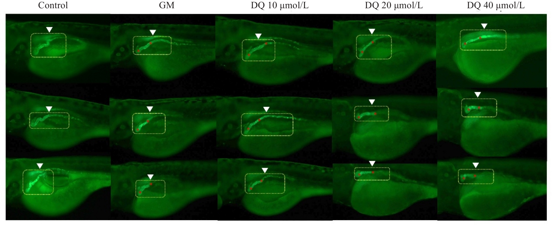
图1 敌草快对斑马鱼肾脏发育的影响
Fig.1 Effect of diquat on kidney development in zebrafish (Original magnification: ×40).Three replicates are shown in each group.
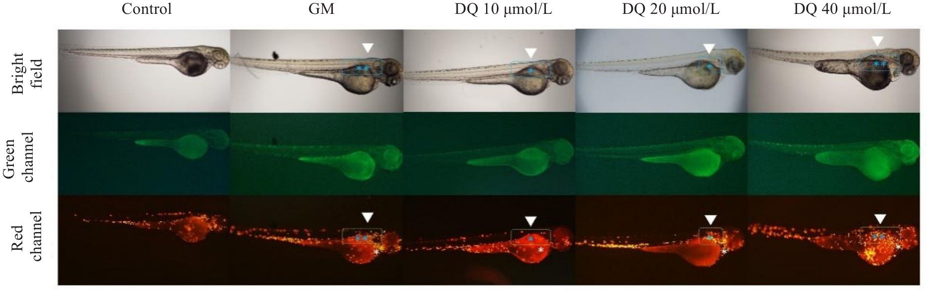
图2 敌草快对斑马鱼体内炎症反应的影响
Fig.2 Inflammatory response induced by diquat in zebrafish (×40).
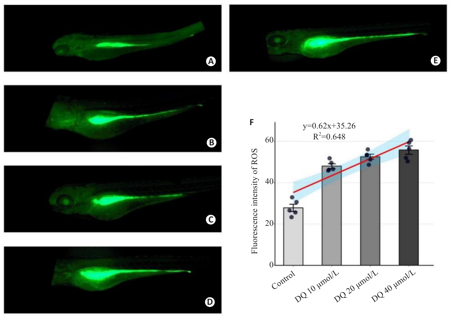
图3 荧光定量分析敌草快暴露对斑马鱼活体ROS水平的影响
Fig.3 Fluorescence quantitative analysis of the effect of diquat on ROS level in live-stained zebrafish (×40). A: Control group. B: Gentamicin-positive group. C: Diquat 10 μmol/L group. D: Diquat 20 μmol/L group. E: Diquat 40 μmol/L group. F: Results of quantitative analysis.
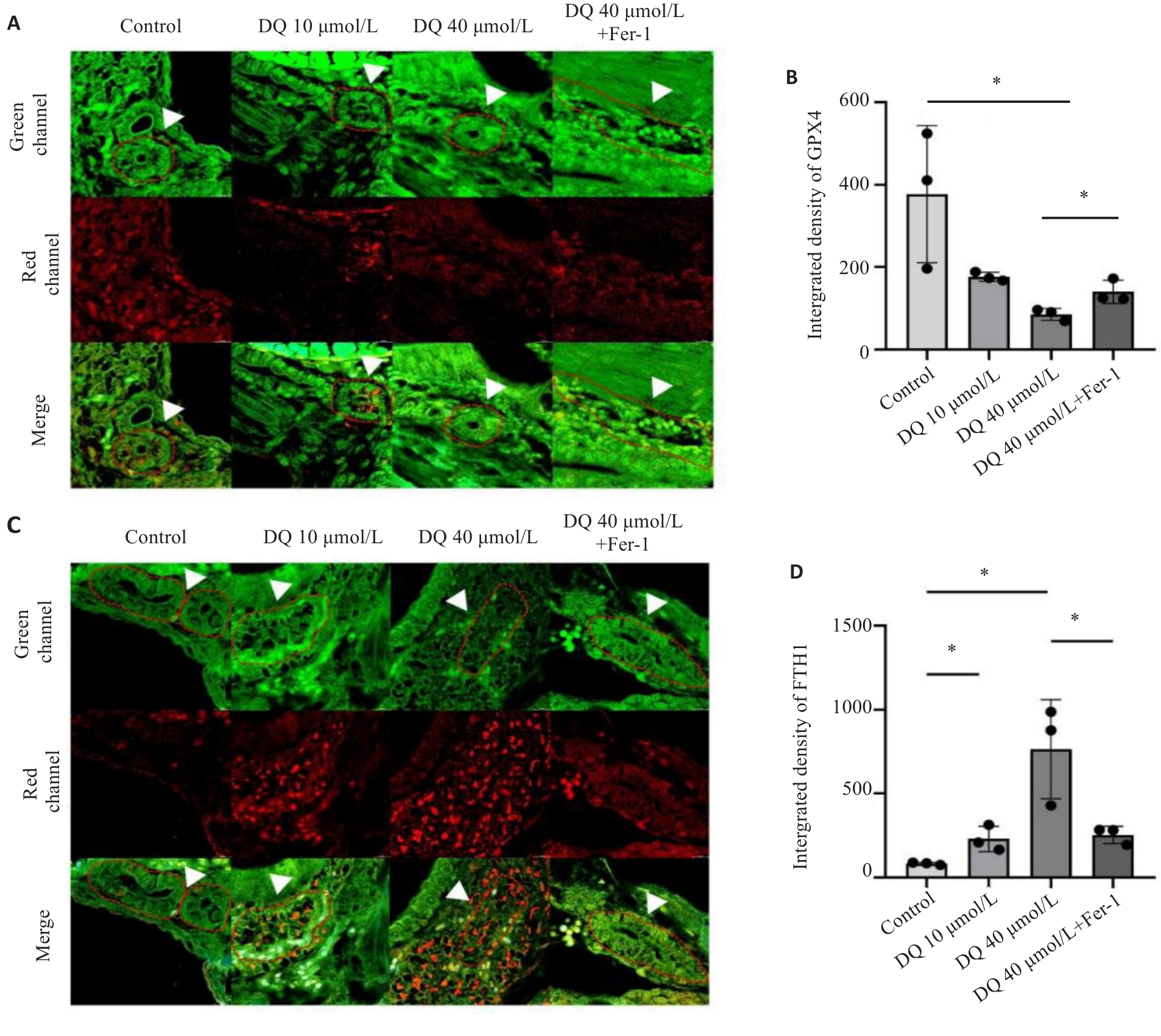
图4 抑制铁死亡对敌草快引起斑马鱼肾组织GPX4和FTH1表达影响
Fig 4 Effect of ferroptosis inhibition on expressions of GPX4 and FTH1 in the kidneys of diquat-exposed zebrafish (×400). A, B: Immunofluorescence and quantitative analysis of GPX4. C,D: Immunofluorescence and quantitative analysis of FTH1. The white arrows indicated the zebrafish kidney tissue. *P<0.05.
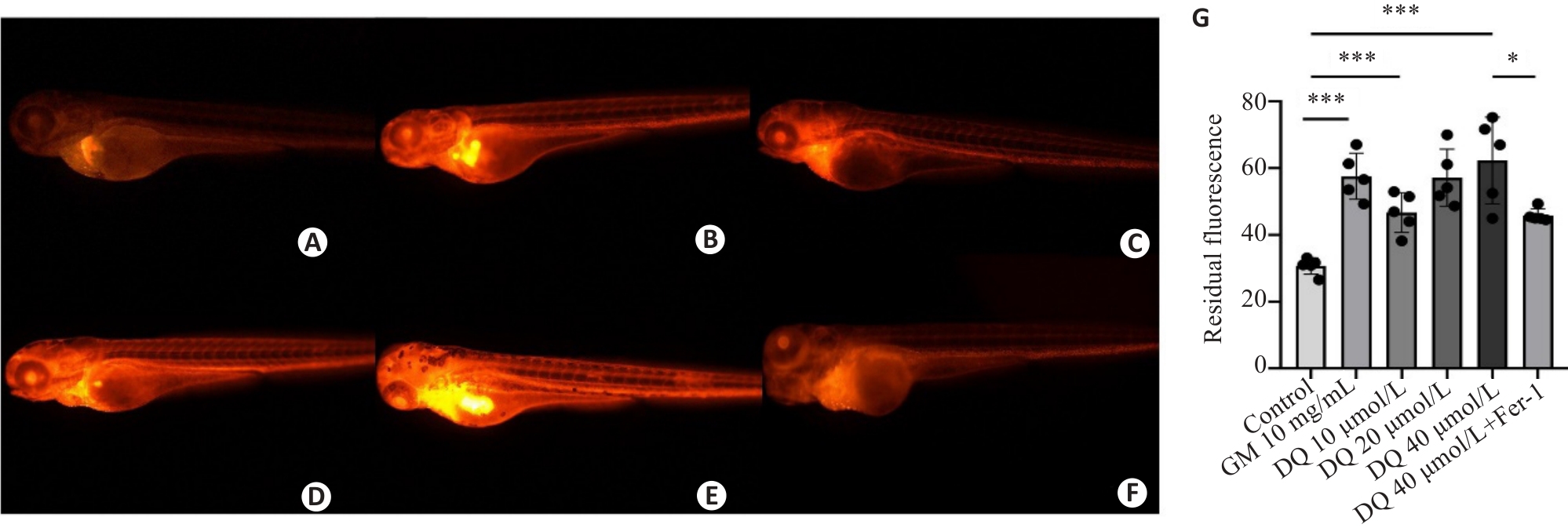
图5 敌草快暴露对斑马鱼肾小球滤过率的影响
Fig.5 Effect of diquat exposure on glomerular filtration rate in zebrafish (×40). The metabolism of rhodamine B-labeled dextran was detected using fluorescence quantification. A: Control group. B: Gentamicin-positive group. C: Diquat 10 μmol/L group. D: Diquat 20 μmol/L group. E: Diquat 40 μmol/L group. F: Diquat 40 μmol/L group+Fer-1 intervention group. G: The results of quantitative analysis. *P<0.05, ***P<0.001.
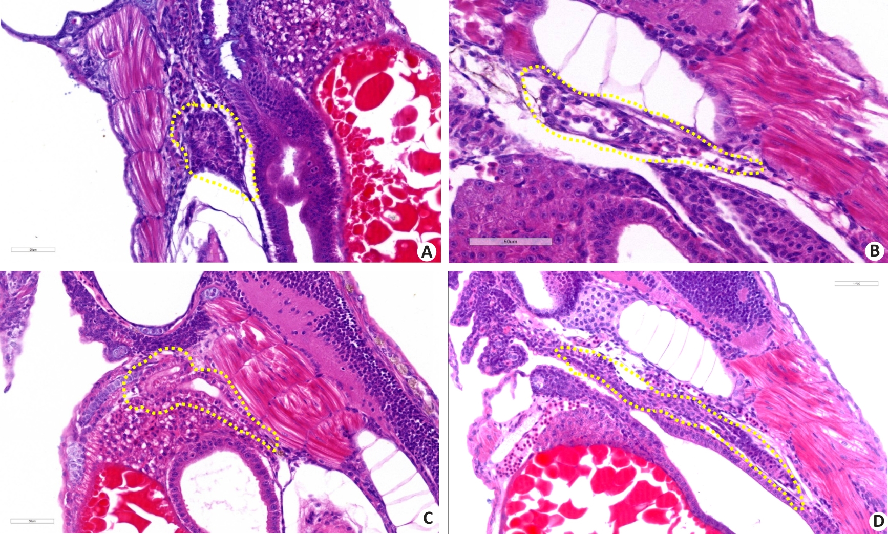
图6 病理学分析敌草快暴露对斑马鱼肾组织损伤的影响
Fig.6 Pathological analysis of the effect of diquat exposure on renal injury in zebrafish (HE staining, ×400). A: Control group. B: Gentamicin-positive group. C: Diquat exposure 40 μmol/L group. D: Diquat 40 μmol/L group+Fer-1 intervention group.
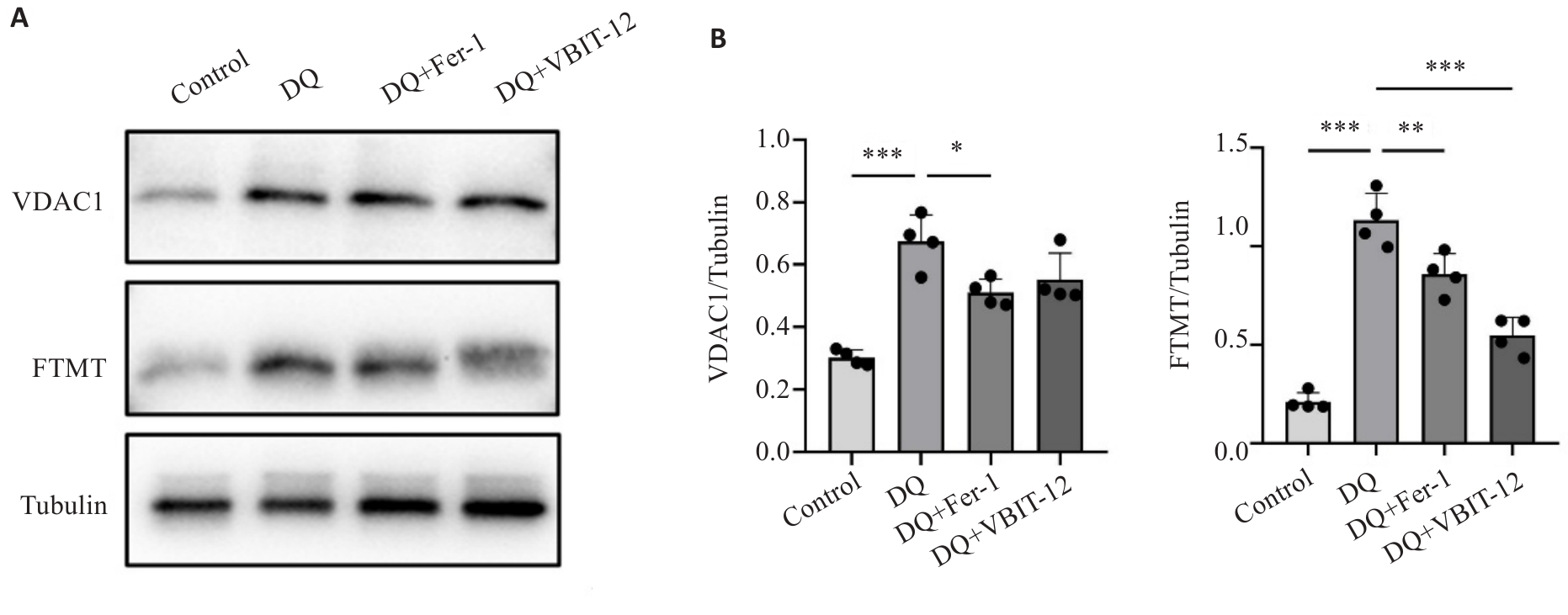
图7 敌草快暴露与干预对VDAC1和FTMT蛋白表达的影响
Fig.7 Effect of diquat exposure and drug interventions on protein expressions of VDAC1 and FTMT. A: Protein expressions of VDAC1 and FTMT detected using Western blotting. B: Quantitative analysis of the protein expressions (n=20). *P<0.05, **P<0.01, ***P<0.001. Each experimental repetition involved 20 larvae.
| [1] | Aloise DM, Memon A, Zaldivar A. Diquat herbicide organo-phosphate poisoning and multi-organ failure: a case report[J]. Cureus, 2022, 14(7): e27241. |
| [2] | 孟 娜, 孙艺青, 刘 亮, 等. 急性敌草快中毒86例临床分析[J]. 中华危重病急救医学, 2022(3): 301-5. doi:10.3760/cma.j.cn121430-20220128-00105 |
| [3] | 敌草快中毒诊断与治疗专家共识组. 急性敌草快中毒诊断与治疗专家共识[J]. 中华急诊医学杂志, 2020, 10(29): 1282-302. |
| [4] | Magalhães N, Carvalho F, Dinis-Oliveira RJ. Human and experimental toxicology of diquat poisoning: Toxicokinetics, mechanisms of toxicity, clinical features, and treatment[J]. Hum Exp Toxicol, 2018, 37(11): 1131-60. doi:10.1177/0960327118765330 |
| [5] | Zeng DH, Chen XH, Li Y, et al. Clinical and pathological characteristics of acute kidney injury caused by diquat poisoning[J]. Clin Toxicol (Phila), 2023, 61(9): 705-8. doi:10.1080/15563650.2023.2262113 |
| [6] | Vohra R, Salazar A, Cantrell FL, et al. The poison pen: bedside diagnosis of urinary diquat[J]. J Med Toxicol, 2010, 6(1): 35-6. doi:10.1007/s13181-010-0033-6 |
| [7] | Chen YC, Ou ZJ, Zhang RC, et al. Case report: Successful outcome of a young patient with rhabdomyolysis and shock caused by diquat poisoning[J]. Front Med (Lausanne), 2023, 10: 1116912. doi:10.3389/fmed.2023.1116912 |
| [8] | Yin J, Liu MF, Ren WK, et al. Effects of dietary supplementation with glutamate and aspartate on diquat-induced oxidative stress in piglets[J]. PLoS One, 2015, 10(4): e0122893. doi:10.1371/journal.pone.0122893 |
| [9] | Jović-Stosić J, Babić G, Todorović V. Fatal diquat intoxication[J]. Vojnosanit Pregl, 2009, 66(6): 477-81. doi:10.2298/vsp0906477j |
| [10] | Suleiman SA, Stevens JB. Bipyridylium herbicide toxicity: effects of paraquat and diquat on isolated rat hepatocytes[J]. J Environ Pathol Toxicol Oncol, 1987, 7(3): 73-84. doi:10.3109/08923978709035231 |
| [11] | Lai KM, Wang JJ, Lin SY, et al. Sensing of mitochondrial DNA by ZBP1 promotes RIPK3-mediated necroptosis and ferroptosis in response to diquat poisoning[J]. Cell Death Differ, 2024, 31(5): 635-50. doi:10.1038/s41418-024-01279-5 |
| [12] | von Mässenhausen A, Tonnus W, Himmerkus N, et al. Phenytoin inhibits necroptosis[J]. Cell Death Dis, 2018, 9(3): 359. doi:10.1038/s41419-018-0394-3 |
| [13] | Qin QY, Yu NJ, Gu YX, et al. Inhibiting multiple forms of cell death optimizes ganglion cells survival after retinal ischemia reperfusion injury[J]. Cell Death Dis, 2022, 13(5): 507. doi:10.1038/s41419-022-04911-9 |
| [14] | Wu YZ, Cui SQ, Wang WJ, et al. Kidney and lung injury in rats following acute diquat exposure[J]. Exp Ther Med, 2022, 23(4): 275. doi:10.3892/etm.2022.11201 |
| [15] | Scindia Y, Dey P, Thirunagari A, et al. Hepcidin mitigates renal ischemia-reperfusion injury by modulating systemic iron homeostasis[J]. J Am Soc Nephrol, 2015, 26(11): 2800-14. doi:10.1681/asn.2014101037 |
| [16] | 黄 奕, 林丽珊, 黄浩华, 等. VDAC1通过诱导气道上皮细胞铁死亡参与屋尘螨诱导的哮喘小鼠气道炎症[J]. 南方医科大学学报, 2023, 43(8): 1333-8. |
| [17] | Kim J, Gupta R, Blanco LP, et al. VDAC oligomers form mitochondrial pores to release mtDNA fragments and promote lupus-like disease[J]. Science, 2019, 366(6472): 1531-6. doi:10.1126/science.aav4011 |
| [18] | Howe K, Clark MD, Torroja CF, et al. The zebrafish reference genome sequence and its relationship to the human genome[J]. Nature, 2013, 496(7446): 498-503. |
| [19] | McCampbell KK, Springer KN, Wingert RA. Analysis of nephron composition and function in the adult zebrafish kidney[J]. J Vis Exp, 2014(90): e51644. doi:10.3791/51644-v |
| [20] | Diep CQ, Peng ZZ, Ukah TK, et al. Development of the zebrafish mesonephros[J]. Genesis, 2015, 53(3/4): 257-69. doi:10.1002/dvg.22846 |
| [21] | Outtandy P, Russell C, Kleta R, et al. Zebrafish as a model for kidney function and disease[J]. Pediatr Nephrol, 2019, 34(5): 751-62. doi:10.1007/s00467-018-3921-7 |
| [22] | Xu X, Wei Y, Hua HW, et al. Glycine alleviated intestinal injury by inhibiting ferroptosis in piglets challenged with diquat[J]. Animals (Basel), 2022, 12(22): 3071. doi:10.3390/ani12223071 |
| [23] | Chen YN, Zhang H, Li Y, et al. Pterostilbene confers protection against diquat-induced intestinal damage with potential regulation of redox status and ferroptosis in broiler chickens[J]. Oxid Med Cell Longev, 2023, 2023: 8258354. doi:10.1155/2023/8258354 |
| [24] | Cheng JY, Yang L, Zhang ZL, et al. Diquat causes mouse testis injury through inducing heme oxygenase-1-mediated ferroptosis in spermatogonia[J]. Ecotoxicol Environ Saf, 2024, 280: 116562. doi:10.1016/j.ecoenv.2024.116562 |
| [25] | Cui SQ, Zhang XX, Wang C, et al. Study on the therapeutic effect of glucocorticoids on acute kidney injury in rats exposed to diquat[J]. Biomed Pharmacother, 2023, 166: 115310. doi:10.1016/j.biopha.2023.115310 |
| [26] | Gorgulho R, Jacinto R, Lopes SS, et al. Usefulness of zebrafish larvae to evaluate drug-induced functional and morphological renal tubular alterations[J]. Arch Toxicol, 2018, 92(1): 411-23. doi:10.1007/s00204-017-2063-1 |
| [27] | Drechsel DA, Patel M. Differential contribution of the mitochondrial respiratory chain complexes to reactive oxygen species production by redox cycling agents implicated in Parkinsonism[J]. Toxicol Sci, 2009, 112(2): 427-34. doi:10.1093/toxsci/kfp223 |
| [28] | Qu J, Pei H, Li XZ, et al. Erythrocyte membrane biomimetic EGCG nanoparticles attenuate renal injury induced by diquat through the NF-κB/NLRP3 inflammasome pathway[J]. Front Pharmacol, 2024, 15: 1414918. doi:10.3389/fphar.2024.1414918 |
| [29] | Huang SF, Lin SR, Zhou SL, et al. Soluble thrombomodulin alleviates Diquat-induced acute kidney injury by inhibiting the HMGB1/IκBα/NF‑κB signalling pathway[J]. Food Chem Toxicol, 2023, 178: 113871. doi:10.1016/j.fct.2023.113871 |
| [30] | Liu Y, Yuan JM, Xi WS, et al. Lactiplantibacillus plantarum ameliorated morphological damage and barrier dysfunction and reduced apoptosis and ferroptosis in the jejunum of oxidatively stressed piglets[J]. Animals (Basel), 2024, 14(22): 3335. doi:10.3390/ani14223335 |
| [31] | Chen KY, Tang YH, Lan LH, et al. Autophagy mediated FTH1 degradation activates gasdermin E dependent pyroptosis contributing to diquat induced kidney injury[J]. Food Chem Toxicol, 2024, 184: 114411. doi:10.1016/j.fct.2023.114411 |
| [32] | Wu LZ, Luo ZW, Luo FL, et al. Edaravone inhibits neuronal ferroptosis and alleviates acute Central nervous system injury induced by diquat via enhancement of METTL14-mediated m6A methylation of Aldh1l1[J]. Free Radic Res, 2025, 59(3): 274-88. doi:10.1080/10715762.2025.2482774 |
| [33] | Linkermann A, Skouta R, Himmerkus N, et al. Synchronized renal tubular cell death involves ferroptosis[J]. Proc Natl Acad Sci USA, 2014, 111(47): 16836-41. doi:10.1073/pnas.1415518111 |
| [34] | Sun MF, Zhu L, Chen X. The role of ferroptosis in renal injury induced by diquat[J]. Chin J Indust Hygiene Occupat Dis, 2025, 43(1): 14-24. |
| [35] | Yang YT, Lin QS, Zhu XY, et al. Activation of lipophagy is required for RAB7 to regulate ferroptosis in sepsis-induced acute kidney injury[J]. Free Radic Biol Med, 2024, 218: 120-31. doi:10.1016/j.freeradbiomed.2024.04.213 |
| [36] | Cao YX, Liu XW, Guo CJ, et al. Biomimetic reactive oxygen/nitrogen nanoscavengers inhibit "ferroptosis storm" and modulate immune targeting for acute kidney injury[J]. J Control Release, 2025, 379: 59-76. doi:10.1016/j.jconrel.2025.01.006 |
| [37] | Karmi O, Marjault HB, Bai F, et al. A VDAC1-mediated NEET protein chain transfers [2Fe-2S] clusters between the mitochondria and the cytosol and impacts mitochondrial dynamics[J]. Proc Natl Acad Sci USA, 2022, 119(7): e2121491119. doi:10.1073/pnas.2121491119 |
| [38] | Shao FY, Han JY, Tian ZY, et al. Synergistic ROS generation and directional overloading of endogenous calcium induce mito-chondrial dysfunction in living cells[J]. Biomaterials, 2023, 301: 122284. doi:10.1016/j.biomaterials.2023.122284 |
| [39] | Niu BL, Lei XH, Xu QL, et al. Protecting mitochondria via inhibiting VDAC1 oligomerization alleviates ferroptosis in acetaminophen-induced acute liver injury[J]. Cell Biol Toxicol, 2022, 38(3): 505-30. doi:10.1007/s10565-021-09624-x |
| [40] | Yang J, Lu X, Hao JL, et al. VSTM2L protects prostate cancer cells against ferroptosis via inhibiting VDAC1 oligomerization and maintaining mitochondria homeostasis[J]. Nat Commun, 2025, 16(1): 1160. doi:10.1038/s41467-025-56494-6 |
| [41] | Drysdale J, Arosio P, Invernizzi R, et al. Mitochondrial ferritin: a new player in iron metabolism[J]. Blood Cells Mol Dis, 2002, 29(3): 376-83. doi:10.1006/bcmd.2002.0577 |
| [42] | Wang YQ, Chang SY, Wu Q, et al. The protective role of mitochondrial ferritin on erastin-induced ferroptosis[J]. Front Aging Neurosci, 2016, 8: 308. doi:10.3389/fnagi.2016.00308 |
| [43] | Qian B, Jiang RJ, Song JL, et al. Organophosphorus flame retardant TDCPP induces neurotoxicity via mitophagy-related ferroptosis in vivo and in vitro [J]. Chemosphere, 2022, 308(Pt 2): 136345. doi:10.1016/j.chemosphere.2022.136345 |
| [1] | 陈鑫源, 吴成挺, 李瑞迪, 潘雪芹, 张耀丹, 陶俊宇, 林才志. 双术汤通过P53/SLC7A11/GPX4通路诱导胃癌细胞铁死亡[J]. 南方医科大学学报, 2025, 45(7): 1363-1371. |
| [2] | 张梦影, 赵晨玲, 田丽伟, 余郭芳, 杨文明, 董婷. 肝豆扶木汤通过GPX4/ACSL4/ALOX15通路抑制铁死亡改善Wilson病小鼠的肝脏脂肪变性[J]. 南方医科大学学报, 2025, 45(7): 1471-1478. |
| [3] | 王子皓, 陶丽丽, 邹碧清, 安胜利. 重症监护病房急性肾损伤患者首次24 h动脉氧分压与死亡率相关:基于MIMIC-IV数据库[J]. 南方医科大学学报, 2025, 45(5): 1056-1062. |
| [4] | 张柳攀, 石晓彤, 李露兰, 师瑞, 安胜利, 曾振华. 合并急性肾损伤的危重症患者输注白蛋白后血清白蛋白水平与28 d死亡率的相关性[J]. 南方医科大学学报, 2025, 45(5): 1074-1081. |
| [5] | 张安邦, 孙秀颀, 庞博, 吴远华, 时靖宇, 张宁, 叶涛. 电针预处理通过调节肠道-大脑轴及Nrf2/HO-1信号通路抑制铁死亡减轻大鼠脑缺血再灌注损伤[J]. 南方医科大学学报, 2025, 45(5): 911-920. |
| [6] | 张林落, 李长青, 皇玲玲, 周学平, 娄媛媛. 梓醇扶正制毒配伍从SLC7A11/GPX4通路抑制铁死亡减轻雷公藤甲素肝毒性[J]. 南方医科大学学报, 2025, 45(4): 810-818. |
| [7] | 季春斐, 左宗超, 王钧, 李妙男. N-乙酰神经氨酸中通过抑制Nrf2轴促进缺氧/复氧损伤的H9C2心肌细胞发生铁死亡[J]. 南方医科大学学报, 2025, 45(1): 72-79. |
| [8] | 陈凯, 孟兆菲, 闵静婷, 王佳慧, 李正红, 高琴, 胡俊锋. 姜黄素通过抑制TXNIP/TRX-1/GPX4通路介导的铁死亡减轻脓毒症小鼠肺损伤[J]. 南方医科大学学报, 2024, 44(9): 1805-1813. |
| [9] | 欧阳明子, 崔佳琦, 王慧, 梁正, 皮大锦, 陈利国, 陈前军, 吴迎朝. 开心散通过减轻前额叶皮质铁死亡缓解小鼠的阿霉素化疗性抑郁[J]. 南方医科大学学报, 2024, 44(8): 1441-1449. |
| [10] | 张银亮, 骆泽谭, 赵睿, 赵娜, 徐志东, 奥迪, 丛古一, 刘新宇, 郑海伦. 血根碱通过调控STUB1/GPX4诱导直肠癌细胞发生铁死亡[J]. 南方医科大学学报, 2024, 44(8): 1537-1544. |
| [11] | 王元国, 张鹏. 铁死亡抑制基因在食管癌中的高表达分析[J]. 南方医科大学学报, 2024, 44(7): 1389-1396. |
| [12] | 何华星, 刘璐琳, 刘颖茵, 陈纳川, 孙素霞. 丁酸钠与索拉非尼可能通过YAP诱导铁死亡协同抑制肝癌细胞增殖[J]. 南方医科大学学报, 2024, 44(7): 1425-1430. |
| [13] | 任智先, 周倍贤, 王林鑫, 李菁, 张荣平, 潘锡平. 5-羟基-6,7-二甲氧基黄酮抑制流感病毒诱导A549细胞炎症反应和铁死亡的作用及机制[J]. 南方医科大学学报, 2024, 44(6): 1070-1078. |
| [14] | 张方圆, 刘刚. 右美托咪定通过激活Nrf2/HO-1/GPX4通路抑制肾小管上皮细胞的铁死亡[J]. 南方医科大学学报, 2024, 44(6): 1135-1140. |
| [15] | 王南, 石斌, 马小兰, 吴伟超, 曹佳. FMRP通过激活RAS/MAPK信号通路抑制结直肠肿瘤细胞的铁死亡[J]. 南方医科大学学报, 2024, 44(5): 885-893. |
| 阅读次数 | ||||||
|
全文 |
|
|||||
|
摘要 |
|
|||||