| 1 |
Suresh D, Li A, Miller MJ, et al. Associations between metabolic hyperferritinaemia, fibrosis-promoting alleles and clinical outcomes in steatotic liver disease[J]. Liver Int, 2024, 44(2): 389-98.
|
| 2 |
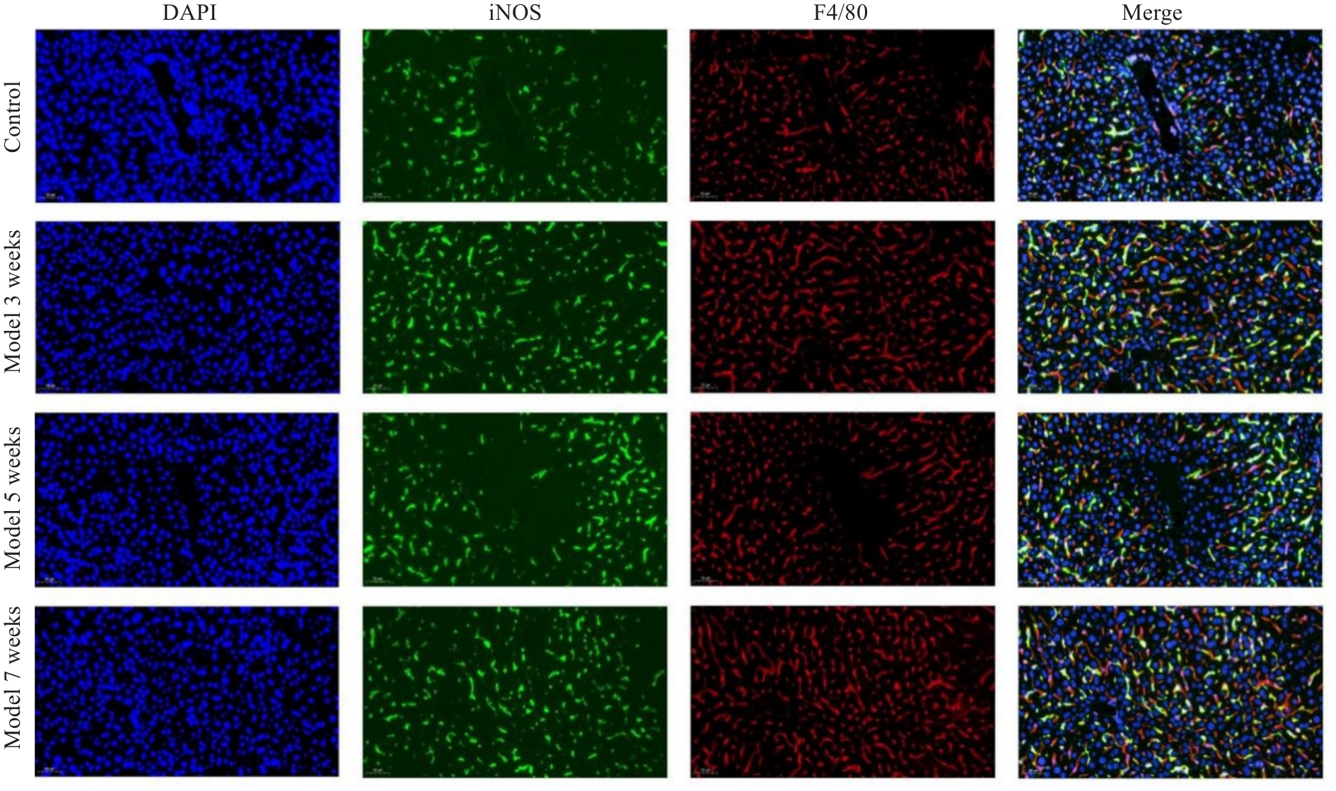
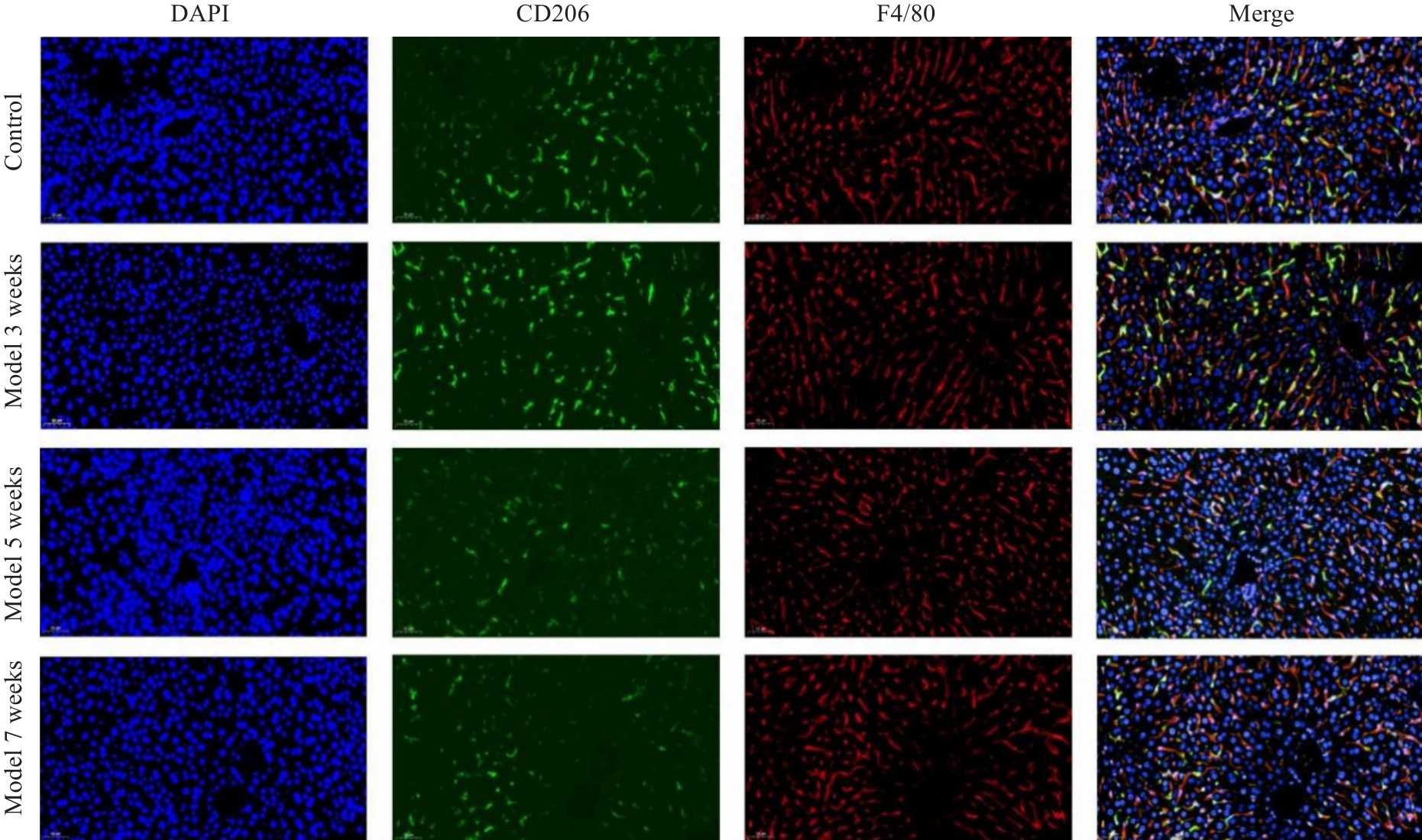
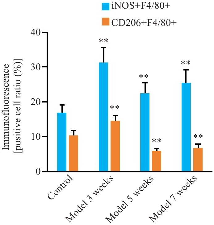
Wang C, Ma C, Gong LH, et al. Macrophage polarization and its role in liver disease[J]. Front Immunol, 2021, 12: 803037.
|
| 3 |
Zanganeh S, Hutter G, Spitler R, et al. Iron oxide nanoparticles inhibit tumour growth by inducing pro-inflammatory macrophage polarization in tumour tissues[J]. Nat Nanotechnol, 2016, 11(11): 986-94.
|
| 4 |
Zhou Y, Que KT, Zhang Z, et al. Iron overloaded polarizes macrophage to proinflammation phenotype through ROS/acetyl-p53 pathway[J]. Cancer Med, 2018, 7(8): 4012-22.
|
| 5 |
Handa P, Thomas S, Morgan-Stevenson V, et al. Iron alters macrophage polarization status and leads to steatohepatitis and fibrogenesis[J]. J Leukoc Biol, 2019, 105(5): 1015-26.
|
| 6 |
Dufrusine B, Di Francesco A, Oddi S, et al. Iron-dependent trafficking of 5-lipoxygenase and impact on human macrophage activation[J]. Front Immunol, 2019, 10: 1347.
|
| 7 |
Liu EN, Li Z, Zhang Y, et al. Hepcidin induces M1 macrophage polarization in monocytes or THP-1 derived macrophages[J]. Iran J Immunol, 2019, 16(3): 190-9.
|
| 8 |
Zhang X, Fan LN, Wu JF, et al. Macrophage p38α promotes nutritional steatohepatitis through M1 polarization[J]. J Hepatol, 2019, 71(1): 163-74.
|
| 9 |
Shendge AK, Panja S, Basu T, et al. Ameliorating effects of white mulberry on iron-overload-induced oxidative stress and liver fibro-sis in Swiss albino mice[J]. Food Chem Toxicol, 2021, 156: 112520.
|
| 10 |
Philippe MA, Ruddell RG, Ramm GA. Role of iron in hepatic fibrosis: one piece in the puzzle[J]. World J Gastroenterol, 2007, 13(35): 4746-54.
|
| 11 |
Wood MJ, Gadd VL, Powell LW, et al. Ductular reaction in hereditary hemochromatosis: the link between hepatocyte senesc-ence and fibrosis progression[J]. Hepatology, 2014, 59(3): 848-57.
|
| 12 |
Pietrangelo A. Iron in NASH, chronic liver diseases and HCC: how much iron is too much[J]? J Hepatol, 2009, 50(2): 249-51.
|
| 13 |
Houglum K, Bedossa P, Chojkier M. TGF-beta and collagen-alpha 1 (I) gene expression are increased in hepatic acinar zone 1 of rats with iron overload[J]. Am J Physiol, 1994, 267(5 Pt 1): G908-13.
|
| 14 |
Carthew P, Edwards RE, Smith AG, et al. Rapid induction of hepatic fibrosis in the gerbil after the parenteral administration of iron-dextran complex[J]. Hepatology, 1991, 13(3): 534-9.
|
| 15 |
Ruddell RG, Hoang-Le D, Barwood JM, et al. Ferritin functions as a proinflammatory cytokine via iron-independent protein kinase C Zeta/nuclear factor kappaB-regulated signaling in rat hepatic stellate cells[J]. Hepatology, 2009, 49(3): 887-900.
|
| 16 |
Bridle KR, Crawford DHG, Ramm GA. Identification and characterization of the hepatic stellate cell transferrin receptor[J]. Am J Pathol, 2003, 162(5): 1661-7.
|
| 17 |
Shi HB, Shi HL, Ren F, et al. Naringin in Ganshuang Granule suppresses activation of hepatic stellate cells for anti-fibrosis effect by inhibition of mammalian target of rapamycin[J]. J Cell Mol Med, 2017, 21(3): 500-9.
|
| 18 |
Kraml P. The role of iron in the pathogenesis of atherosclerosis[J]. Physiol Res, 2017, 66(): S55-67.
|
| 19 |
Mesquita G, Silva T, Gomes AC, et al. H-Ferritin is essential for macrophages' capacity to store or detoxify exogenously added iron[J]. Sci Rep, 2020, 10(1): 3061.
|
| 20 |
Gan ZS, Wang QQ, Li JH, et al. Iron reduces M1 macrophage polarization in RAW264.7 macrophages associated with inhibition of STAT1[J]. Mediators Inflamm, 2017, 2017: 8570818.
|
| 21 |
Ward RJ, Wilmet S, Legssyer R, et al. Effects of marginal iron overload on iron homeostasis and immune function in alveolar macrophages isolated from pregnant and normal rats[J]. Biometals, 2009, 22(2): 211-23.
|
| 22 |
Lee CJ, Jeong H, Bae Y, et al. Targeting of M2-like tumor-associated macrophages with a melittin-based pro-apoptotic peptide[J]. J Immunother Cancer, 2019, 7(1): 147.
|
| 23 |
Kao JK, Wang SC, Ho LW, et al. M2-like polarization of THP-1 monocyte-derived macrophages under chronic iron overload[J]. Ann Hematol, 2020, 99(3): 431-41.
|
| 24 |
Kroner A, Greenhalgh AD, Zarruk JG, et al. TNF and increased intracellular iron alter macrophage polarization to a detrimental M1 phenotype in the injured spinal cord[J]. Neuron, 2014, 83(5): 1098-116.
|
| 25 |
Sumitomo R, Hirai T, Fujita M, et al. M2 tumor-associated macrophages promote tumor progression in non-small-cell lung cancer[J]. Exp Ther Med, 2019, 18(6): 4490-8.
|
| 26 |
Harhaji L, Vuckovic O, Miljkovic D, et al. Iron down-regulates macrophage anti-tumour activity by blocking nitric oxide production[J]. Clin Exp Immunol, 2004, 137(1): 109-16.
|
| 27 |
Yamaguchi T, Fushida S, Yamamoto Y, et al. Tumor-associated macrophages of the M2 phenotype contribute to progression in gastric cancer with peritoneal dissemination[J]. Gastric Cancer, 2016, 19(4): 1052-65.
|
| 28 |
Hoeft K, Bloch DB, Graw JA, et al. Iron loading exaggerates the inflammatory response to the toll-like receptor 4 ligand lipopo-lysaccharide by altering mitochondrial homeostasis[J]. Anesthesiology, 2017, 127(1): 121-35.
|
| 29 |
Sindrilaru A, Peters T, Wieschalka S, et al. An unrestrained proinflammatory M1 macrophage population induced by iron impairs wound healing in humans and mice[J]. J Clin Invest, 2011, 121(3): 985-97.
|
| 30 |
Agoro R, Taleb M, Quesniaux VFJ, et al. Cell iron status influences macrophage polarization[J]. PLoS One, 2018, 13(5): e0196921.
|
 ), 周薏2, 钱春美3, 穆蓝2, 阙任烨1(