南方医科大学学报 ›› 2025, Vol. 45 ›› Issue (2): 359-370.doi: 10.12122/j.issn.1673-4254.2025.02.17
宾禹1( ), 李子雯1, 左素微1, 孙思诺1, 李敏2, 宋佳茵2, 林旭1, 薛刚2(), 吴靖芳1()
收稿日期:2024-11-06
出版日期:2025-02-20
发布日期:2025-03-03
通讯作者:
薛刚,吴靖芳
E-mail:binyuyouxiang@163.com;641426767@qq.com;wjfxg@163.com
作者简介:宾 禹,在读硕士研究生,E-mail: binyuyouxiang@163.com
基金资助:
Yu BIN1(), Ziwen LI1, Suwei ZUO1, Sinuo SUN1, Min LI2, Jiayin SONG2, Xu LIN1, Gang XUE2(), Jingfang WU1()
Received:2024-11-06
Online:2025-02-20
Published:2025-03-03
Contact:
Gang XUE, Jingfang WU
E-mail:binyuyouxiang@163.com;641426767@qq.com;wjfxg@163.com
摘要:
目的 探讨载脂蛋白C1(APOC1)在甲状腺乳头状癌(PTC)组织中的表达及对PTC细胞增殖、凋亡及关键信号通路的影响。 方法 通过GEPIA 2及K-M数据库分析APOC1在PTC的表达及对预后的影响。采用免疫组织化学、Western blotting检测APOC1在PTC和癌旁组织中的表达以及3株PTC细胞和正常甲状腺Nthyori 3-1细胞的表达。采用细胞转染技术,构建APOC1敲低和过表达PTC细胞模型(TPC-1细胞和BCPAP细胞);采用Western blotting和RT-qPCR分别在蛋白水平和mRNA水平检验细胞模型构建是否成功。通过生长曲线、集落形成实验检测敲低和过表达APOC1后细胞的生长情况和集落形成能力;通过流式细胞术检测敲低和过表达APOC1对细胞周期、凋亡的影响。采用RT-qPCR和Western blotting检测增殖及凋亡相关靶基因P21,P27,CDK4,CyclinD1,BCL-2,Bax,caspase-3/caspase-9 mRNA和蛋白以及JAK2/STAT3信号通路关键蛋白的变化。 结果 成功构建APOC1敲低和过表达PTC细胞模型;PTC组织及3个PTC细胞株APOC1表达高于癌旁组织和Nthyori 3-1细胞(P<0.001)。与对照组相比,沉默组细胞增殖能力减弱(P<0.05);G0/G1期的细胞比例增高(P<0.01),S和G2期的细胞比例下降(P<0.05);细胞凋亡率明显增高(P<0.05);CDK4,CyclinD1,BCL-2 mRNA和蛋白表达量下调(P<0.05);p-JAK2,p-STAT3蛋白表达下调(P<0.001);增强组与沉默组结果相反:P21,P27,Bax,caspase-3/caspase-9 mRNA和蛋白表达下调(P<0.05);p-JAK2,p-STAT3蛋白表达上调(P<0.001)。JAK2抑制剂AG490显著减弱过表达APOC1对BCPAP细胞JAK2/STAT3信号通路的激活效应(P<0.01)。 结论 APOC1可能通过激活JAK2/STAT3信号通路,缩短G0/G1期进程,促进PTC细胞增殖并抑制凋亡。
宾禹, 李子雯, 左素微, 孙思诺, 李敏, 宋佳茵, 林旭, 薛刚, 吴靖芳. 载脂蛋白C1高表达通过激活JAK2/STAT3信号通路促进甲状腺乳头状癌细胞的增殖并抑制凋亡[J]. 南方医科大学学报, 2025, 45(2): 359-370.
Yu BIN, Ziwen LI, Suwei ZUO, Sinuo SUN, Min LI, Jiayin SONG, Xu LIN, Gang XUE, Jingfang WU. High expression of apolipoprotein C1 promotes proliferation and inhibits apoptosis of papillary thyroid carcinoma cells by activating the JAK2/STAT3 signaling pathway[J]. Journal of Southern Medical University, 2025, 45(2): 359-370.
| Gene | Sequence |
|---|---|
| APOC1 | F:5'-CTGGTGGTGGTTCTGTCGAT-3' |
| R:5'-TCACTCTGTTTGATGCGGCT-3' | |
| β-actin | F:5'-CCTGGGCATGGAGTCCTGTG-3' |
| R:5'-AGGGGCCGGACTCGTCATAC-3' |
表1 引物序列
Tab.1 Primer sequences for RT-qPCR
| Gene | Sequence |
|---|---|
| APOC1 | F:5'-CTGGTGGTGGTTCTGTCGAT-3' |
| R:5'-TCACTCTGTTTGATGCGGCT-3' | |
| β-actin | F:5'-CCTGGGCATGGAGTCCTGTG-3' |
| R:5'-AGGGGCCGGACTCGTCATAC-3' |
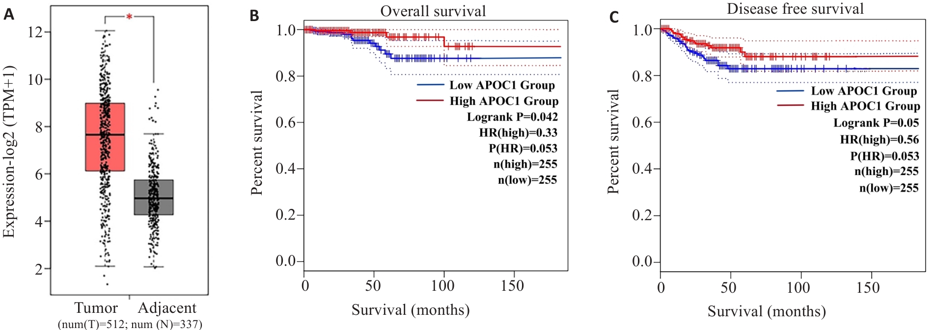
图1 生物信息学网站分析APOC1在患者PTC组织及Adjacent组织的表达
Fig.1 Bioinformatic analysis of APOC1 expression in PTC and Adjacent tissues. A: Analysis of expression levels of APOC1 in PTC tissues and adjacent tissues using GEPIA 2 database (*P<0.05). B, C: Analysis of the relationship between APOC1 mRNA expression and patient prognosis using Kaplan-Meier database.
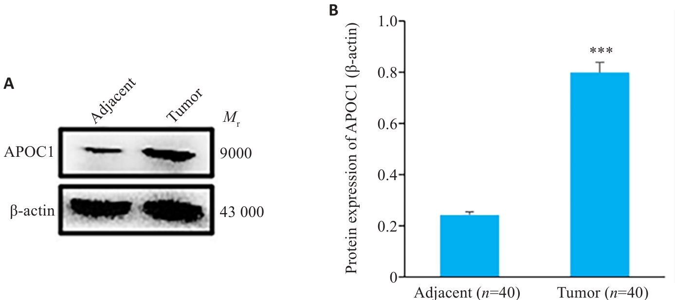
图2 APOC1在PTC及癌旁组织中的表达
Fig.2 Expression of APOC1 in clinical samples of PTC and adjacent tissues. A: Western blotting for detecting APOC1 expression in PTC tissues and adjacent tissues. B: Quantitative analysis of expression level. ***P<0.001 vs adjacent tissue.
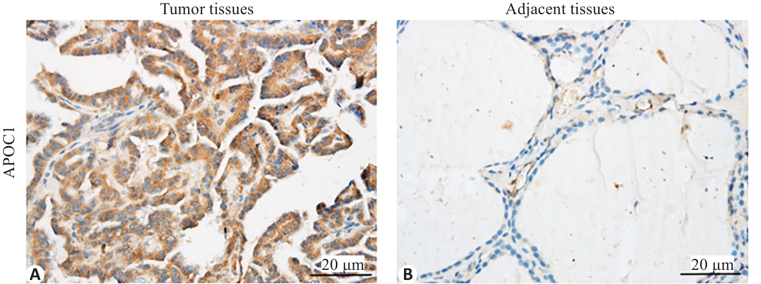
图3 APOC1在PTC及癌旁组织中的表达
Fig.3 Immunohistochemical detection of APOC1 expression in PTC and Adjacent tissue. A: APOC1 positive signal in PTC tissue (brown) in the cytoplasm. B: Negative expression of APOC1 in adjacent tissue.
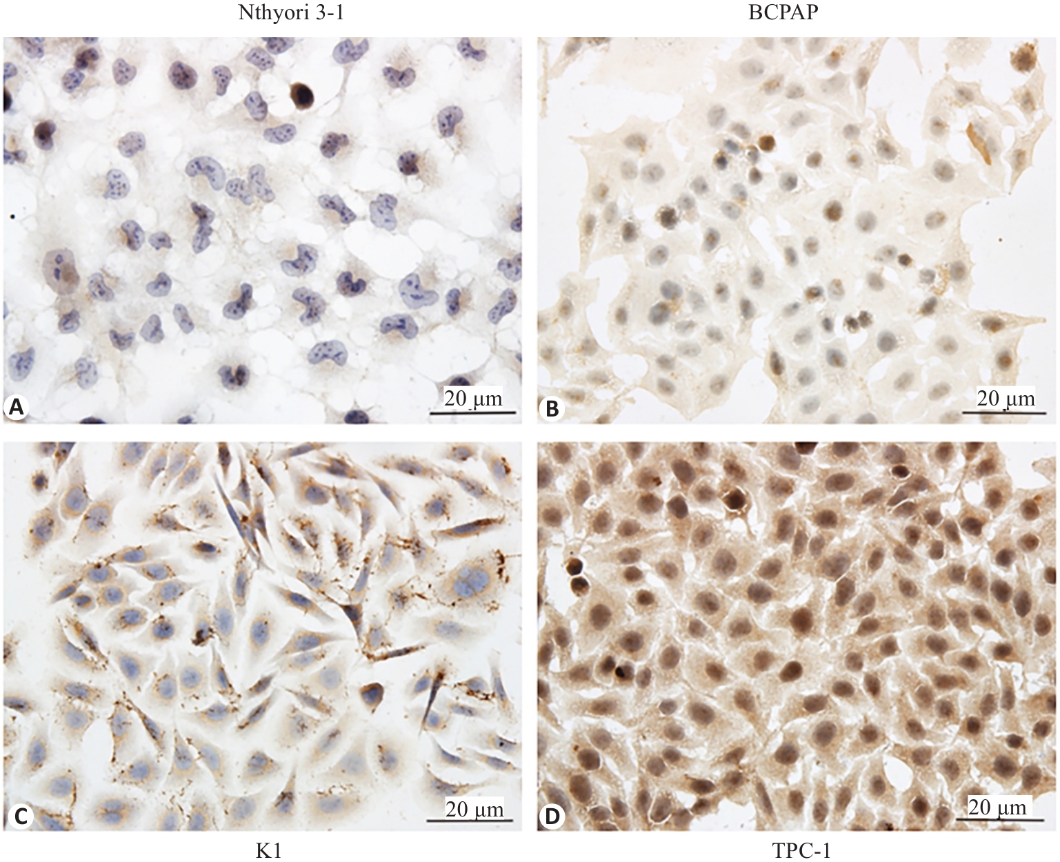
图4 ICC结果显示APOC1表达量TPC-1>K1>BCPAP> Nthyori 3-1
Fig.4 Immunocytochemical detection of APOC1 protein in TPC-1, K1, BCPAP and Nthyori 3-1 cells. A: Negative expression of APOC1 in normal thyiod follicular epithelial Nthyori 3-1 cells. B-D: Positive signals of APOC1 in the 3 PTC cell lines located in the cytoplasm.
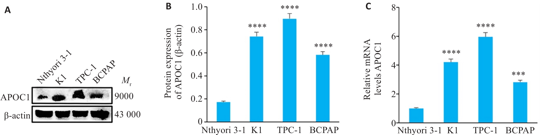
图5 APOC1在K1, TPC-1, BCPAP及Nthyori 3-1中的表达
Fig.5 Western blotting and RT-qPCR for detecting APOC1 expression in K1, TPC-1, BCPAP and Nthyori 3-1 cells. A,B: Expression levels of APOC1 protein in different cell lines detected by Western blotting. C: Expression levels of APOC1 mRNA in different cell lines. ***P<0.001, ****P<0.0001 vs Nthyori 3-1 cells (n=3).
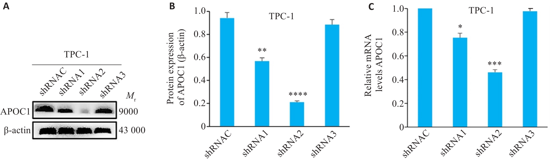
图6 沉默对照组及不同沉默组中APOC1蛋白及mRNA的表达水平
Fig.6 Expression of APOC1 protein and mRNA in TPC-1 cells with APOC1 knockdown. A,B: Expression levels of APOC1 protein in TPC-1 cells with APOC1 knockdown detected by Western blotting. C: Expression levels of APOC1 mRNA in TPC-1 cells with APOC1 knockdown. *P<0.05, **P<0.01, ***P<0.001, ****P<0.0001 vs shRNAC (n=3).
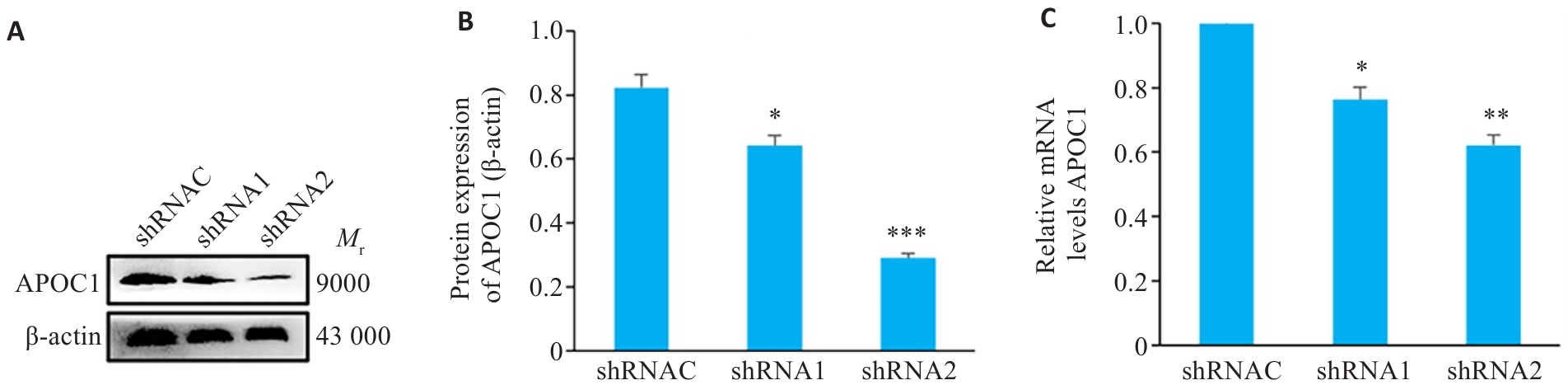
图 7 TPC-1细胞沉默APOC1后的蛋白及mRNA表达含量
Fig.7 Decreased APOC1 expressions at both protein and mRNA levels in TPC-1 cells with APOC1 knockdown. A, B: Expression levels of APOC1 protein in shRNAC group, shRNA1 group, and shRNA2 group. C: Expression levels of APOC1 mRNA in the shRNAC group, shRNA1 group, and shRNA2 group. *P<0.05, **P<0.01, ***P<0.001 vs shRNAC (n=3).
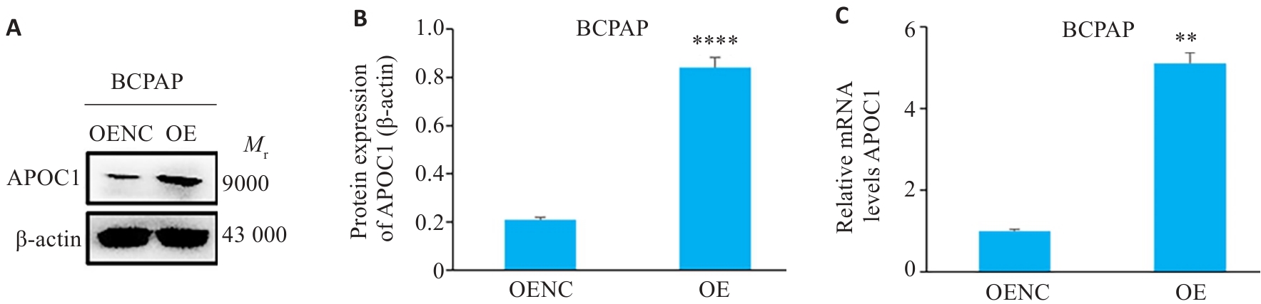
图8 BCPAP细胞增强APOC1后的蛋白及mRNA表达含量
Fig.8 Increased APOC1 protein and mRNA levels in BCPAP cells with APOC1 overexpression. A, B: Expression levels of APOC1 protein in OENC and OE groups. C: Expression levels of APOC1 mRNA in OENC and OE groups. **P<0.01, ****P<0.0001 vs OENC (n=3).
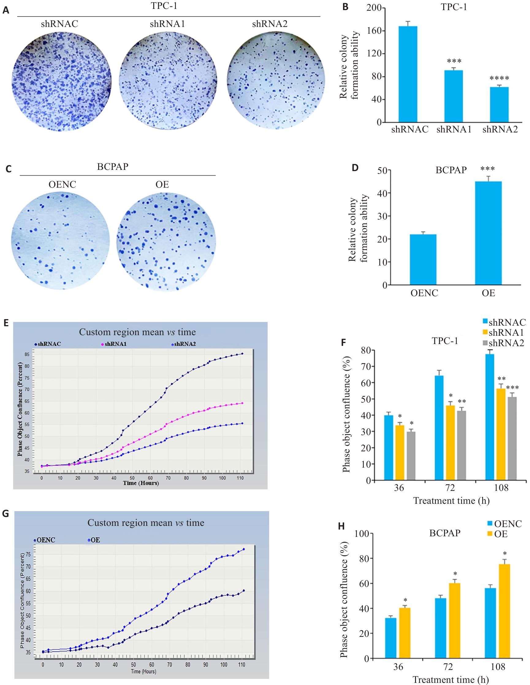
图9 沉默及增强APOC1对细胞增殖的影响
Fig.9 Effect of APOC1 knockdown and overexpresison on PTC cell proliferation. A, C: Colony formation assay of the transfected cells. B,D: Number of cell clusters in the transfected cells. E,G: Growth curves of the transfected cells. F,H: Cell confluence of the transfected cells. *P<0.05,**P<0.01,***P<0.001, ****P<0.001 vs shRNAC/OENC (n=3).
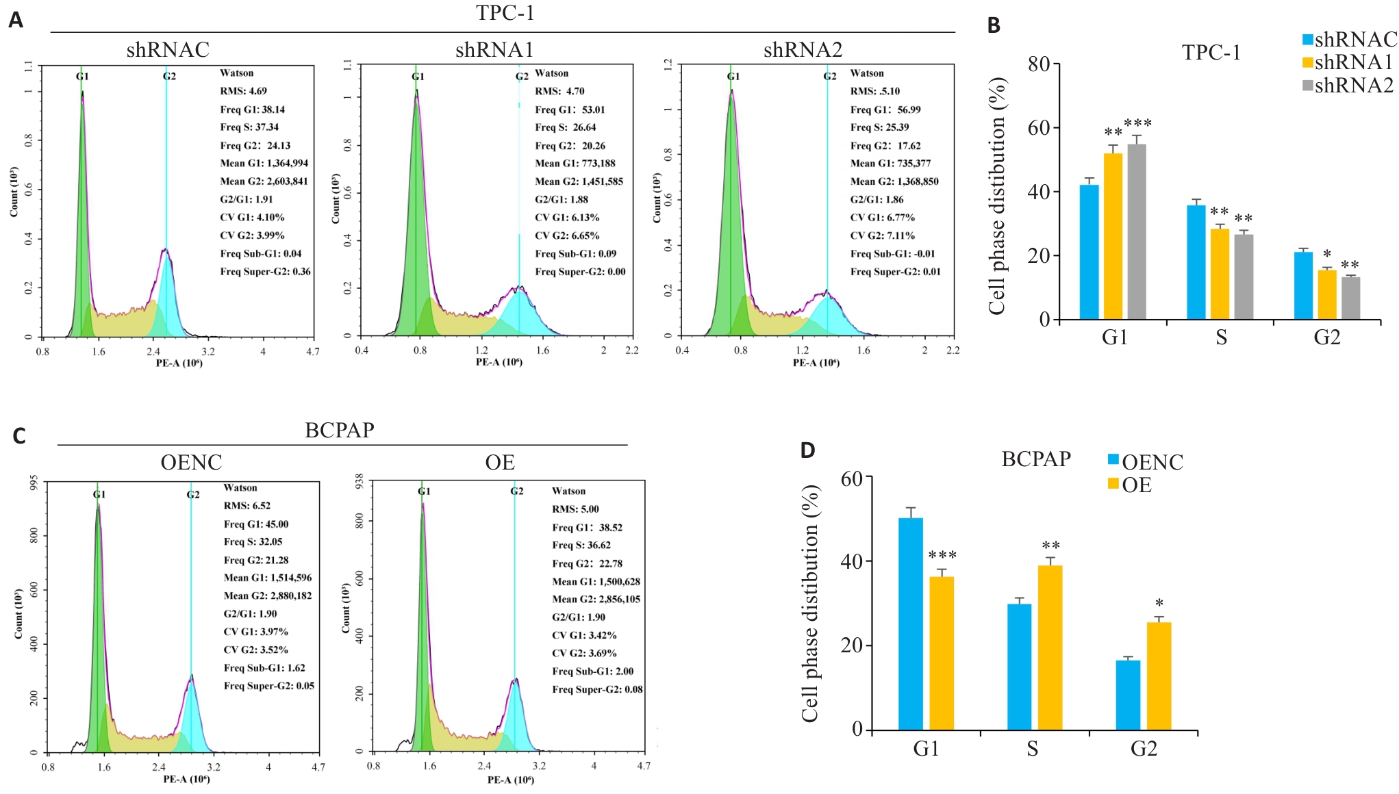
图10 沉默及增强APOC1对细胞周期的影响
Fig.10 Effect of APOC1 knockdown and overexpresison on cell cycle. A, C: Cell cycle diagrams of the cells with APOC1 knockdown and overexpresison. B, D: Analysis of cell cycle percentage. *P<0.05, **P<0.01, ***P<0.001 vs shRNAC/OENC (n=3).
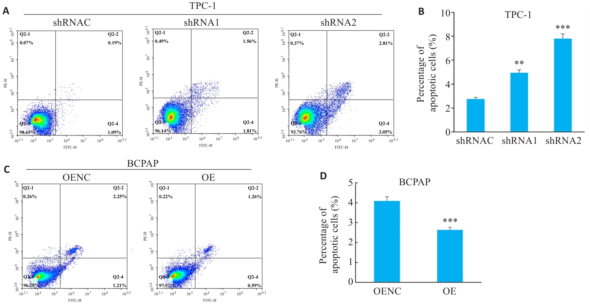
图11 沉默及增强APOC1对细胞凋亡的影响
Fig.11 Effect of APOC1 knockdown and overexpresison on cell apoptosis. A,C: Cell apoptosis maps of cells with APOC1 knockdown and overexpresison. B,D: Percentages of apoptotic cells each group.**P<0.01, ***P<0.001 vs shRNAC/OENC (n=3).
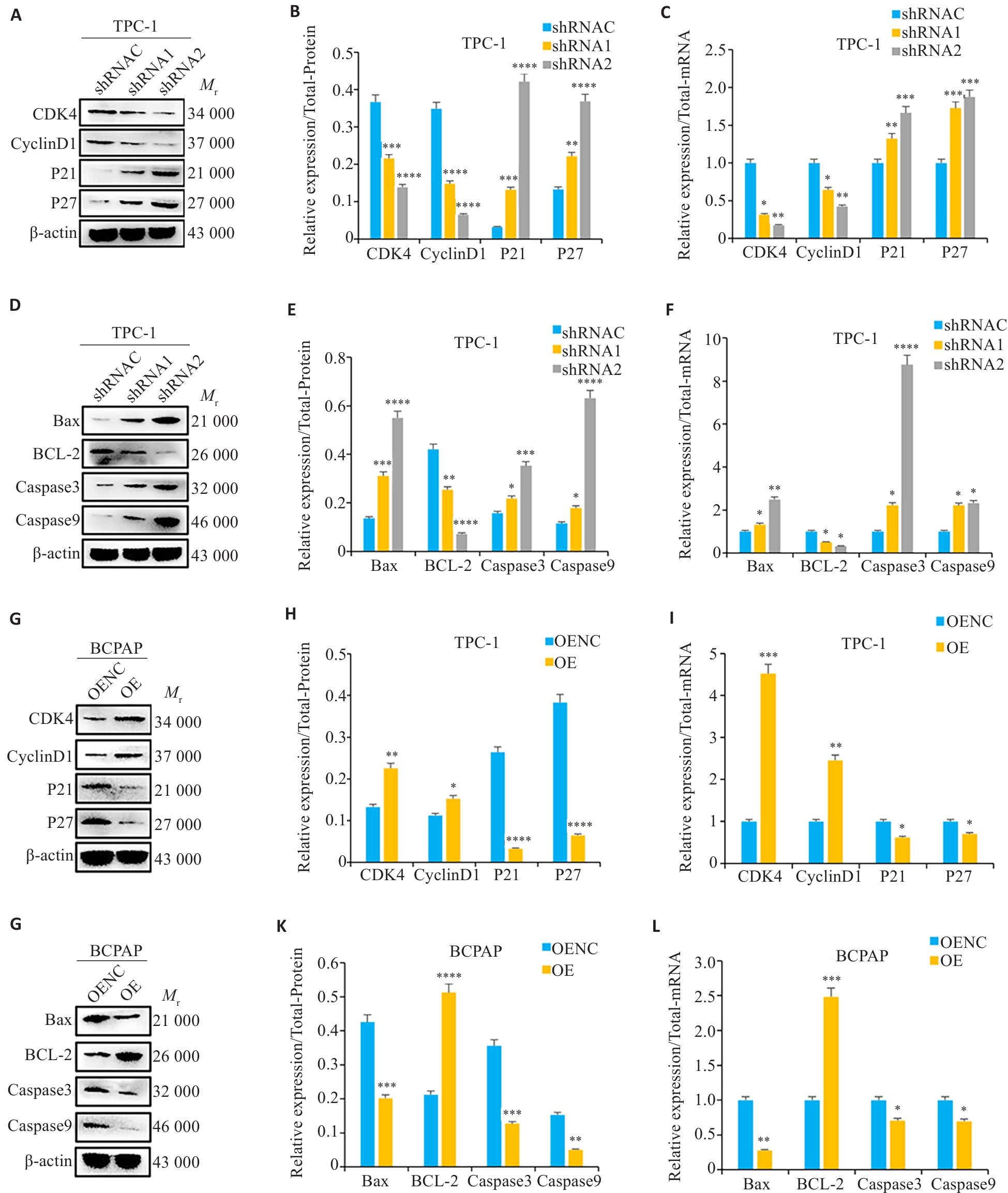
图12 沉默及增强APOC1对周期、凋亡蛋白及mRNA的影响
Fig.12 Effect of APOC1 knockdown and overexpresison on expresisons of cell cycle and apoptosis-related proteins and their mRNA levels. A-C: Cell cycle-related protein and mRNA expression levels of in cells with APOC1 knockdown. D-F: Cell cycle-related protein and mRNA expression levels in APOC1 knockdown group. G-I: Apoptosis-related protein and mRNA expression levels in APOC1 overexpression group. J-L: Apoptosis-related protein and mRNA expression levels in APOC1 knockdown group. *P<0.05, **P<0.01,***P<0.001, ****P<0.0001 vs shRNAC/OENC (n=3).
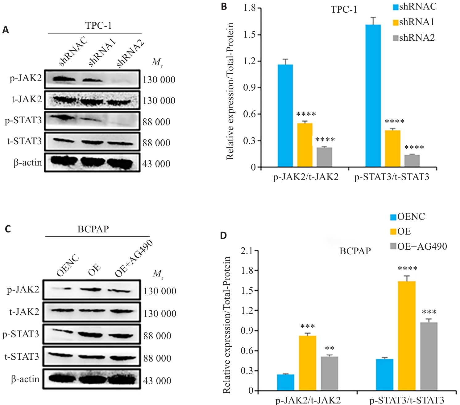
图13 APOC1的表达对JAK2/STAT3信号通路的影响
Fig.13 APOC1 knockdown and overexpresison on expressions of key proteins in the JAK2/STAT3 signaling pathway detected by Western blotting. A,B: Expression levels of key proteins in the JAK2/STAT3 signaling pathway in APOC1 knockdown group. C,D: Expression levels of key proteins in the JAK2/STAT3 signaling pathway in APOC1 overexpression group. **P<0.01, ***P<0.001, ****P<0.0001 vs shRNAC/OENC (n=3).
| 1 | 陈一峰, 郑泽荣, 林进吉, 等. 甲状腺乳头状癌合并滤泡癌10例临床病理分析[J]. 临床与实验病理学杂志, 2020, 36(4): 464-6. |
| 2 | Li R, Zhang YJ, Xiang RQ, et al. Analysis of the mechanism of maslinic acid on papillary thyroid carcinoma based on RNA-seq technology[J]. Evid Based Complement Alternat Med, 2022, 2022: 7000531. |
| 3 | Jong MC, Hofker MH, Havekes LM. Role of ApoCs in lipoprotein metabolism: functional differences between ApoC1, ApoC2, and ApoC3[J]. Arterioscler Thromb Vasc Biol, 1999, 19(3): 472-84. |
| 4 | Fuior EV, Gafencu AV. Apolipoprotein C1: its pleiotropic effects in lipid metabolism and beyond[J]. Int J Mol Sci, 2019, 20(23): 5939. |
| 5 | Zhou XP, Chen Y, Mok KY, et al. Non-coding variability at the APOE locus contributes to the Alzheimer's risk[J]. Nat Commun, 2019, 10(1): 3310. |
| 6 | Cao XY, Wu BQ, Guo SM, et al. APOC1 predicts a worse prognosis for esophageal squamous cell carcinoma and is associated with tumor immune infiltration during tumorigenesis[J]. Pathol Oncol Res, 2023, 29: 1610976. |
| 7 | Mei LH, Long J, Wu SE, et al. APOC1 reduced anti-PD-1 immunotherapy of nonsmall cell lung cancer via the transformation of M2 into M1 macrophages by ferroptosis by NRF2/HO-1[J]. Anticancer Drugs, 2024, 35(4): 333-43. |
| 8 | Hao XP, Zheng ZY, Liu HY, et al. Inhibition of APOC1 promotes the transformation of M2 into M1 macrophages via the ferroptosis pathway and enhances anti-PD1 immunotherapy in hepatocellular carcinoma based on single-cell RNA sequencing[J]. Redox Biol, 2022, 56: 102463. |
| 9 | Jiang H, Tang JY, Xue D, et al. Apolipoprotein C1 stimulates the malignant process of renal cell carcinoma via the Wnt3a signaling[J]. Cancer Cell Int, 2021, 21(1): 41. |
| 10 | Yang SM, Du JX, Wang W, et al. APOC1 is a prognostic biomarker associated with M2 macrophages in ovarian cancer[J]. BMC Cancer, 2024, 24(1): 364. |
| 11 | Ren H, Chen ZH, Yang L, et al. Apolipoprotein C1 (APOC1) promotes tumor progression via MAPK signaling pathways in colorectal cancer[J]. Cancer Manag Res, 2019, 11: 4917-30. |
| 12 | Yi J, Ren LW, Wu J, et al. Apolipoprotein C1 (APOC1) as a novel diagnostic and prognostic biomarker for gastric cancer[J]. Ann Transl Med, 2019, 7(16): 380. |
| 13 | Yan Y, Zhou YH, Wang K, et al. Apolipoprotein C1 (APOC1), A candidate diagnostic serum biomarker for breast cancer identified by serum proteomics study[J]. Crit Rev Eukaryot Gene Expr, 2022, 32(4): 1-9. |
| 14 | Lei B, Qian L, Zhang YP, et al. MLAA-34 knockdown shows enhanced antitumor activity via JAK2/STAT3 signaling pathway in acute monocytic leukemia[J]. J Cancer, 2020, 11(23): 6768-81. |
| 15 | 李超友, 罗亚星, 孙新民, 等. 载脂蛋白C1在甲状腺乳头状癌中的临床表达及生物信息学分析[J]. 中国耳鼻咽喉颅底外科杂志, 2021, 27(4): 445-51. |
| 16 | 王培宇, 黄 祺, 王少东, 等. 《全球癌症统计数据2022》要点解读[J]. 中国胸心血管外科临床杂志, 2024, 31(7): 933-54. |
| 17 | Miranda-Filho A, Lortet-Tieulent J, Bray F, et al. Thyroid cancer incidence trends by histology in 25 countries: a population-based study[J]. Lancet Diabetes Endocrinol, 2021, 9(4): 225-34. |
| 18 | Vaccarella S, Franceschi S, Bray F, et al. Worldwide thyroid-cancer epidemic? the increasing impact of overdiagnosis[J]. N Engl J Med, 2016, 375(7): 614-7. |
| 19 | Li MM, Meheus F, Polazzi S, et al. The economic cost of thyroid cancer in France and the corresponding share associated with treatment of overdiagnosed cases[J]. Value Health, 2023, 26(8): 1175-82. |
| 20 | Ishijima T, Nakajima K. Mechanisms of microglia proliferation in a rat model of facial nerve anatomy[J]. Biology, 2023, 12(8): 1121. |
| 21 | Cai ZJ, Wang JR, Li YD, et al. Overexpressed Cyclin D1 and CDK4 proteins are responsible for the resistance to CDK4/6 inhibitor in breast cancer that can be reversed by PI3K/mTOR inhibitors[J]. Sci China Life Sci, 2023, 66(1): 94-109. |
| 22 | Coqueret O. New roles for p21 and p27 cell-cycle inhibitors: a function for each cell compartment[J]? Trends Cell Biol, 2003, 13(2): 65-70. |
| 23 | 王钦磊. APOC1通过Wnt/β-catenin信号通路诱导G0/G1期阻滞抑制胆管癌细胞增殖的机制研究[D]. 青岛: 青岛大学, 2024. |
| 24 | Pu HC, Qian Q, Wang FL, et al. Schizandrin A induces the apoptosis and suppresses the proliferation, invasion and migration of gastric cancer cells by activating endoplasmic reticulum stress[J]. Mol Med Rep, 2021, 24(5): 787. |
| 25 | Zhang Y, Yang X, Ge XH, et al. Puerarin attenuates neurological deficits via Bcl-2/Bax/cleaved caspase-3 and Sirt3/SOD2 apoptotic pathways in subarachnoid hemorrhage mice[J]. Biomed Pharmacother, 2019, 109: 726-33. |
| 26 | Guan HM, Li WQ, Liu J, et al. LncRNA HIF1A-AS2 modulated by HPV16 E6 regulates apoptosis of cervical cancer cells via P53/caspase9/caspase3 axis[J]. Cell Signal, 2022, 97: 110390. |
| 27 | Takano S, Yoshitomi H, Togawa A, et al. Apolipoprotein C-1 maintains cell survival by preventing from apoptosis in pancreatic cancer cells[J]. Oncogene, 2008, 27(20): 2810-22. |
| 28 | 宋慧娟, 徐振华, 何东宁. 载脂蛋白C1表达对人肝癌HepG2细胞增殖和凋亡的影响及其机制[J]. 吉林大学学报: 医学版, 2024, 50(1): 128-35. |
| 29 | Li RJ, He HX, He XX. APOC1 promotes the progression of osteosarcoma by binding to MTCH2[J]. Exp Ther Med, 2023, 25(4): 163. |
| 30 | 余以珊. APOC1通过MAPK/ERK通路促进食管鳞状细胞癌增殖及转移的机制研究[D]. 济南: 山东大学, 2023. |
| 31 | Gao FF, Chen JL, Zhang TT, et al. LPCAT1 functions as an oncogene in cervical cancer through mediating JAK2/STAT3 signaling[J]. Exp Cell Res, 2022, 421(1): 113360. |
| 32 | Judd LM, Menheniott TR, Ling H, et al. Inhibition of the JAK2/STAT3 pathway reduces gastric cancer growth in vitro and in vivo [J]. PLoS One, 2014, 9(5): e95993. |
| 33 | Fu LX, Lian QW, Pan JD, et al. JAK2 tyrosine kinase inhibitor AG490 suppresses cell growth and invasion of gallbladder cancer cells via inhibition of JAK2/STAT3 signaling[J]. J Biol Regul Homeost Agents, 2017, 31(1): 51-8. |
| 34 | Brooks AJ, Putoczki T. JAK-STAT signalling pathway in cancer[J]. Cancers, 2020, 12(7): 1971. |
| 35 | Xing ZY, Wang X, Liu JQ, et al. Effect of miR-210 on the chemosensitivity of breast cancer by regulating JAK-STAT signaling pathway[J]. Biomed Res Int, 2021, 2021: 7703159. |
| 36 | Cheng X, PKKYeung, Zhong K, et al. Astrocytic endothelin-1 overexpression promotes neural progenitor cells proliferation and differentiation into astrocytes via the Jak2/Stat3 pathway after stroke[J]. J Neuroinflammation, 2019, 16(1): 227. |
| 37 | Yin Z, Ma TT, Lin Y, et al. IL-6/STAT3 pathway intermediates M1/M2 macrophage polarization during the development of hepatocellular carcinoma[J]. J Cell Biochem, 2018, 119(11): 9419-32. |
| 38 | Kim MS, Lee WS, Jeong J, et al. Induction of metastatic potential by TrkB via activation of IL6/JAK2/STAT3 and PI3K/AKT signaling in breast cancer[J]. Oncotarget, 2015, 6(37): 40158-71. |
| 39 | Zhou JM, Wu A, Yu XT, et al. SIRT6 inhibits growth of gastric cancer by inhibiting JAK2/STAT3 pathway[J]. Oncol Rep, 2017, 38(2): 1059-66. |
| 40 | Carpenter RL, Lo HW. STAT3 target genes relevant to human cancers[J]. Cancers, 2014, 6(2): 897-925. |
| 41 | Wang JQ, Zhang Y, Song H, et al. The circular RNA circSPARC enhances the migration and proliferation of colorectal cancer by regulating the JAK/STAT pathway[J]. Mol Cancer, 2021, 20(1): 81. |
| 42 | Li YJ, Wei J, Sun Y, et al. DLGAP5 regulates the proliferation, migration, invasion, and cell cycle of breast cancer cells via the JAK2/STAT3 signaling axis[J]. Int J Mol Sci, 2023, 24(21): 15819. |
| [1] | 常笑语, 张瀚文, 曹红亭, 侯玲, 孟鑫, 陶虹, 罗彦, 李光华. 热应激对大鼠胸主动脉内皮细胞生物钟基因 Bmal1和细胞周期蛋白表达水平的影响[J]. 南方医科大学学报, 2025, 45(7): 1353-1362. |
| [2] | 谢婷, 王云云, 郭婷, 袁春华. 雷氏大疣蛛多肽毒素组分通过激活促凋亡通路和协同作用抑制癌细胞增殖[J]. 南方医科大学学报, 2025, 45(7): 1460-1470. |
| [3] | 杨毓甲, 杨丽芳, 吴雅玲, 段兆达, 于春泽, 吴春云, 于建云, 杨力. 大麻二酚经PERK-eIF2α-ATF4-CHOP通路减轻多重脑震荡大鼠的神经元内质网应激和凋亡[J]. 南方医科大学学报, 2025, 45(6): 1240-1250. |
| [4] | 曾玉梅, 李继科, 黄仲曦, 周毅波. 绒毛样蛋白VILL通过与LMO7蛋白相互作用抑制鼻咽癌细胞的增殖[J]. 南方医科大学学报, 2025, 45(5): 954-961. |
| [5] | 陈悦, 肖林雨, 任侣, 宋雪, 李静, 胡建国. 水晶兰苷通过抑制PI3K/AKT信号通路减少神经元凋亡改善脊髓损伤后小鼠的运动功能[J]. 南方医科大学学报, 2025, 45(4): 774-784. |
| [6] | 储菲, 陈孝华, 宋博文, 杨晶晶, 左芦根. 苏荠宁黄酮通过抑制PI3K/AKT信号通路拮抗肠上皮细胞凋亡改善小鼠实验性结肠炎[J]. 南方医科大学学报, 2025, 45(4): 819-828. |
| [7] | 岳雅清, 牟召霞, 王希波, 刘艳. Aurora-A过表达通过激活NF-κBp65/ARPC4信号轴促进宫颈癌细胞的侵袭和转移[J]. 南方医科大学学报, 2025, 45(4): 837-843. |
| [8] | 张毅, 沈昱, 万志强, 陶嵩, 柳亚魁, 王栓虎. CDKN3高表达促进胃癌细胞的迁移和侵袭:基于调控p53/NF-κB信号通路和抑制胃癌细胞凋亡[J]. 南方医科大学学报, 2025, 45(4): 853-861. |
| [9] | 庆顺杰, 沈智勇. 过表达己糖激酶2通过激活JAK/STAT途径促进结直肠癌细胞的增殖、迁移和侵袭并调节肿瘤免疫微环境[J]. 南方医科大学学报, 2025, 45(3): 542-553. |
| [10] | 黄晴晴, 张文静, 张小凤, 王炼, 宋雪, 耿志军, 左芦根, 王月月, 李静, 胡建国. 高表达MYO1B促进胃癌细胞增殖、迁移和侵袭并与患者的不良预后有关[J]. 南方医科大学学报, 2025, 45(3): 622-631. |
| [11] | 陈镝, 吕莹, 郭怡欣, 张怡荣, 王蕊璇, 周小若, 陈雨欣, 武晓慧. 双氢青蒿素可显著增强阿霉素诱导的三阴性乳腺癌细胞凋亡:基于负向调控STAT3/HIF-1α通路[J]. 南方医科大学学报, 2025, 45(2): 254-260. |
| [12] | 邹金华, 王惠, 张冬艳. SLC1A5通过促进M2型巨噬细胞极化促进肝癌进展[J]. 南方医科大学学报, 2025, 45(2): 269-284. |
| [13] | 曹周芳, 汪元, 王梦娜, 孙玥, 刘菲菲. LINC00837/miR-671-5p/SERPINE2功能轴促进类风湿关节炎成纤维细胞样滑膜细胞的恶性病理学过程[J]. 南方医科大学学报, 2025, 45(2): 371-378. |
| [14] | 陈孝华, 鲁辉, 王子良, 王炼, 夏勇生, 耿志军, 张小凤, 宋雪, 王月月, 李静, 胡建国, 左芦根. ABI2在胃癌进展和预后中的作用及其调控机制[J]. 南方医科大学学报, 2024, 44(9): 1653-1661. |
| [15] | 薛良军, 谈秋瑜, 许静文, 冯璐, 李文锦, 颜亮, 李玉磊. MiR-6838-5p过表达下调DDR1基因表达抑制乳腺癌MCF-7细胞的增殖[J]. 南方医科大学学报, 2024, 44(9): 1677-1684. |
| 阅读次数 | ||||||
|
全文 |
|
|||||
|
摘要 |
|
|||||