| [1] |
Cross M, Smith E, Hoy D, et al. The global burden of hip and knee osteoarthritis: estimates from the Global Burden of Disease 2010 study[J]. Ann Rheum Dis, 2014, 73(7): 1323-30. doi:10.1136/annrheumdis-2013-204763
|
| [2] |
Racine J, Aaron RK. Pathogenesis and epidemiology of osteoarthritis[J]. R I Med J, 2013,96(3):19-22.
|
| [3] |
Hwang HS, Kim HA. Chondrocyte apoptosis in the pathogenesis of osteoarthritis[J]. Int J Mol Sci, 2015, 16(11): 26035-54. doi:10.3390/ijms161125943
|
| [4] |
Miao GQ, Zang XH, Hou HG, et al. Bax targeted by miR-29a regulates chondrocyte apoptosis in osteoarthritis[J]. BioMed Res Int, 2019, 2019: 1434538. doi:10.1155/2019/1434538
|
| [5] |
Scanzello CR, Plaas A, Crow MK. Innate immune system activation in osteoarthritis: is osteoarthritis a chronic wound?[J]. Curr Opin Rheumatol, 2008, 20(5): 565-72. doi:10.1097/bor.0b013e32830aba34
|
| [6] |
杨琪琪, 姜天鑫, 李春宁, 等. 电针治疗膝关节骨关节炎慢性疼痛患者的疗效及对中枢敏化的影响[J/OL].中国针灸, [2026-05-11].
|
| [7] |
Melzack R, Wall PD. Pain mechanisms: a new theory[J]. Science, 1965, 150(3699): 971-9. doi:10.1126/science.150.3699.971
|
| [8] |
Duan B, Cheng LZ, Bourane S, et al. Identification of spinal circuits transmitting and gating mechanical pain[J]. Cell, 2014, 159(6): 1417-32. doi:10.1016/j.cell.2014.11.003
|
| [9] |
李杨乐, 杨 锋, 樊静杰, 等. 电针对膝骨关节炎大鼠脊髓背角CB2R、p-P38和p-ERK表达的影响[J]. 中国中医药信息杂志, 2022, 29(7): 60-6.
|
| [10] |
鲍哲明, 单 青, 李 晨, 等. 碘乙酸钠大鼠骨关节炎模型骨微结构研究[J]. 中国骨质疏松杂志, 2022, 28(2): 210-4, 219.
|
| [11] |
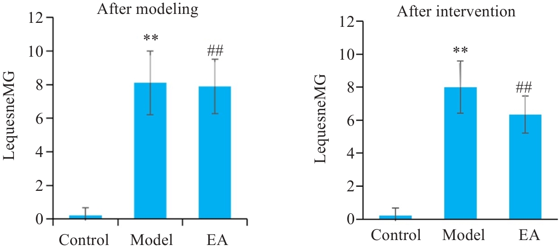
Lequesne MG, Samson M. Indices of severity in osteoarthritis for weight bearing joints[J]. J Rheumatol Suppl, 1991, 27: 16-8.
|
| [12] |
中国针灸学会. 实验动物常用穴位名称与定位第2部分: 大鼠[J]. 针刺研究, 2021, 46(4): 351-2.
|
| [13] |
Lequesne MG. The algofunctional indices for hip and knee osteoarthritis[J]. J Rheumatol, 1997, 24 4:779-81.
|
| [14] |
Dong JB, Liao Y, Wu BH. TAK-242 ameliorates epileptic symptoms in mice by inhibiting the TLR4/NF‑κB signaling pathway[J]. Ann Transl Med, 2022, 10(14): 795. doi:10.21037/atm-22-2707
|
| [15] |
李益军, 梁兴森, 方细霞, 等. 电针与关节液炎症因子及关节运动功能恢复的相关性研究[J]. 时珍国医国药, 2024, 35(6): 1415-9. doi:10.3969/j.issn.1008-0805.2024.06.31
|
| [16] |
郑 祥, 高颂爱, 尤 浩, 等. 电针可改善骨关节炎大鼠的关节炎症和运动功能: 基于调控Wnt-7B/β-catenin信号通路[J]. 南方医科大学学报, 2023, 43(4): 590-6.
|
| [17] |
陈 蔚, 李恒聪, 万红叶, 等. 电针对膝关节骨关节炎大鼠滑膜炎性反应期自发痛及后期触诱发痛的影响[J]. 中国针灸, 2022, 42(12): 1385-93. doi:10.13703/j.0255-2930.20220526-k0006
|
| [18] |
周欣华, 杨琪琪, 李蔚然, 等. 电针调控PI3K/Akt/mTOR通路对膝关节骨关节炎大鼠膝关节滑膜组织自噬的影响[J]. 针刺研究, 2026, 51(1): 59-67.
|
| [19] |
周 巧, 刘 健, 万 磊, 等. 新风胶囊抑制软骨细胞炎症和细胞外基质降解: 基于调控miR-502-5p/TRAF2/NF-κB轴[J]. 南方医科大学学报, 2024, 44(1): 108-18. doi:10.12122/j.issn.1673-4254.2024.01.13
|
| [20] |
Jørgensen AEM, Agergaard J, Schjerling P, et al. The regional turnover of cartilage collagen matrix in late-stage human knee osteoarthritis[J]. Osteoarthritis Cartilage, 2022, 30(6): 886-95. doi:10.1016/j.joca.2022.03.007
|
| [21] |
Yang JZ, Chen DL, He Q, et al. Arctiin alleviates knee osteoarthritis by suppressing chondrocyte oxidative stress induced by accumulated iron via AKT/NRF2/HO-1 signaling pathway[J]. Sci Rep, 2024, 14: 31935. doi:10.1038/s41598-024-83383-7
|
| [22] |
Zhang JM, Hao XX, Chi RM, et al. Moderate mechanical stress suppresses the IL-1β-induced chondrocyte apoptosis by regulating mitochondrial dynamics[J]. J Cell Physiol, 2021, 236(11): 7504-15. doi:10.1002/jcp.30386
|
| [23] |
Millerand M, Berenbaum F, Jacques C. Danger signals and inflammaging in osteoarthritis[J]. Clin Exp Rheumatol, 2019, 37 Suppl 120(5): 48-56.
|
| [24] |
Kapoor M, Martel-Pelletier J, Lajeunesse D, et al. Role of proinflammatory cytokines in the pathophysiology of osteoarthritis[J]. Nat Rev Rheumatol, 2011, 7(1): 33-42. doi:10.1038/nrrheum.2010.196
|
| [25] |
Zhang X, Hsueh MF, Huebner JL, et al. TNF-α carried by plasma extracellular vesicles predicts knee osteoarthritis progression[J]. Front Immunol, 2021, 12: 758386. doi:10.3389/fimmu.2021.758386
|
| [26] |
Tseng HC, Wu MR, Lee CH, et al. Differentiation capacity of bone marrow-derived rat mesenchymal stem cells from DsRed and cre transgenic cre/loxP models[J]. Cells, 2022, 11(17): 2769. doi:10.3390/cells11172769
|
| [27] |
Chen L, Wei K, Li J, et al. Integrated analysis of LncRNA-mediated CeRNA network in calcific aortic valve disease[J]. Cells, 2022, 11(14): 2204. doi:10.3390/cells11142204
|
| [28] |
Zhang Y, Zeng Y. Curcumin reduces inflammation in knee osteoarthritis rats through blocking TLR4/MyD88/NF-κB signal pathway[J]. Drug Dev Res, 2019, 80(3): 353-9. doi:10.1002/ddr.21509
|
| [29] |
Xu XT, Li NP, Wu YR, et al. Zhuifeng tougu capsules inhibit the TLR4/MyD88/NF‑κB signaling pathway and alleviate knee osteoarthritis: in vitro and in vivo experiments[J]. Front Pharmacol, 2022, 13: 951860. doi:10.3389/fphar.2022.951860
|
| [30] |
庄丽华, 詹松华, 杨烁慧, 等. 电针通过调节TLR4/MyD88/NF-κb信号通路改善缺血性脑卒中损伤研究现状[J]. 中华中医药学刊, 2019, 37(9): 2182-5.
|
| [31] |
晏 黎, 饶亚华, 黄 伟, 等. 损伤相关分子模式与组织修复[J]. 中国免疫学杂志, 2022, 38(7): 872-7.
|
| [32] |
邵建彬, 王 霞, 徐华洲, 等. 芪苓通络方抑制TLR4/MyD88/NF-κB信号通路改善特发性膜性肾病大鼠肾损伤[J]. 中国实验方剂学杂志, 2022, 28(4): 100-8.
|
| [33] |
Sun FF, Hu PF, Xiong Y, et al. Tricetin protects rat chondrocytes against IL-1β-induced inflammation and apoptosis[J]. Oxid Med Cell Longev, 2019, 2019(1): 4695381. doi:10.1155/2019/4695381
|
| [34] |
D'Arcy MS. Cell death: a review of the major forms of apoptosis, necrosis and autophagy[J]. Cell Biol Int, 2019, 43(6): 582-92. doi:10.1002/cbin.11137
|
| [35] |
李亚楠, 倪 娟, 方禹舜, 等. 姜黄素通过调控脂多糖诱导的炎症抑制软骨细胞凋亡[J]. 中成药, 2021, 43(7): 1904-8. doi:10.3969/j.issn.1001-1528.2021.07.042
|
| [36] |
Zhang YB, Cai WS, Han GT, et al. Panax notoginseng saponins prevent senescence and inhibit apoptosis by regulating the PI3K-AKT-mTOR pathway in osteoarthritic chondrocytes[J]. Int J Mol Med, 2020, 49(3):30.
|

 ), 杨琪琪2, 张振丰1, 鲍亮1, 姜天鑫2, 张玉3, 李飞2(