
南方医科大学学报 ›› 2026, Vol. 46 ›› Issue (7): 1474-1486.doi: 10.12122/j.issn.1673-4254.2026.07.02
• • 上一篇
董旻昱1( ), 方泽扬1, 张芸芸1, 吴友谅1, 郑煌傑1, 刘泳君1, 阳琼1, 孔德钱2(), 陈科全1(), 王斯琪1()
收稿日期:2025-11-24
出版日期:2026-07-20
发布日期:2026-07-20
通讯作者:
孔德钱,陈科全,王斯琪
E-mail:dongminyu99@126.com;2451498539@qq.com;yinyuedegushi@126.com;2023681011@gzhmu.edu.cn
作者简介:董旻昱,在读博士研究生,医师,E-mail: dongminyu99@126.com
基金资助:
Minyu DONG1(), Zeyang FANG1, Yunyun ZHANG1, Youliang WU1, Huangjie ZHENG1, Yongjun LIU1, Qiong YANG1, Deqian KONG2(), Kequan CHEN1(), Siqi WANG1()
Received:2025-11-24
Online:2026-07-20
Published:2026-07-20
Contact:
Deqian KONG, Kequan CHEN, Siqi WANG
E-mail:dongminyu99@126.com;2451498539@qq.com;yinyuedegushi@126.com;2023681011@gzhmu.edu.cn
Supported by:摘要:
目的 研究肥胖加重艰难梭菌感染(CDI)的作用机制,探讨肠道菌群代谢产物丁酸作为替代疗法的潜在价值。 方法 利用CDI小鼠模型,分别饲喂高脂饲料和普通饲料构建肥胖CDI模型(HFD)和正常CDI模型(NCD)进行对比,同时构建体外高脂CDI模型,6只/组。通过检测小鼠肝脏、附睾白色脂肪组织质量,肝功能指标及油红O染色评估肥胖小鼠模型;超氧化物歧化酶(SOD)及丙二醛(MDA)含量测定氧化应激水平;HE染色评估结肠损伤程度;RT-PCR和免疫组化检测小鼠肠黏膜屏障(ZO-1、Occludin)表达水平;RT-PCR和ELISA检测小鼠血清和粪便的艰难梭菌毒素A(TcdA)和毒素B(TcdB)含量;气相色谱-质谱(GC-MS)法检测小鼠粪便中短链脂肪酸(SCFAs)含量;免疫荧光检测高脂细胞膜型的肠黏膜屏障(ZO-1、Occludin)表达水平;流式细胞术检测高脂细胞模型凋亡情况。 结果 与NCD组相比,接种后第2天,HFD组小鼠生存率下降至70%,显著低于NCD组的90%;血清TcdA和TcdB分别由7.5 ng/L和6.5 ng/L升高至15.3 ng/L和15.0 ng/L,粪便TcdA和TcdB分别由2.8l g fold和3.9l g fold升高至5.8l g fold和6.5l g fold。HFD组AST、ALT分别由2.3 U/mL和5.2 U/mL升高至9.8 U/mL和20.0 U/mL,TG、TC分别由0.6 U/mL和1.5 U/mL升高至2.9 U/mL和10.5 U/mL;粪便丁酸盐浓度由3.20±0.62 ng/mL降至1.05±0.55 ng/mL。与此同时,HFD组结肠组织损伤加重,ZO-1和Occludin表达下调,SOD降低、MDA升高。丁酸盐干预可减轻体质量下降和死亡,降低毒素负荷,改善结肠及肝脏损伤,恢复紧密连接蛋白表达并抑制细胞凋亡。FMT亦具有类似保护作用,自感染后第2天起使生存率较HFD组提高约10%,并改善肠屏障功能、肝功能损伤及氧化应激水平。上述差异均具有统计学意义(P<0.05)。 结论 高脂饮食显著加重CDI疾病进程,丁酸在改善肥胖相关CDI中具有潜在替代治疗作用。
董旻昱, 方泽扬, 张芸芸, 吴友谅, 郑煌傑, 刘泳君, 阳琼, 孔德钱, 陈科全, 王斯琪. 丁酸缓解高脂饮食诱导肥胖小鼠艰难梭菌感染的机制研究[J]. 南方医科大学学报, 2026, 46(7): 1474-1486.
Minyu DONG, Zeyang FANG, Yunyun ZHANG, Youliang WU, Huangjie ZHENG, Yongjun LIU, Qiong YANG, Deqian KONG, Kequan CHEN, Siqi WANG. Butyrate alleviates Clostridium difficile infection in high-fat diet-induced obese mice[J]. Journal of Southern Medical University, 2026, 46(7): 1474-1486.
| Primer name | Sequences |
|---|---|
| Toxin A | Forward:GGACATGGTAAAGATGAATTC |
| Reverse: CCCAATAGAAGATTCAATATTAAGCTT | |
| Toxin B | Forward: TCTGATGCACTATGTGACTTA |
| Reverse: ATCTACGAGCGTAGTCAC | |
| GAPDH (Mouse) | Forward: GGAGAAACCTGCCAAGTATGA |
| Reverse: TCCTCAGTGTAGCCCAAGA | |
| ZO-1 (Mouse) | Forward: TGAACGTCCCTGACCTTTCG |
| Reverse: CTGTGGAGACTGCGTGGAAT | |
| Occludin (Mouse) | Forward: CTTTGGCTACGGAGGTGGCTAT |
| Reverse: CTTTGGCTGCTCTTGGGTCTG |
表1 RT-qPCR引物序列
Tab.1 Primer sequences for RT-qPCR
| Primer name | Sequences |
|---|---|
| Toxin A | Forward:GGACATGGTAAAGATGAATTC |
| Reverse: CCCAATAGAAGATTCAATATTAAGCTT | |
| Toxin B | Forward: TCTGATGCACTATGTGACTTA |
| Reverse: ATCTACGAGCGTAGTCAC | |
| GAPDH (Mouse) | Forward: GGAGAAACCTGCCAAGTATGA |
| Reverse: TCCTCAGTGTAGCCCAAGA | |
| ZO-1 (Mouse) | Forward: TGAACGTCCCTGACCTTTCG |
| Reverse: CTGTGGAGACTGCGTGGAAT | |
| Occludin (Mouse) | Forward: CTTTGGCTACGGAGGTGGCTAT |
| Reverse: CTTTGGCTGCTCTTGGGTCTG |
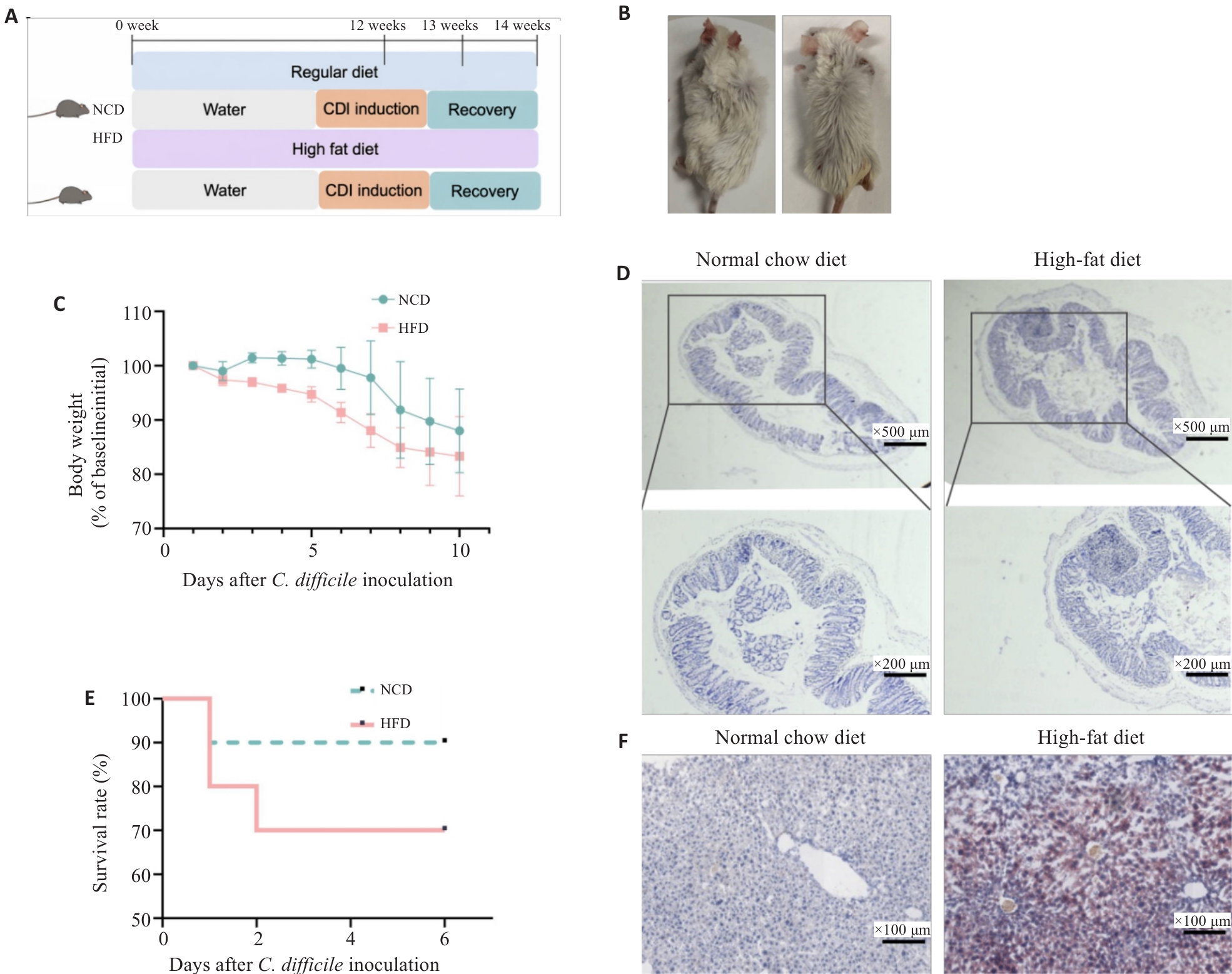
图1 高脂饮食较正常饮食加重小鼠艰难梭菌感染
Fig.1 Mice fed with HFD exacerbate CDI compared with NCD. A: Schematic diagram of establishment of HFD+CDI and normal chow diet (NCD)+CDI mouse models. B: Appearance of the mice in the HFD and NCD groups. C: Changes in body weight after inoculation with Clostridioides difficile. D: HE staining of colon tissues. E: Survival rate after inoculation with Clostridioides difficile. F: Oil Red O staining of liver tissues. NCD: Normal Chow Diet, HFD: High-Fat Diet.
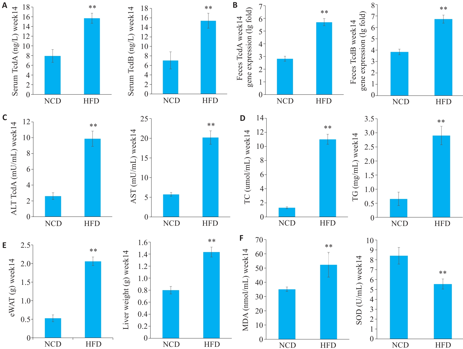
图2 高脂饮食较正常饮食加重小鼠艰难梭菌感染
Fig. 2 Mice fed with HFD exacerbate CDI compared with NCD. A: Serum TcdA and TcdB levels. B: Feces TcdA and TcdB levels. C: Serum ALT and AST levels. D: Serum TG and TC concentrations. E: eWAT weight and liver weight. F: SOD activity and MDA levels. Data are presented as Mean±SD(n=6). **P<0.01 vs NCD.
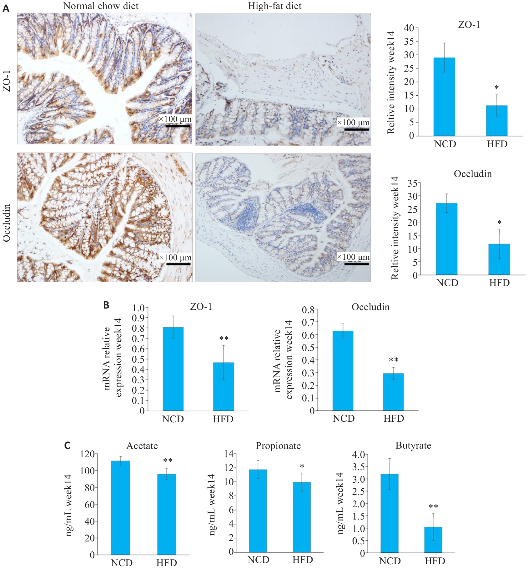
图3 高脂饮食较正常饮食加重小鼠肠道屏障损伤
Fig.3 HFD aggravates intestinal barrier function impairment in CID mice. A: Immunohistochemical staining of ZO-1 and occluding. B: RT-PCR results of ZO-1 and Occludin. C: Concentrations of short-chain fatty acids (SCFAs) in mice of the HFD and NCD groups. Data are presented as Mean±SD (n=6), *P<0.05, **P<0.01 vs NCD.
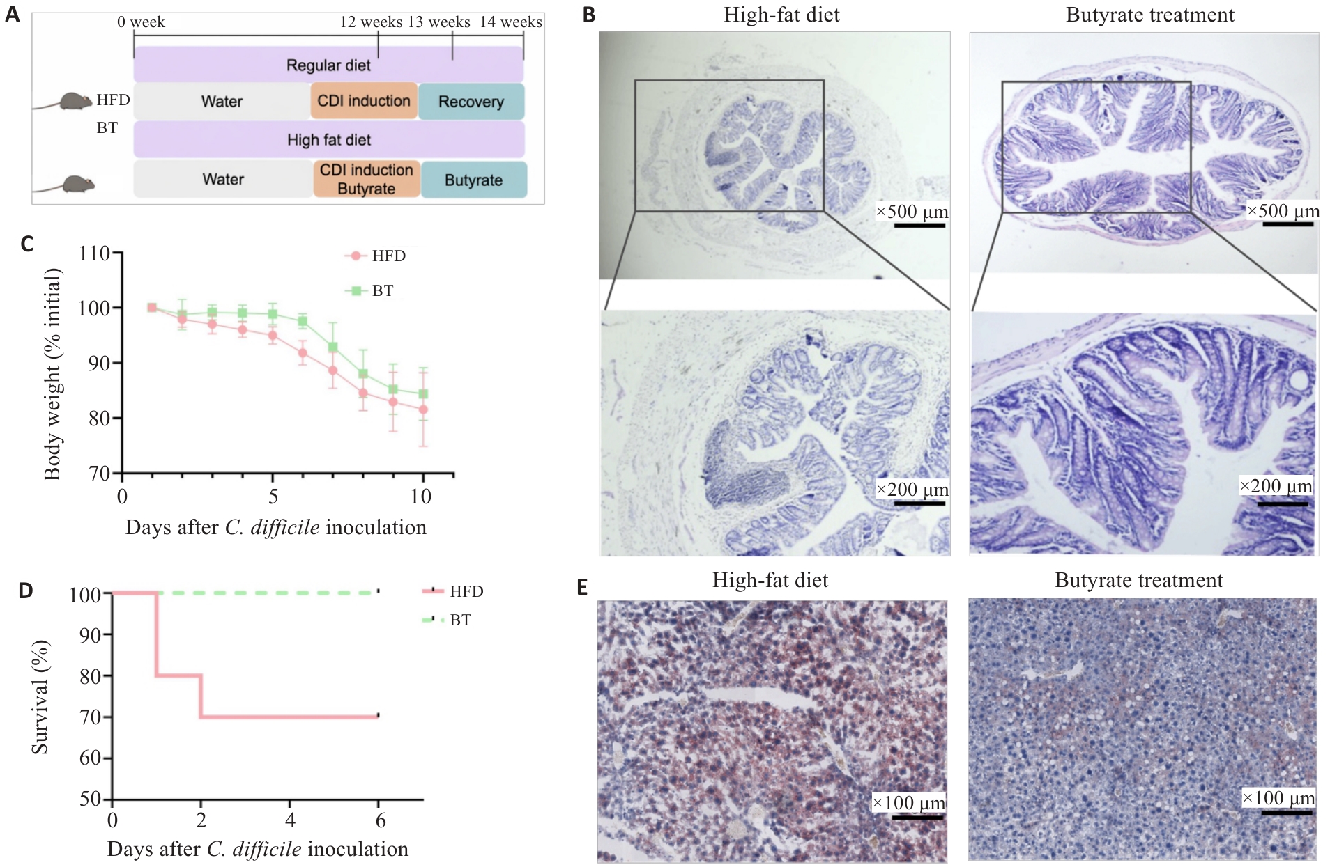
图4 丁酸盐缓解高脂饮食小鼠的艰难梭菌感染
Fig.4 Butyrate treatment alleviates symptoms of CDI mice with HFD feeding. A: Schematic diagram of butyrate intervention in HFD+CDI mice. B: HE staining of colon tissues. C: Changes in body weight after inoculation with Clostridioides difficile. D: Survival rate after inoculation with Clostridioides difficile. E: Oil Red O staining of liver tissues. BT: Butyrate treatment.
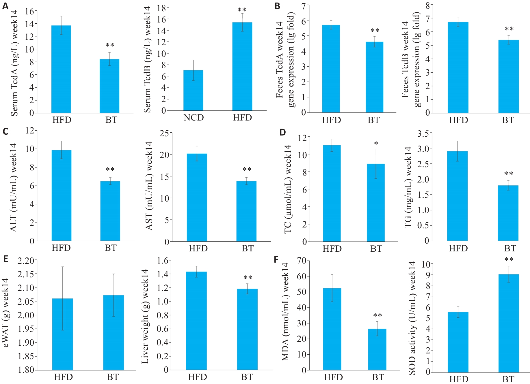
图5 丁酸盐缓解高脂饮食小鼠的艰难梭菌感染
Fig.5 Butyrate treatment alleviates symptoms of CDI mice with HFD feeding. A: Serum levels of TcdA and TcdB. B: Fecal levels TcdA and TcdB. C: Serum ALT and AST levels. D: Serum TG and TC concentrations. E: eWAT weight and liver weight. F: SOD activity and MDA levels. Data are presented as Mean±SD(n=6). *P<0.05, **P<0.01 vs HFD.
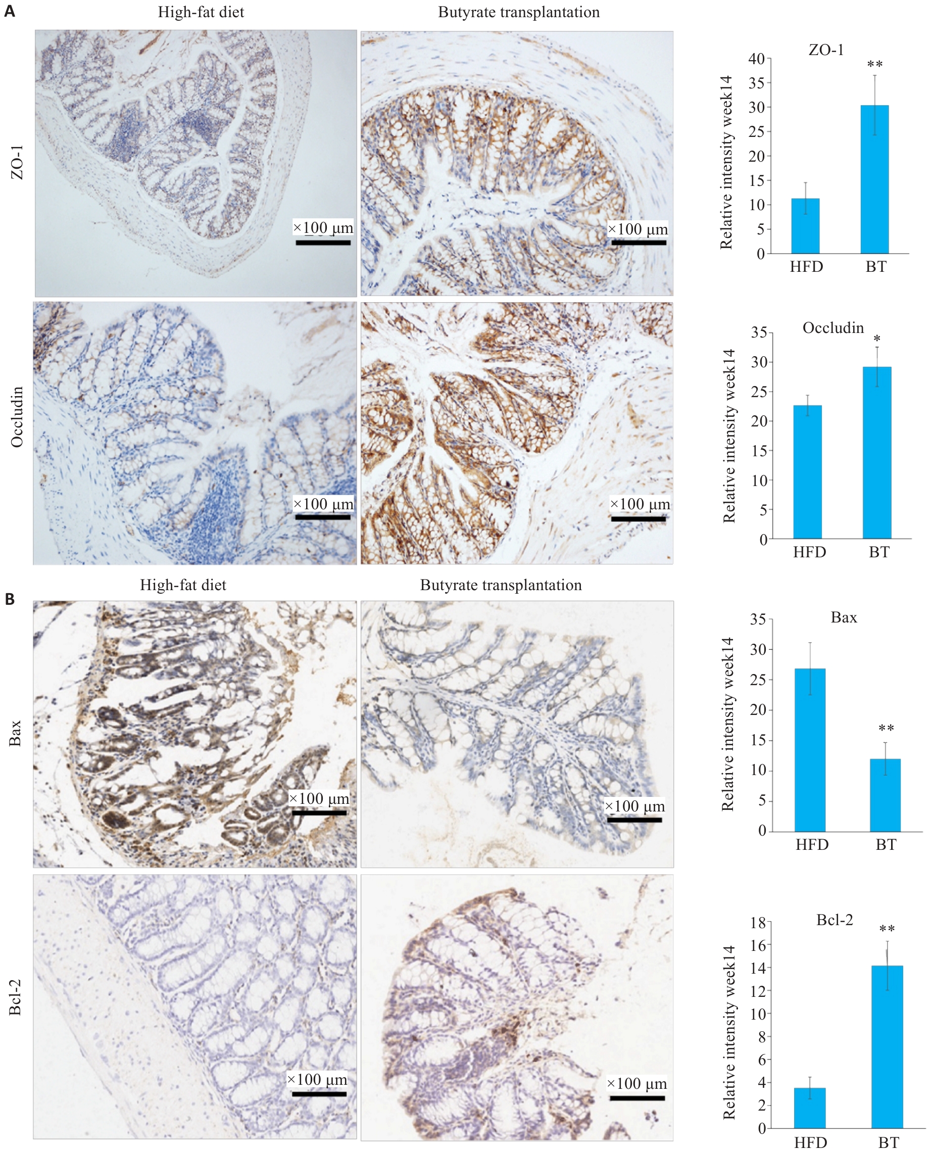
图6 丁酸盐改善高脂饮食合并艰难梭菌感染小鼠的肠道屏障功能损伤
Fig.6 Butyrate improves intestinal barrier function impairment in HFD mice with CDI. A: Immunohistochemical staining results of intestinal barrier markers in colon tissues. B: Immunohistochemical analysis of Bax and Bcl-2. Data are presented as Mean±SD.*P<0.05, **P<0.01 vs HFD.
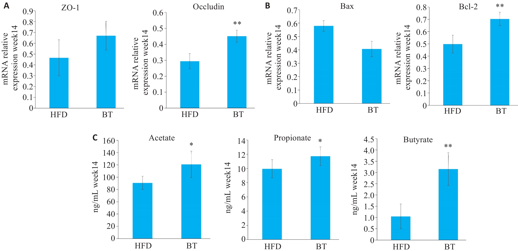
图7 丁酸盐改善高脂饮食合并艰难梭菌感染小鼠的肠道屏障功能损伤
Fig.7 Butyrate improves intestinal barrier function impairment in HFD mice with CDI. A: RT-PCR results of intestinal barrier-related genes. B: RT-PCR results of apoptosis-related genes. C: SCFA concentrations in HFD+CDI mice after butyrate treatment. Data are presented as Mean±SD. *P<0.05, **P<0.01 vs HFD.
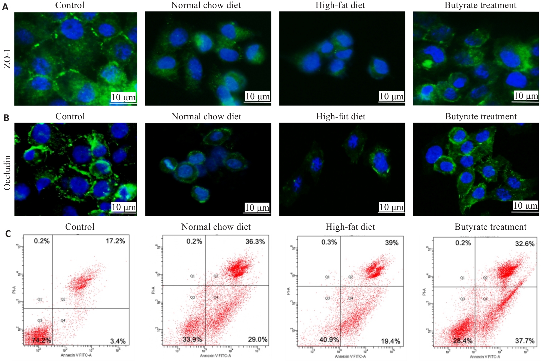
图8 丁酸盐减轻高脂模型细胞凋亡
Fig.8 Butyrate alleviates palmitic acid-induced cell apoptosis. A: Immunofluorescence staining results of ZO-1. B: Immunofluorescence staining results of occludin. C: Flow cytometric analysis of cell apoptosis.
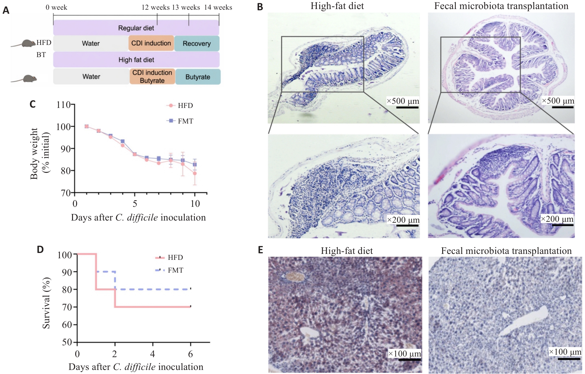
图9 粪菌移植改善高脂饮食合并艰难梭菌感染小鼠的病情
Fig.9 Fecal microbiota transplantation (FMT) ameliorates severity of CDI in HFD mice. A: Schematic diagram of FMT intervention experiment in HFD-CDI mice. B: HE staining of colon tissues. C: Changes in body weight after inoculation with Clostridioides difficile. D: Survival rate after inoculation with Clostridioides difficile. E: Oil Red O staining of liver tissues. FMT: Fecal microbiota transplantation.
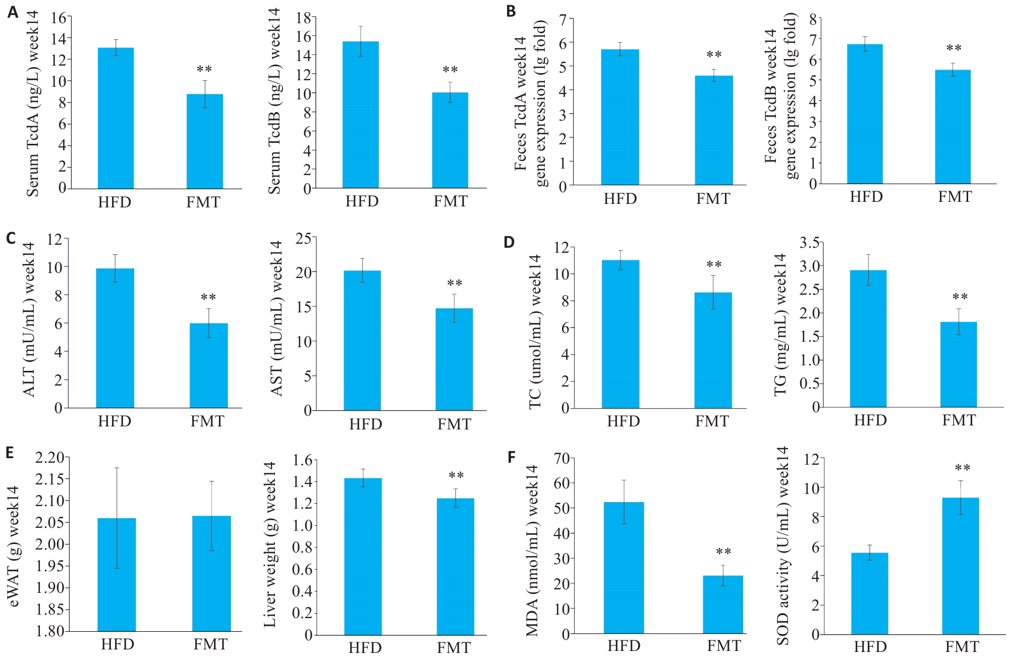
图10 粪菌移植改善高脂饮食合并艰难梭菌感染小鼠的病情
Fig.10 Fecal microbiota transplantation (FMT) ameliorates severity of CDI in HFD mice. A: Serum levels of TcdA and TcdB. B: Feces levels of TcdA and TcdB. C: Serum ALT and AST levels. D: Serum TG and TC concentrations. E: eWAT weight and liver weight. F: SOD activity and MDA levels. Data are presented as Mean±SD, n=6. **P<0.01 vs HFD.
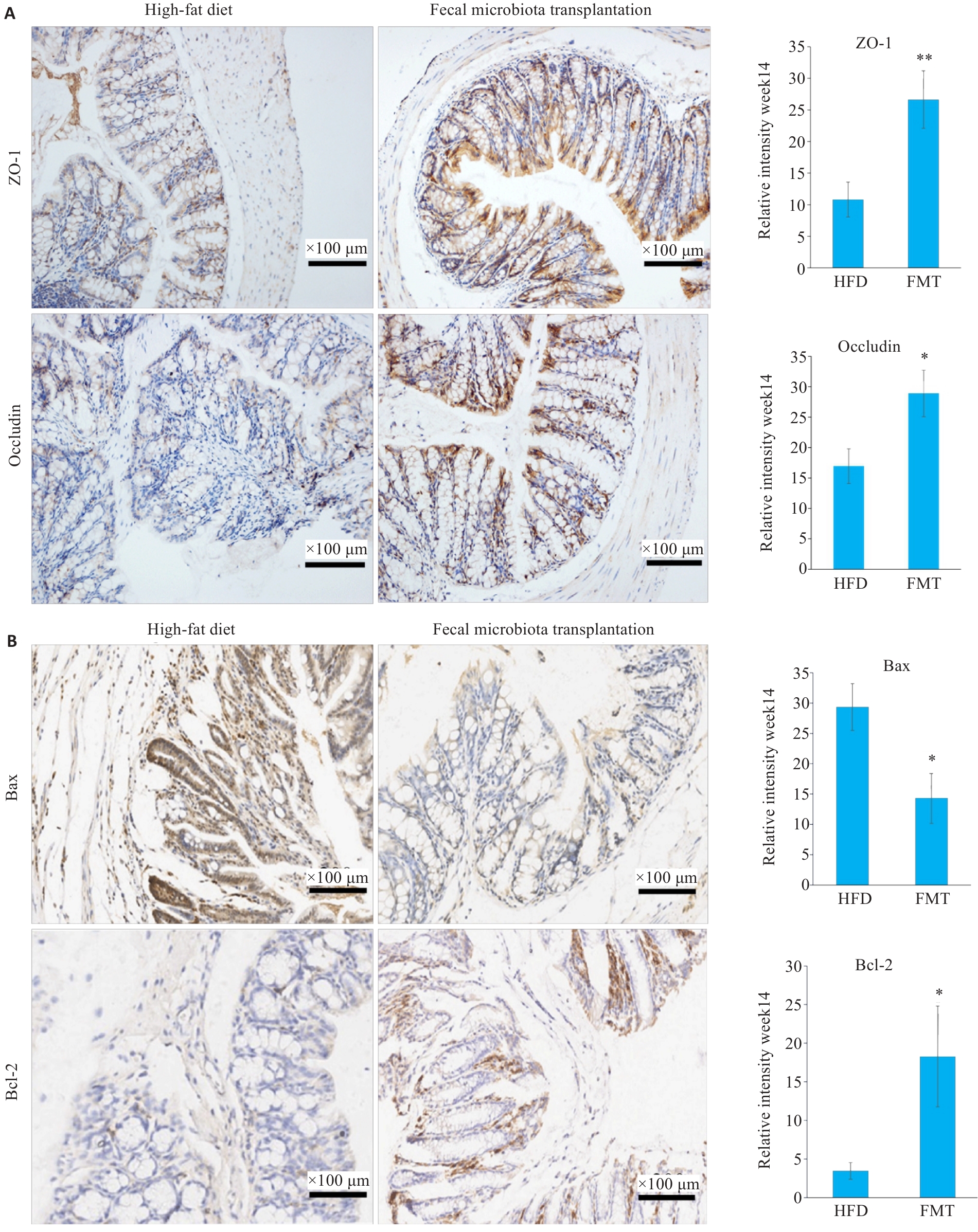
图11 粪菌移植减轻高脂饮食合并艰难梭菌感染小鼠肠道屏障功能损伤
Fig.11 FMT ameliorates intestinal barrier function impairment in HFD mice with CDI. A: Immunohistochemical analysis of intestinal barrier markers in mouse colon tissues. B Immunohistochemical analysis of Bax and Bcl-2. Data are presented as Mean±SD(n=6). *P<0.05, **P<0.01 vs HFD.
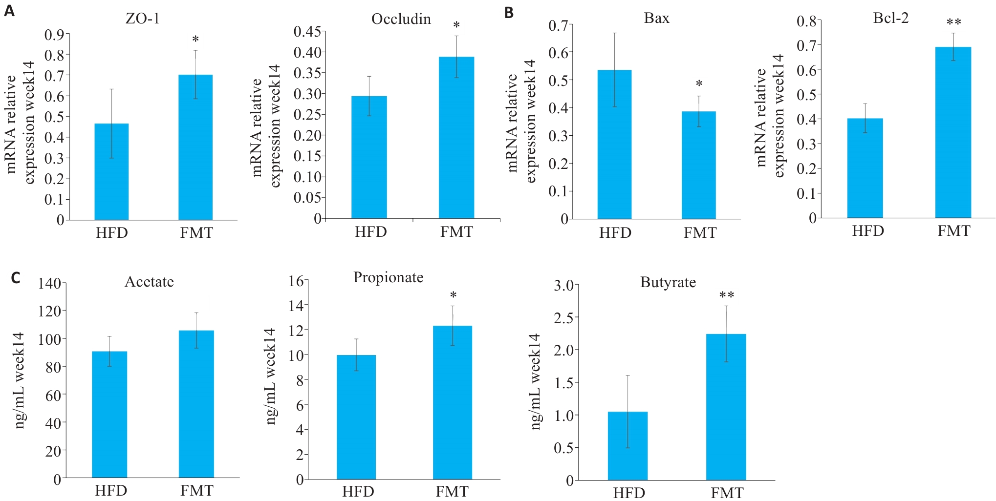
图12 粪菌移植减轻高脂饮食合并艰难梭菌感染小鼠肠道屏障功能损伤
Fig. 12 MT ameliorates intestinal barrier function impairment in HFD mice with CDI. A: RT-PCR results of intestinal barrier-related genes. B: RT-PCR results of apoptosis-related genes. C: SCFA concentrations in HFD+CDI mice after FMT treatment. Data are presented as Mean±SD (n=6). *P<0.05, **P<0.01 vs HFD.
| [1] | Turner NA, Anderson DJ. Hospital infection control: Clostridioides difficile [J]. Clin Colon Rectal Surg, 2020, 33(2): 98-108. doi:10.1055/s-0040-1701234 |
| [2] | Bishara J, Farah R, Mograbi J, et al. Obesity as a risk factor for Clostridium difficile infection[J]. Clin Infect Dis, 2013, 57(4): 489-93. doi:10.1093/cid/cit280 |
| [3] | Mulki R, Baumann AJ, Alnabelsi T, et al. Body mass index greater than 35 is associated with severe Clostridium difficile infection[J]. Aliment Pharmacol Ther, 2017, 45(1): 75-81. doi:10.1111/apt.13832 |
| [4] | Nathanson BH, Higgins TL, McGee WT. The dangers of extreme body mass index values in patients with Clostridium difficile [J]. Infection, 2017, 45(6): 787-93. doi:10.1007/s15010-017-1036-x |
| [5] | Le Chatelier E, Nielsen T, Qin JJ, et al. Richness of human gut microbiome correlates with metabolic markers[J]. Nature, 2013, 500(7464): 541-6. |
| [6] | Turnbaugh PJ, Hamady M, Yatsunenko T, et al. A core gut microbiome in obese and lean twins[J]. Nature, 2009, 457(7228): 480-4. doi:10.1038/nature07540 |
| [7] | Cotillard A, Kennedy SP, Kong LC, et al. Dietary intervention impact on gut microbial gene richness[J]. Nature, 2013, 500(7464): 585-8. doi:10.1038/nature12738 |
| [8] | Boulangé CL, Neves AL, Chilloux J, et al. Impact of the gut microbiota on inflammation, obesity, and metabolic disease[J]. Genome Med, 2016, 8(1): 42. doi:10.1186/s13073-016-0303-2 |
| [9] | Kelly CR, Fischer M, Allegretti JR, et al. ACG clinical guidelines: prevention, diagnosis, and treatment of Clostridioides difficile infections[J]. Am J Gastroenterol, 2021, 116(6): 1124-47. doi:10.14309/ajg.0000000000001278 |
| [10] | Chiu CW, Tsai PJ, Lee CC, et al. Application of microbiome management in therapy for Clostridioides difficile infections: from fecal microbiota transplantation to probiotics to microbiota-preserving antimicrobial agents[J]. Pathogens, 2021, 10(6): 649. doi:10.3390/pathogens10060649 |
| [11] | de Vos WM, Tilg H, Van Hul M, et al. Gut microbiome and health: mechanistic insights[J]. Gut, 2022, 71(5): 1020-32. doi:10.1136/gutjnl-2021-326789 |
| [12] | Qin JJ, Li YR, Cai ZM, et al. A metagenome-wide association study of gut microbiota in type 2 diabetes[J]. Nature, 2012, 490(7418): 55-60. |
| [13] | Fei N, Zhao LP. An opportunistic pathogen isolated from the gut of an obese human causes obesity in germfree mice[J]. ISME J, 2013, 7(4): 880-4. doi:10.1038/ismej.2012.153 |
| [14] | Hazleton KZ, Martin CG, Orlicky DJ, et al. Dietary fat promotes antibiotic-induced Clostridioides difficile mortality in mice[J]. NPJ Biofilms Microbiomes, 2022, 8(1): 15. doi:10.1038/s41522-022-00276-1 |
| [15] | Mefferd CC, Bhute SS, Phan JR, et al. A high-fat/high-protein, atkins-type diet exacerbates Clostridioides (Clostridium) difficile infection in mice, whereas a high-carbohydrate diet protects[J]. mSystems, 2020, 5(1): e00765-19. doi:10.1128/msystems.00765-19 |
| [16] | Blaak EE, Canfora EE, Theis S, et al. Short chain fatty acids in human gut and metabolic health[J]. Benef Microbes, 2020, 11(5): 411-55. doi:10.3920/bm2020.0057 |
| [17] | Litvak Y, Byndloss MX, Bäumler AJ. Colonocyte metabolism shapes the gut microbiota[J]. Science, 2018, 362(6418): eaat9076. doi:10.1126/science.aat9076 |
| [18] | Tian HL, Cui JQ, Ye C, et al. Depletion of butyrate-producing microbes of the Firmicutes predicts nonresponse to FMT therapy in patients with recurrent Clostridium difficile infection[J]. Gut Microbes, 2023, 15(1): 2236362. doi:10.1080/19490976.2023.2236362 |
| [19] | Stoeva MK, Garcia-So J, Justice N, et al. Butyrate-producing human gut symbiont, Clostridium butyricum, and its role in health and disease[J]. Gut Microbes, 2021, 13(1): 1907272. doi:10.1080/19490976.2021.1907272 |
| [20] | Wang SQ, Xiang LY, Li F, et al. Butyrate protects against Clostridium difficile infection by regulating bile acid metabolism[J]. Microbiol Spectr, 2023, 11(4): e04479-22. doi:10.1128/spectrum.04479-22 |
| [21] | Fachi JL, de Souza Felipe J, Pral LP, et al. Butyrate protects mice from Clostridium difficile-induced colitis through an HIF-1-dependent mechanism[J]. Cell Rep, 2019, 27(3): 750-61.e7. doi:10.1016/j.celrep.2019.03.054 |
| [22] | Wang SQ, Lv D, Jiang SH, et al. Quantitative reduction in short-chain fatty acids, especially butyrate, contributes to the progression of chronic kidney disease[J]. Clin Sci, 2019, 133(17): 1857-70. doi:10.1042/cs20190171 |
| [23] | Qiu JX, Ma YH, Qiu J. Regulation of intestinal immunity by dietary fatty acids[J]. Mucosal Immunol, 2022, 15(5): 846-56. doi:10.1038/s41385-022-00547-2 |
| [24] | Sun PH, Wang ML, Liu YX, et al. High-fat diet-disturbed gut microbiota-colonocyte interactions contribute to dysregulating peripheral tryptophan-kynurenine metabolism[J]. Microbiome, 2023, 11(1): 154. doi:10.1186/s40168-023-01606-x |
| [25] | Schaeffler A, Gross P, Buettner R, et al. Fatty acid-induced induction of Toll-like receptor-4/nuclear factor-kappaB pathway in adipocytes links nutritional signalling with innate immunity[J]. Immunology, 2009, 126(2): 233-45. doi:10.1111/j.1365-2567.2008.02892.x |
| [26] | Hryckowian AJ, Van Treuren W, Smits SA, et al. Microbiota-accessible carbohydrates suppress Clostridium difficile infection in a murine model[J]. Nat Microbiol, 2018, 3(6): 662-9. doi:10.1038/s41564-018-0150-6 |
| [27] | Pensinger DA, Fisher AT, Dobrila HA, et al. Butyrate differentiates permissiveness to Clostridioides difficile infection and influences growth of diverse C. difficile isolates[J]. Infect Immun, 2023, 91(2): e00570-22. doi:10.1128/iai.00570-22 |
| [28] | Pensinger DA, Dobrila HA, Stevenson DM, et al. Exogenous butyrate inhibits butyrogenic metabolism and alters virulence phenotypes in Clostridioides difficile [J]. mBio, 2024, 15(3): e02535-23. doi:10.1128/mbio.02535-23 |
| [29] | Karlsson S, Lindberg A, Norin E, et al. Toxins, butyric acid, and other short-chain fatty acids are coordinately expressed and down-regulated by cysteine in Clostridium difficile [J]. Infect Immun, 2000, 68(10): 5881-8. doi:10.1128/iai.68.10.5881-5888.2000 |
| [30] | Baldassare MA, Bhattacharjee D, Coles JD, et al. Butyrate enhances Clostridioides difficile sporulation in vitro [J]. J Bacteriol, 2023, 205(9): e00138-23. doi:10.1128/jb.00138-23 |
| [31] | Li GF, Lin J, Zhang C, et al. Microbiota metabolite butyrate constrains neutrophil functions and ameliorates mucosal inflammation in inflammatory bowel disease[J]. Gut Microbes, 2021, 13(1): 1968257. doi:10.1080/19490976.2021.1968257 |
| [32] | Salvi PS, Cowles RA. Butyrate and the intestinal epithelium: modulation of proliferation and inflammation in homeostasis and disease[J]. Cells, 2021, 10(7): 1775. doi:10.3390/cells10071775 |
| [33] | Zhou D, Pan Q, Xin FZ, et al. Sodium butyrate attenuates high-fat diet-induced steatohepatitis in mice by improving gut microbiota and gastrointestinal barrier[J]. World J Gastroenterol, 2017, 23(1): 60-75. doi:10.3748/wjg.v23.i1.60 |
| [34] | Coppola S, Avagliano C, Calignano A, et al. The protective role of butyrate against obesity and obesity-related diseases[J]. Molecules, 2021, 26(3): 682. doi:10.3390/molecules26030682 |
| [35] | Luceri C, Femia AP, Fazi M, et al. Effect of butyrate enemas on gene expression profiles and endoscopic/histopathological scores of diverted colorectal mucosa: a randomized trial[J]. Dig Liver Dis, 2016, 48(1): 27-33. doi:10.1016/j.dld.2015.09.005 |
| [36] | Barnes D, Ng K, Smits S, et al. Competitively selected donor fecal microbiota transplantation: butyrate concentration and diversity as measures of donor quality[J]. J Pediatr Gastroenterol Nutr, 2018, 67(2): 185-7. doi:10.1097/mpg.0000000000001940 |
| [37] | Facchin S, Vitulo N, Calgaro M, et al. Microbiota changes induced by microencapsulated sodium butyrate in patients with inflammatory bowel disease[J]. Neurogastroenterol Motil, 2020, 32(10): e13914. doi:10.1111/nmo.13914 |
| [38] | Cao SJ, Budina E, Raczy MM, et al. A serine-conjugated butyrate prodrug with high oral bioavailability suppresses autoimmune arthritis and neuroinflammation in mice[J]. Nat Biomed Eng, 2024, 8(5): 611-27. doi:10.1038/s41551-024-01190-x |
| [1] | 杨佳瑶, 何玉莲, 郭延垒, 尚芳红, 花雷, 阳勇, 张小梅, 魏江平. 党参-茯苓配伍调控ERα/PI3K/Akt信号通路改善单侧颈总动脉永久结扎痴呆大鼠的认知障碍[J]. 南方医科大学学报, 2026, 46(2): 247-258. |
| [2] | 许嘉蓉, 黄傲, 丁祉恺, 鲍宇, 赵仓焕, 蔡文智. 子午流注针法对失眠大鼠Glu/GABA-Gln代谢环路异常的调节作用[J]. 南方医科大学学报, 2025, 45(8): 1616-1624. |
| [3] | 李晓丰, 叶桃春, 席露, 李春桥, 刘慧慧. 针刺降低Vnn1/FAM126B基因表达进而减轻肥胖哮喘小鼠的气道慢性炎症[J]. 南方医科大学学报, 2025, 45(12): 2573-2584. |
| [4] | 董梦璐, 朱恬, 马俊文, 杜晓红, 冯媛. 复氧改善间歇性缺氧所致的肥胖大鼠下丘脑瘦素反应性降低[J]. 南方医科大学学报, 2024, 44(9): 1696-1703. |
| [5] | 肖静, 李盈, 方敏, 巩红, 李文, 张春艳, 陈方尧, 张岩, 韩拓. 甘油三酯-葡萄糖指数与非肥胖型非酒精性脂肪性肝病的相关性[J]. 南方医科大学学报, 2024, 44(7): 1266-1271. |
| [6] | 何华星, 刘璐琳, 刘颖茵, 陈纳川, 孙素霞. 丁酸钠与索拉非尼可能通过YAP诱导铁死亡协同抑制肝癌细胞增殖[J]. 南方医科大学学报, 2024, 44(7): 1425-1430. |
| [7] | 王飞霞, 张政, 孙艳, 杨柳菁, 郭桐彤, 潘夜厅, 丁嵩涛, 蒋林, 刘含登. Bmal1可能介导了丁酸钠对帕金森病小鼠的神经保护作用[J]. 南方医科大学学报, 2024, 44(5): 876-884. |
| [8] | 包汉生, 王苏童, 吕穆杰, 王永成, 姜 萍, 李 晓. 激活α7nAchR促进肥胖小鼠的脂肪稳态和米色脂肪生成及产热作用[J]. 南方医科大学学报, 2024, 44(3): 499-506. |
| [9] | 曹福羊, 郭永馨, 郭舒婷, 周志康, 曹江北, 仝 黎, 米卫东. 激活小鼠ZI 区GABA 能神经元可促进七氟醚和丙泊酚的麻醉诱导而对麻醉维持及觉醒无影响[J]. 南方医科大学学报, 2023, 43(5): 718-726. |
| [10] | 周 蓓, 李 静, 方晨圆, 黄亚楠, 桑贵蕊, 陶少平, 何春玲. 二甲双胍/维格列汀和利拉鲁肽对肥胖合并2型糖尿病患者的临床疗效比较[J]. 南方医科大学学报, 2023, 43(3): 436-442. |
| [11] | 叶 潇, 宋迎香, 赵 瑜, 朱大龙. 脂肪组织黏膜相关恒定T细胞通过分泌白介素4调节小鼠脂肪棕色化[J]. 南方医科大学学报, 2023, 43(11): 1881-1885. |
| [12] | 黄云龙, 朱玉峰, 石 瑾, 刘 蓉, 曾 婷, 韩良辅. β-羟基丁酸抑制肺腺癌细胞增殖、迁移及侵袭的作用机制[J]. 南方医科大学学报, 2023, 43(10): 1744-1751. |
| [13] | 吴江豪, 吴永鑫, 杨韵菲, 余 靖, 傅 饶, 孙 悦, 肖 谦. Mibefradil可改善肥胖小鼠骨骼肌的质量、功能和结构[J]. 南方医科大学学报, 2022, 42(7): 1032-1037. |
| [14] | 杨鑫宇, 朱苏月, 靳 娜, 李 妍, 甄 骋, 张环环, 徐爱萍, 汪萌芽, 郑 超. Orexin-A通过激活OX1R、OX2R和非Ca2+依赖的PKC抑制新生大鼠脊髓腹角神经元的γ-氨基丁酸电流[J]. 南方医科大学学报, 2021, 41(5): 694-701. |
| [15] | 丁孟汝, 王国栋, 袁平川, 何曙光, 邵太丽, 柳春燕, 孔 祥. 多糖调控糖脂代谢的作用及其机制研究进展[J]. 南方医科大学学报, 2021, 41(3): 471-474. |
| 阅读次数 | ||||||
|
全文 |
|
|||||
|
摘要 |
|
|||||
