南方医科大学学报 ›› 2026, Vol. 46 ›› Issue (1): 113-121.doi: 10.12122/j.issn.1673-4254.2026.01.12
李艳宇( ), 李晨(), 戴传君, 郭润之, 韩浩宇, 卢林明, 周芳芳, 支慧()
收稿日期:2025-06-04
出版日期:2026-01-20
发布日期:2026-01-16
通讯作者:
支慧
E-mail:liy5658@163.com;2468391839@qq.com;Zhi_hui01@hotmail.com
作者简介:李艳宇,在读硕士研究生,E-mail: liy5658@163.com基金资助:
Yanyu LI(), Chen LI(), Chuanjun DAI, Runzhi GUO, Haoyu HAN, Linming LU, Fangfang ZHOU, Hui ZHI()
Received:2025-06-04
Online:2026-01-20
Published:2026-01-16
Contact:
Hui ZHI
E-mail:liy5658@163.com;2468391839@qq.com;Zhi_hui01@hotmail.com
Supported by:摘要:
目的 探讨皖南蝮蛇抑瘤组分-Ι(AHVAC-I)对顺铂耐药胃癌细胞增殖、侵袭能力的影响及其可能的作用机制。 方法 药物浓度递增法构建顺铂耐药胃癌细胞(MKN-28/DDP),细胞克隆实验和CCK-8实验观察AHVAC-I对MKN-28/DDP增殖能力的影响;将细胞分为对照组(用不含药物的培养基处理)和AHVAC-I处理组(含2、4、8 μg/mL AHVAC-I的培养基)。细胞划痕实验和Transwell实验检测AHVAC-I对MKN-28/DDP侵袭能力的影响;Western blot检测AHVAC-I对MKN-28/DDP上皮-间充质转化的影响;定量PCR与Western blotting实验检测视黄酸诱导蛋白14(RAI14)的表达水平。 结果 相较于对照组,2、4、8 μg/mL AHVAC-I可显著抑制MKN-28/DDP细胞的增殖能力与侵袭能力,差异有统计学意义(P<0.05);与MKN-28细胞相比,MKN-28/DDP细胞RAI14的表达水平增加了2.15倍(P<0.01);与对照组相比,1 μg/mL AHVAC-I处理后RAI14在MKN-28/DDP细胞的表达即降低了25%(P<0.01);提高RAI14的表达可恢复因AHVAC-I而抑制的MKN-28/DDP细胞的增殖与侵袭。 结论 AHVAC-I可通过下调RAI14的表达抑制胃癌顺铂耐药细胞的增殖与侵袭。
李艳宇, 李晨, 戴传君, 郭润之, 韩浩宇, 卢林明, 周芳芳, 支慧. 皖南蝮蛇抑瘤组分-Ι通过调控RAI14抑制顺铂耐药胃癌细胞的增殖与侵袭[J]. 南方医科大学学报, 2026, 46(1): 113-121.
Yanyu LI, Chen LI, Chuanjun DAI, Runzhi GUO, Haoyu HAN, Linming LU, Fangfang ZHOU, Hui ZHI. Antitumor component-Ι in Agkistrodon halys venom inhibits proliferation and migration of cisplatin-resistant gastric cancer cells by downregulating RAI14[J]. Journal of Southern Medical University, 2026, 46(1): 113-121.
| Group | AHVAC-I (μg/mL) | |||||
|---|---|---|---|---|---|---|
| 1 | 2 | 4 | 8 | 16 | 32 | |
| Cell viability | 104.37±2.68 | 100.8±5.45 | 85.65±4.67 | 85.65±4.67 | 46.23±4.74 | 3.01±1.87 |
| t | 3.09 | 0.28 | 5.82 | 8.26 | 23.07 | 89.64 |
| P | >0.05 | >0.05 | <0.05 | <0.01 | <0.001 | <0.0001 |
表1 AHVAC-I对MKN-28/DDP细胞的毒性作用
Tab.1 Toxic effects of AHVAC-I on MKN-28/DDP cells (Mean±SD, %)
| Group | AHVAC-I (μg/mL) | |||||
|---|---|---|---|---|---|---|
| 1 | 2 | 4 | 8 | 16 | 32 | |
| Cell viability | 104.37±2.68 | 100.8±5.45 | 85.65±4.67 | 85.65±4.67 | 46.23±4.74 | 3.01±1.87 |
| t | 3.09 | 0.28 | 5.82 | 8.26 | 23.07 | 89.64 |
| P | >0.05 | >0.05 | <0.05 | <0.01 | <0.001 | <0.0001 |
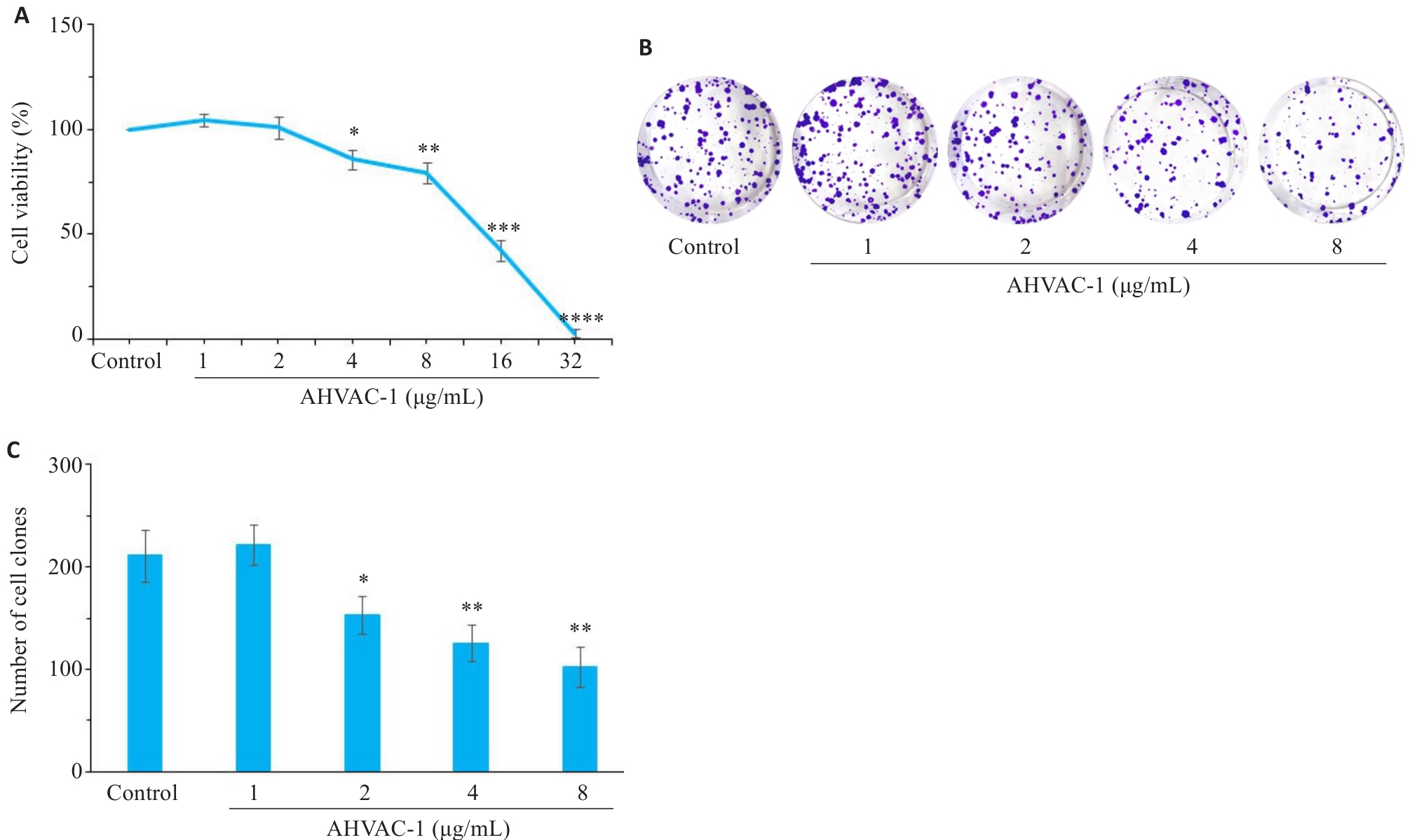
图1 CCK8检测不同浓度AHVAC-I作用MKN-28/DDP细胞72 h后细胞增殖率变化
Fig.1 Cytotoxicity of AHVAC-I in MKN-28/DDP cells after treatment for 72 h. A: Cells viability assessed by CCK8 assay. B: Colony formation assay of MKN-28/DDP cells at 3 weeks after AHVAC-I treatment. C: Quantitative analysis of colonys in each group. Con: Blank control. n=5, *P<0.05, **P<0.01, ***P<0.001, ****P<0.0001 vs Control.
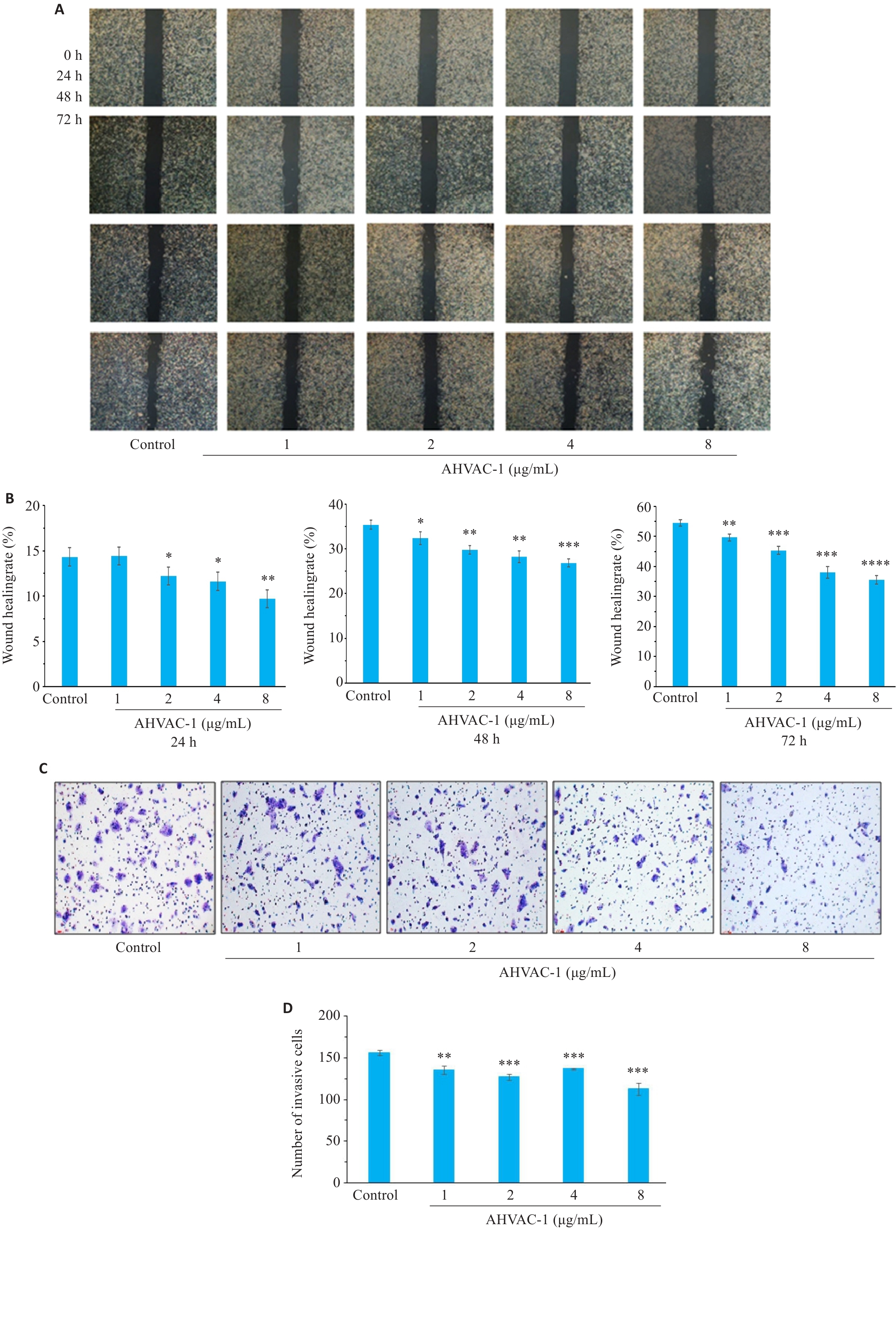
图2 AHVAC-I抑制MKN-28/DDP细胞迁移与侵袭
Fig.2 Inhibitory effects of AHVAC-I on migration and invasion of MKN-28/DDP cells. A: Wounding-healing assay for assessing migration ability of MKN-28/DDP cells (Original magnification:×40). B: Quantitative analysis of migration ability of MKN-28/DDP cells in each group. C: Transwell assay for assessing invasion ability of MKN-28/DDP cells (×100). D: Quantitative analysis of invading cells in each group, n=5, *P<0.05, **P<0.01, ***P<0.001, ****P<0.0001 vs Control.
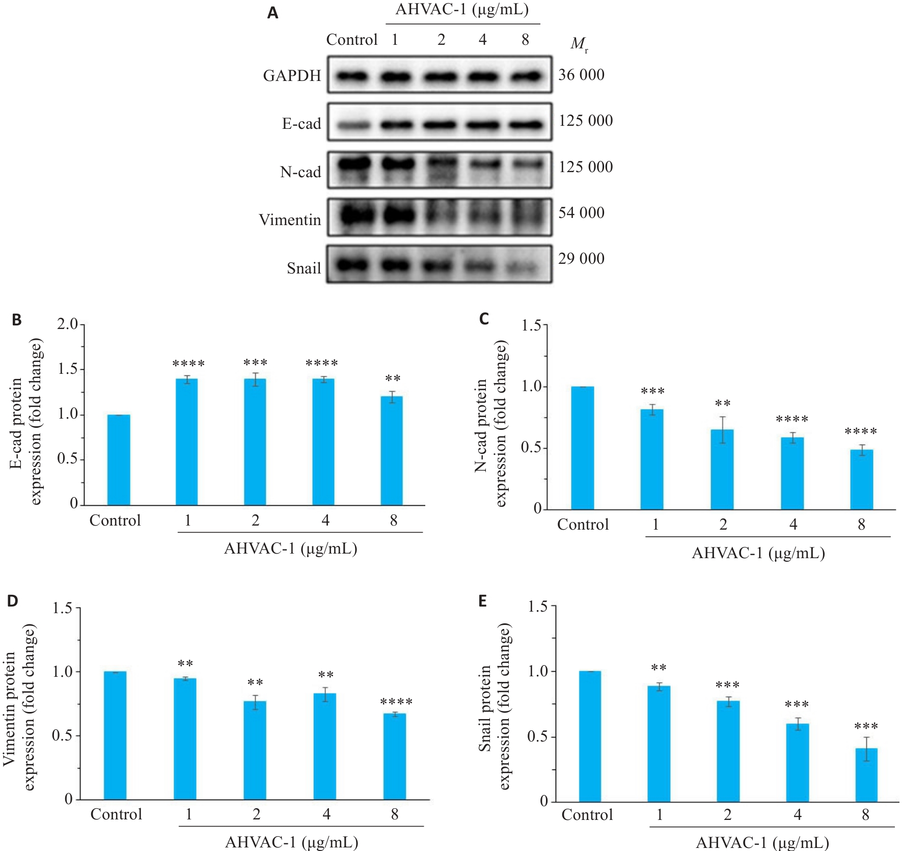
图3 AHVAC-I抑制MKN-28/DDP细胞上皮间质化
Fig.3 AHVAC-I inhibits epithelial-mesenchymal transition (EMT) of MKN-28/DDP cells. A: Immunoblots of E-cad, N-cad, vimentin and snail in MKN-28/DDP cells treated with different concentrations of AHVAC-I. B-E: Protein expression levels of E-cad, N-cad, vimentin and snail, n=3, **P<0.01, ***P<0.001, ****P<0.0001 vs Control.
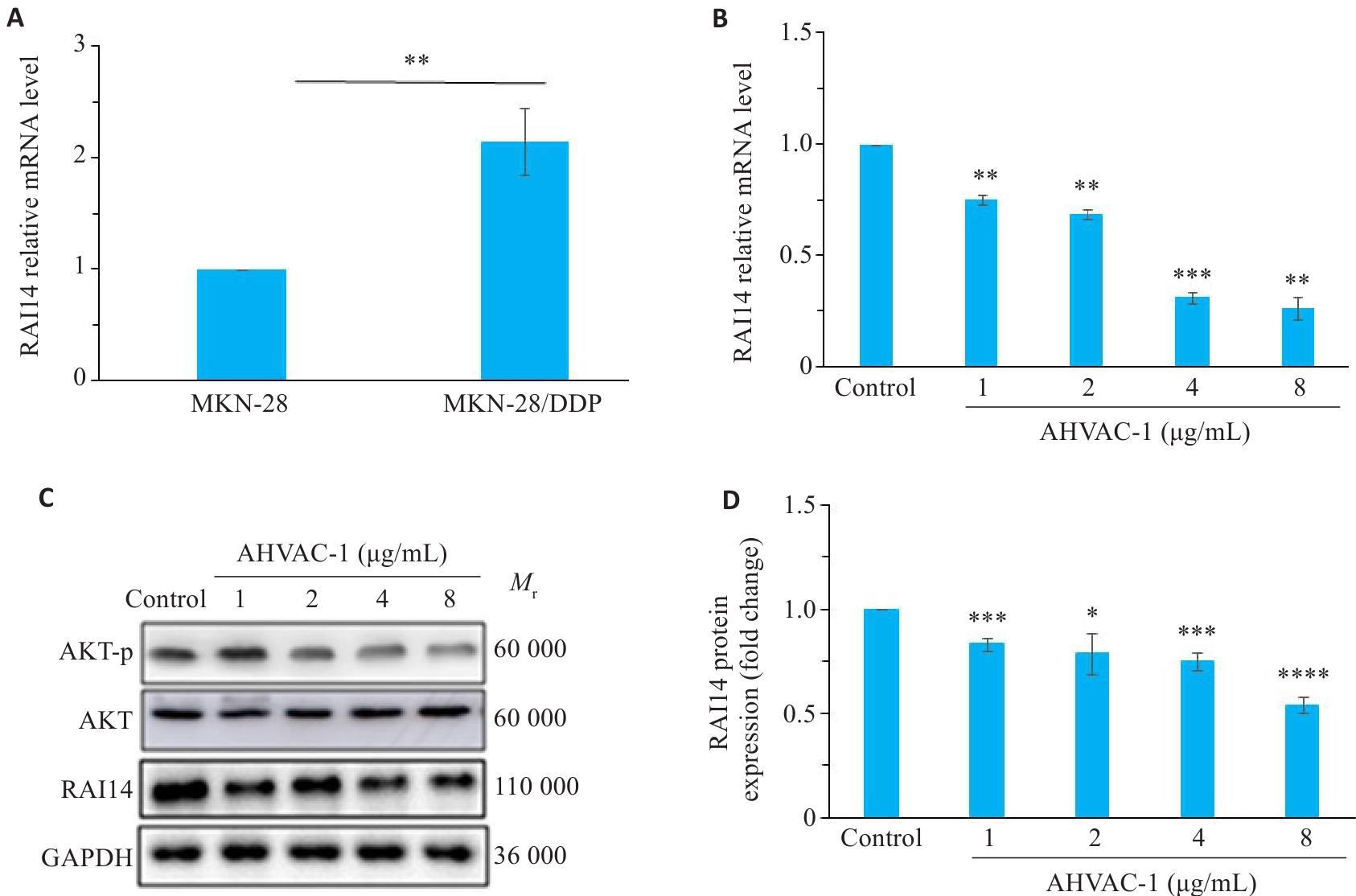
图4 AHVAC-I抑制MKN-28/DDP细胞RAI14表达
Fig.4 AHVAC-I down-regulates RAI14 expression in MKN-28/DDP cells. A: qRT-PCR for detecting RAI14 mRNA expression in MKN-28 cells and MKN-28/DDP cells (n=6, **P<0.01 vs MKN-28 cells). B: qRT-PCR of RAI14 expression in MKN-28/DDP cells treated with different concentrations of AHVAC-I (n=6, **P<0.01, ***P<0.001 vs blank Control). C: Immunoblots of RAI14, AKT and AKT-p in MKN-28/DDP cells treated with different concentrations of AHVAC-I. D: Protein expression levels of RAI14 in different groups (n=3). *P<0.05, **P<0.01, ***P<0.001, ****P<0.0001 vs Control.
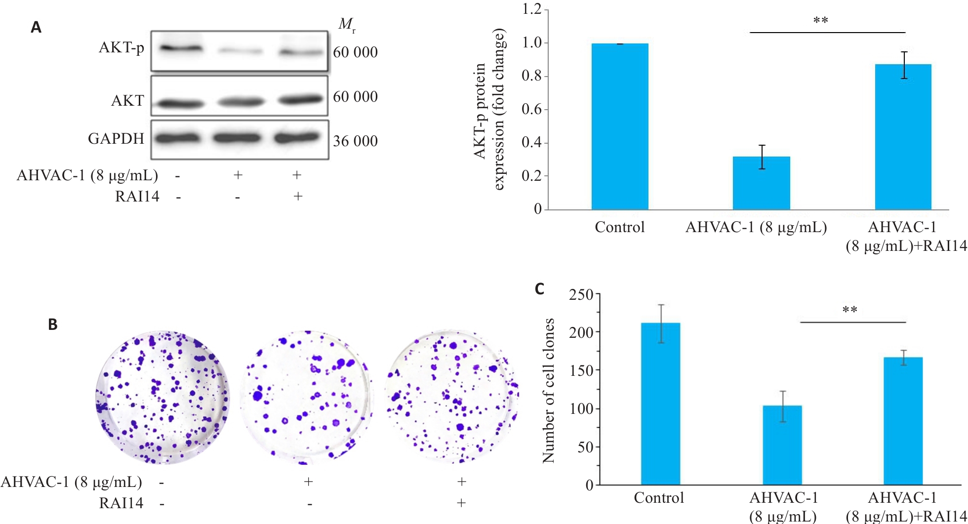
图5 增加RAI14可有效抑制AHVAC-I对 MKN-28/DDP细胞增殖能力的抑制效果
Fig.5 RAI14 supplementation promotes proliferation of AHVAC-I-treated MKN-20/DDP (MKN-20/DDPAHVAC-I) cells. A: Immunoblots of AKT and AKT-p in MKN-20/DDPAHVAC-I cells treated with RAI14 (n=3). B, C: Colony-forming assay showing restored proliferation ability of MKN-20/DDPAHVAC-I cells after RAI14 treatment (n=5). **P<0.01 vs AHVAC-1.
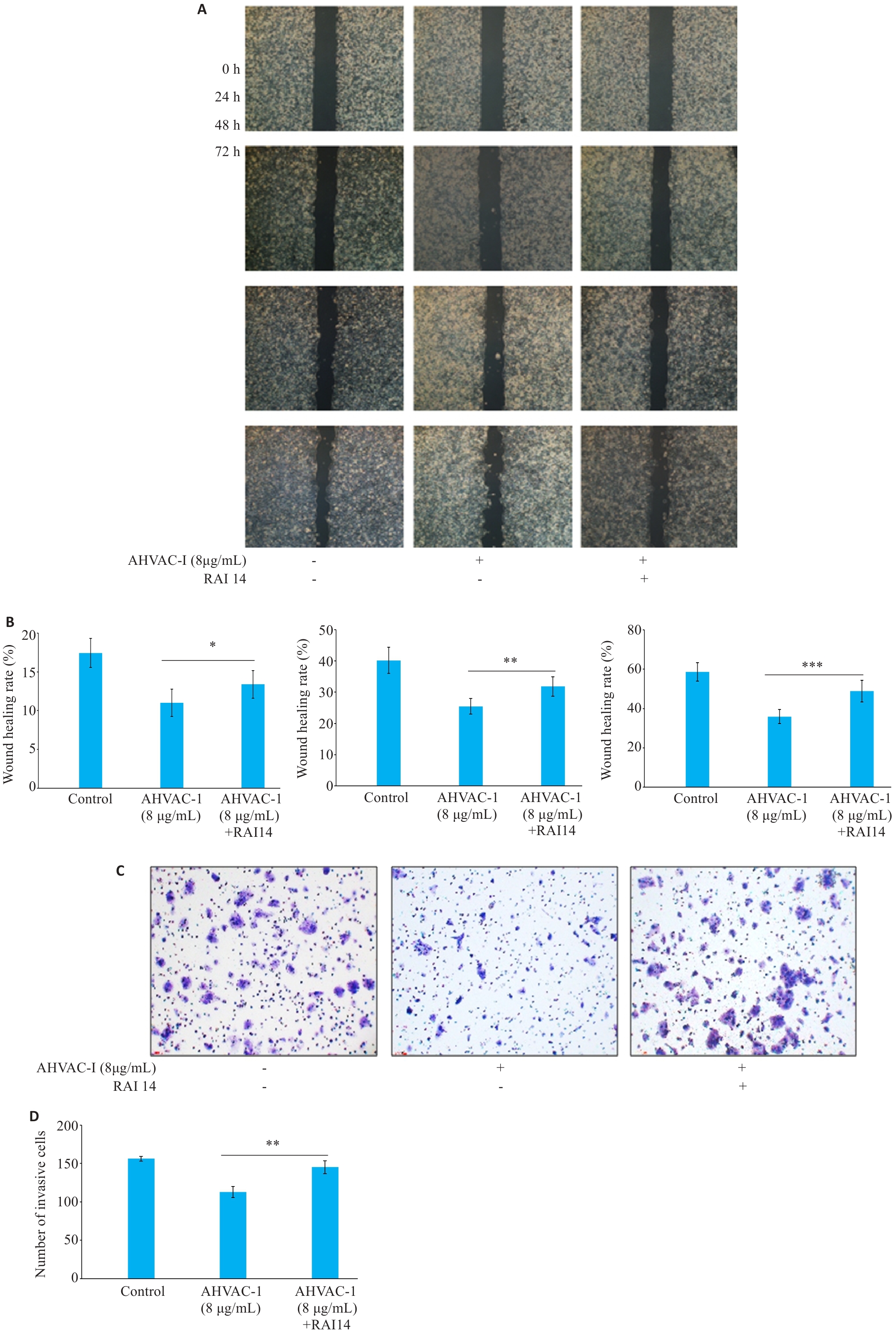
图6 增加RAI14可有效抑制AHVAC-I对 MKN-28/DDP细胞迁移、侵袭能力的抑制效果
Fig.6 RAI14 supplementation promotes migration and invasion ability of MKN-20/DDPAHVAC-I cells. A: Migration ability of MKN-28/DDP cells analyzed by wounding-healing assay (×40). B: Quantitative analysis of migration ability of MKN-28/DDP cells. C: Invasion ability of MKN-28/DDP cells was analyzed by Transwell assay (×100). D: Quantitative analysis of number of invaded cells.n=5, *P<0.05, **P<0.01, ***P<0.001 vs AHVAC-I.
| [1] | Bray F, Laversanne M, Sung H, et al. Global cancer statistics 2022: GLOBOCAN estimates of incidence and mortality worldwide for 36 cancers in 185 countries[J]. CA Cancer J Clin, 2024, 74(3): 229-263. doi:10.3322/caac.21834 |
| [2] | Van Cutsem E, Sagaert X, Topal B, et al. Gastric cancer[J]. Lancet, 2016, 388(10060): 2654-2664. doi:10.1016/s0140-6736(16)30354-3 |
| [3] | Cheng J, Cai M, Shuai XM, et al. First-line systemic therapy for advanced gastric cancer: a systematic review and network meta-analysis[J]. Ther Adv Med Oncol, 2019, 11: 1758835919877726. doi:10.1177/1758835919877726 |
| [4] | Lordick F, Lorenzen S, Yamada Y, et al. Optimal chemotherapy for advanced gastric cancer: is there a global consensus?[J]. Gastric Cancer, 2014, 17(2): 213-25. doi:10.1007/s10120-013-0297-z |
| [5] | Kulus M, Farzaneh M, Bryja A, et al. Phenotypic transitions the processes involved in regulation of growth and proangiogenic properties of stem cells, cancer stem cells and circulating tumor cells[J]. Stem Cell Rev Rep, 2024, 20(4): 967-79. doi:10.1007/s12015-024-10691-w |
| [6] | Wang J, Ma YZ, Guo M, et al. Salvianolic acid B suppresses EMT and apoptosis to lessen drug resistance through AKT/mTOR in gastric cancer cells[J]. Cytotechnology, 2021, 73(1): 49-61. doi:10.1007/s10616-020-00441-4 |
| [7] | Gu M, Zheng W, Zhang M, et al. Downregulation of RAI14 inhibits the proliferation and invasion of breast cancer cells[J]. J Cancer. 2019, 10(25): 6341- 8. doi:10.7150/jca.34910 |
| [8] | Zhang R, Hu M, Chen HN, et al. Phenotypic heterogeneity analysis of APC-mutant colon cancer by proteomics and phosphoproteomics identifies RAI14 as a key prognostic determinant in east asians and westerners[J]. Mol Cell Proteomics, 2023, 22(5): 100532. doi:10.1016/j.mcpro.2023.100532 |
| [9] | Gold BS, Dart RC, Barish RA. Bites of venomous snakes[J]. N Engl J Med, 2002, 347(5): 347-56. doi:10.1056/nejmra013477 |
| [10] | Sasovsky DJ, Angelina E, Leiva LC, et al. Comparative in vitro and in silico analysis of the ability of basic Asp49 phospholipase A2 and Lys49-phospholipase A2-like myotoxins from Bothrops diporus venom to inhibit the metastatic potential of murine mammary tumor cells and endothelial cell tubulogenesis: Asp49 vs Lys49 phospholipases A2: Inhibition of metastasis and angiogenesis[J]. Chem Biol Interact, 2024, 402: 111217. doi:10.1016/j.cbi.2024.111217 |
| [11] | Giordano G, Pancione M. MHC class III lymphocyte antigens 6 as endogenous immunotoxins: Unlocking immunotherapy in proficient mismatch repair colorectal cancer[J]. WIREs Mech Dis, 2024, 16(1): e1631. doi:10.1002/wsbm.1631 |
| [12] | Vyas VK, Brahmbhatt K, Bhatt H, et al. Therapeutic potential of snake venom in cancer therapy: current perspectives[J]. Asian Pac J Trop Biomed, 2013, 3(2): 156-62. doi:10.1016/s2221-1691(13)60042-8 |
| [13] | 王晓庆, 支 慧, 胡浩然, 等. 皖南蝮蛇抑瘤组分Ⅰ急性毒理研究[J]. 牡丹江医学院学报, 2022, 43(3): 44-8. |
| [14] | 黄小梅, 支 慧, 陈 浩, 等. 皖南蝮蛇毒抑瘤组分-Ⅰ对胃癌MKN-28细胞增殖、迁移及凋亡的影响[J]. 中国临床药理学与治疗学, 2024, 29(3): 270-6. |
| [15] | Zhang C, Li D, Yu R, et al. Immune landscape of gastric carcinoma tumor microenvironment identifies a peritoneal relapse relevant immune signature[J]. Front Immunol, 2021, 12: 651033. doi:10.3389/fimmu.2021.651033 |
| [16] | Kang YK, Chin K, Chung HC, et al. S-1 plus leucovorin and oxaliplatin versus S-1 plus cisplatin as first-line therapy in patients with advanced gastric cancer (SOLAR): a randomised, open-label, phase 3 trial[J]. Lancet Oncol, 2020, 21(8): 1045-56. doi:10.1016/s1470-2045(20)30315-6 |
| [17] | 皇甫娟, 张 强, 魏祯瑶, 等. 基于p53介导自噬通路探讨臭椿酮对顺铂耐药胃癌细胞株耐药性的影响[J]. 现代药物与临床, 2021, 36(6): 1112-8. |
| [18] | Hong YS, Ham YA, Choi JH, et al. Effects of allyl sulfur compounds and garlic extract on the expression of Bcl-2, Bax, and p53 in non small cell lung cancer cell lines[J]. Exp Mol Med, 2000, 32(3): 127-34. doi:10.1038/emm.2000.22 |
| [19] | 鲁 珏, 徐飞鹏. 白眉蝮蛇蛇毒细胞毒素对胃癌细胞的杀伤作用及对细胞超微结构的影响[J]. 细胞与分子免疫学杂志, 2009, 25(4): 335-7. |
| [20] | 鲁 珏, 徐飞鹏. 白眉蝮蛇蛇毒细胞毒素H1对实验性大鼠胃癌抑制作用的实验研究[J]. 第四军医大学学报, 2009(11): 975-7. |
| [21] | 韦 敏, 宋 慧, 班建东, 等. 神经生长因子的提取及其对MGC-803细胞的凋亡作用[J]. 广西医科大学学报, 2008, 25(4): 526-9. |
| [22] | Ebrahimi N, Manavi MS, Faghihkhorasani F, et al. Harnessing function of EMT in cancer drug resistance: a metastasis regulator determines chemotherapy response[J]. Cancer Metastasis Rev, 2024, 43(1): 457-79. doi:10.1007/s10555-023-10162-7 |
| [23] | Kutty RK, Kutty G, Samuel W, et al. Molecular characterization and developmental expression of NORPEG a novel gene induced by retinoic acid[J]. J Biol Chem, 2001, 276(4): 2831-40. doi:10.1074/jbc.m007421200 |
| [24] | Xiao Y, Zhang H, Du G, et al. RAI14 Is a Prognostic Biomarker and Correlated With Immune Cell Infiltrates in Gastric Cancer[J]. Technol Cancer Res Treat. 2020, 19: 1533033820970684. doi:10.1177/1533033820970684 |
| [25] | Hawkins SM, Loomans HA, Wan YW, et al. Expression and functional pathway analysis of nuclear receptor NR2F2 in ovarian cancer[J]. J Clin Endocrinol Metab, 2013, 98(7): E1152-62. doi:10.1210/jc.2013-1081 |
| [26] | Paez AV, Pallavicini C, Schuster F, et al. Heme oxygenase-1 in the forefront of a multi-molecular network that governs cell-cell contacts and filopodia-induced zippering in prostate cancer[J]. Cell Death Dis, 2016, 7(12): e2570. doi:10.1038/cddis.2016.420 |
| [27] | Yuan CZ, Hu H, Kuang MY, et al. Super enhancer associated RAI14 is a new potential biomarker in lung adenocarcinoma[J]. Oncotarget, 2017, 8(62): 105251-61. doi:10.18632/oncotarget.22165 |
| [28] | Wang JL, Cai Y, Luo JD, et al. RAI14 silencing suppresses progression of esophageal cancer via the STAT3 pathway[J]. Aging (Albany NY), 2020, 12(18): 18084-98. doi:10.18632/aging.103613 |
| [29] | Xu J, Shi PF, Xia FW, et al. RAI14 promotes melanoma progression by regulating the FBXO32/c-MYC pathway[J]. Int J Mol Sci, 2022, 23(19): 12036. doi:10.3390/ijms231912036 |
| [30] | Zhou J, Yong WP, Yap CS, et al. An integrative approach identified genes associated with drug response in gastric cancer[J]. Carcinogenesis, 2015, 36(4): 441-51. doi:10.1093/carcin/bgv014 |
| [31] | O’Donnell KA, Keng VW, York B, et al. A Sleeping Beauty mutagenesis screen reveals a tumor suppressor role for Ncoa2/Src-2 in liver cancer[J]. Proc Natl Acad Sci USA, 2012, 109(21): E1377-86. doi:10.1073/pnas.1115433109 |
| [32] | Ono K, Demchak B, Ideker T. Cytoscape tools for the web age: D3.js and cytoscape.js exporters[J]. F1000Res, 2014, 3: 143. doi:10.12688/f1000research.4510.2 |
| [33] | Tang Y, Li M, Wang JX, et al. CytoNCA: a cytoscape plugin for centrality analysis and evaluation of protein interaction networks[J]. Biosystems, 2015, 127: 67-72. doi:10.1016/j.biosystems.2014.11.005 |
| [34] | Cui RL, Zou J, Zhao Y, et al. The dual-crosslinked prospective values of RAI14 for the diagnosis and chemosurveillance in triple negative breast cancer[J]. Ann Med, 2023, 55(1): 820-36. doi:10.1080/07853890.2023.2177722 |
| [1] | 赵锦燕, 彭娇, 林明和, 朱晓勤, 黄彬, 林久茂. 清解扶正颗粒通过抑制线粒体依赖的凋亡、激活AMPK-PGC-1α通路缓解5-氟尿嘧啶引起的骨骼肌损伤[J]. 南方医科大学学报, 2026, 46(1): 94-103. |
| [2] | 杨子为, 吕畅, 董柱, 计书磊, 毕生辉, 张雪花, 王晓武. 金樱子通过调控Src-AKT1轴抑制肺动脉高压平滑肌增殖[J]. 南方医科大学学报, 2025, 45(9): 1889-1902. |
| [3] | 王子良, 陈孝华, 杨晶晶, 严晨, 张志郅, 黄炳轶, 赵萌, 刘嵩, 葛思堂, 左芦根, 陈德利. 高表达SURF4通过抑制紧密连接蛋白表达促进胃癌细胞的恶性生物学行为[J]. 南方医科大学学报, 2025, 45(8): 1732-1742. |
| [4] | 陈鑫源, 吴成挺, 李瑞迪, 潘雪芹, 张耀丹, 陶俊宇, 林才志. 双术汤通过P53/SLC7A11/GPX4通路诱导胃癌细胞铁死亡[J]. 南方医科大学学报, 2025, 45(7): 1363-1371. |
| [5] | 谢婷, 王云云, 郭婷, 袁春华. 雷氏大疣蛛多肽毒素组分通过激活促凋亡通路和协同作用抑制癌细胞增殖[J]. 南方医科大学学报, 2025, 45(7): 1460-1470. |
| [6] | 龚秀莹, 侯顺福, 赵苗苗, 王晓娜, 张致涵, 刘清华, 尹崇高, 李洪利. LncRNA SNHG15通过miR-30b-3p调控COX6B1轴促进肺腺癌细胞增殖、迁移和侵袭的分子机制[J]. 南方医科大学学报, 2025, 45(7): 1498-1505. |
| [7] | 吴璇, 方家敏, 韩玮玮, 陈琳, 孙菁, 金齐力. 高表达PRELID1促进胃癌细胞上皮间质转化并与不良预后相关[J]. 南方医科大学学报, 2025, 45(7): 1535-1542. |
| [8] | 李嘉豪, 冼瑞婷, 李荣. 下调ACADM介导的脂毒性抑制雌激素受体阳性乳腺癌细胞的侵袭与转移[J]. 南方医科大学学报, 2025, 45(6): 1163-1173. |
| [9] | 侯鑫睿, 张振东, 曹明远, 杜予心, 王小平. 红景天苷靶向miR-1343-3p-OGDHL/PDHB糖代谢轴抑制胃癌细胞的体内外增殖[J]. 南方医科大学学报, 2025, 45(6): 1226-1239. |
| [10] | 曾玉梅, 李继科, 黄仲曦, 周毅波. 绒毛样蛋白VILL通过与LMO7蛋白相互作用抑制鼻咽癌细胞的增殖[J]. 南方医科大学学报, 2025, 45(5): 954-961. |
| [11] | 岳雅清, 牟召霞, 王希波, 刘艳. Aurora-A过表达通过激活NF-κBp65/ARPC4信号轴促进宫颈癌细胞的侵袭和转移[J]. 南方医科大学学报, 2025, 45(4): 837-843. |
| [12] | 张毅, 沈昱, 万志强, 陶嵩, 柳亚魁, 王栓虎. CDKN3高表达促进胃癌细胞的迁移和侵袭:基于调控p53/NF-κB信号通路和抑制胃癌细胞凋亡[J]. 南方医科大学学报, 2025, 45(4): 853-861. |
| [13] | 庆顺杰, 沈智勇. 过表达己糖激酶2通过激活JAK/STAT途径促进结直肠癌细胞的增殖、迁移和侵袭并调节肿瘤免疫微环境[J]. 南方医科大学学报, 2025, 45(3): 542-553. |
| [14] | 黄晴晴, 张文静, 张小凤, 王炼, 宋雪, 耿志军, 左芦根, 王月月, 李静, 胡建国. 高表达MYO1B促进胃癌细胞增殖、迁移和侵袭并与患者的不良预后有关[J]. 南方医科大学学报, 2025, 45(3): 622-631. |
| [15] | 宋雪, 陈悦, 张敏, 张诺, 左芦根, 李静, 耿志军, 张小凤, 王月月, 王炼, 胡建国. GPSM2在胃癌组织中高表达并通过促进肿瘤细胞的增殖影响患者预后[J]. 南方医科大学学报, 2025, 45(2): 229-238. |
| 阅读次数 | ||||||
|
全文 |
|
|||||
|
摘要 |
|
|||||