Journal of Southern Medical University ›› 2024, Vol. 44 ›› Issue (8): 1497-1507.doi: 10.12122/j.issn.1673-4254.2024.08.08
Previous Articles Next Articles
Yidan PANG1( ), Ya LIU1, Siai CHEN1, Jinglei ZHANG1, Jin ZENG1, Yuanming PAN2(), Juan AN1()
Received:2024-05-07
Online:2024-08-20
Published:2024-09-06
Contact:
Yuanming PAN, Juan AN
E-mail:15546261023@163.com;peter.f.pan@hsc.pku.edu.cn;anjuan@qhu.edu.cn
Supported by:Yidan PANG, Ya LIU, Siai CHEN, Jinglei ZHANG, Jin ZENG, Yuanming PAN, Juan AN. Biological role of SPAG5 in the malignant proliferation of gastric cancer cells[J]. Journal of Southern Medical University, 2024, 44(8): 1497-1507.
Add to citation manager EndNote|Ris|BibTeX
URL: https://www.j-smu.com/EN/10.12122/j.issn.1673-4254.2024.08.08
| Gene | Upstream primer sequences | Downstream primer sequences | Length (bp) |
|---|---|---|---|
| GAPDH | TGACTTCAACAGCGACACCCA | CACCCTGTTGCTGTAGCCAAA | 121 |
| SPAG5 | TTGAGGCCCGTTTAGATACCA | GCTTTCCTTGGAGCAATGTAGTT | 227 |
Tab.1 Primer sequences for RT-qPCR
| Gene | Upstream primer sequences | Downstream primer sequences | Length (bp) |
|---|---|---|---|
| GAPDH | TGACTTCAACAGCGACACCCA | CACCCTGTTGCTGTAGCCAAA | 121 |
| SPAG5 | TTGAGGCCCGTTTAGATACCA | GCTTTCCTTGGAGCAATGTAGTT | 227 |
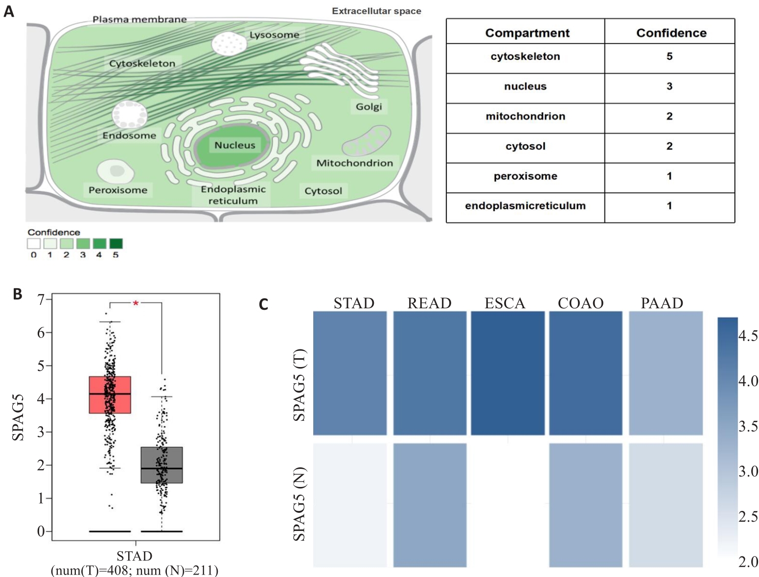
Fig.1 Bioinformatic analysis of SPAG5 expression patterns. A:Proteinatlas database analysis showing SPAG5 distribution in both the cell nucleus and cytoplasm. B: TCGA database analysis showing significantly higher expression of SPAG5 in gastric cancer than in normal tissues (*P<0.05). C: Proteinatlas database analysis showing SPAG5 expressions in gastric, rectal adenocarcinoma, esophageal, colon adenocarcinoma, and pancreatic cancer.
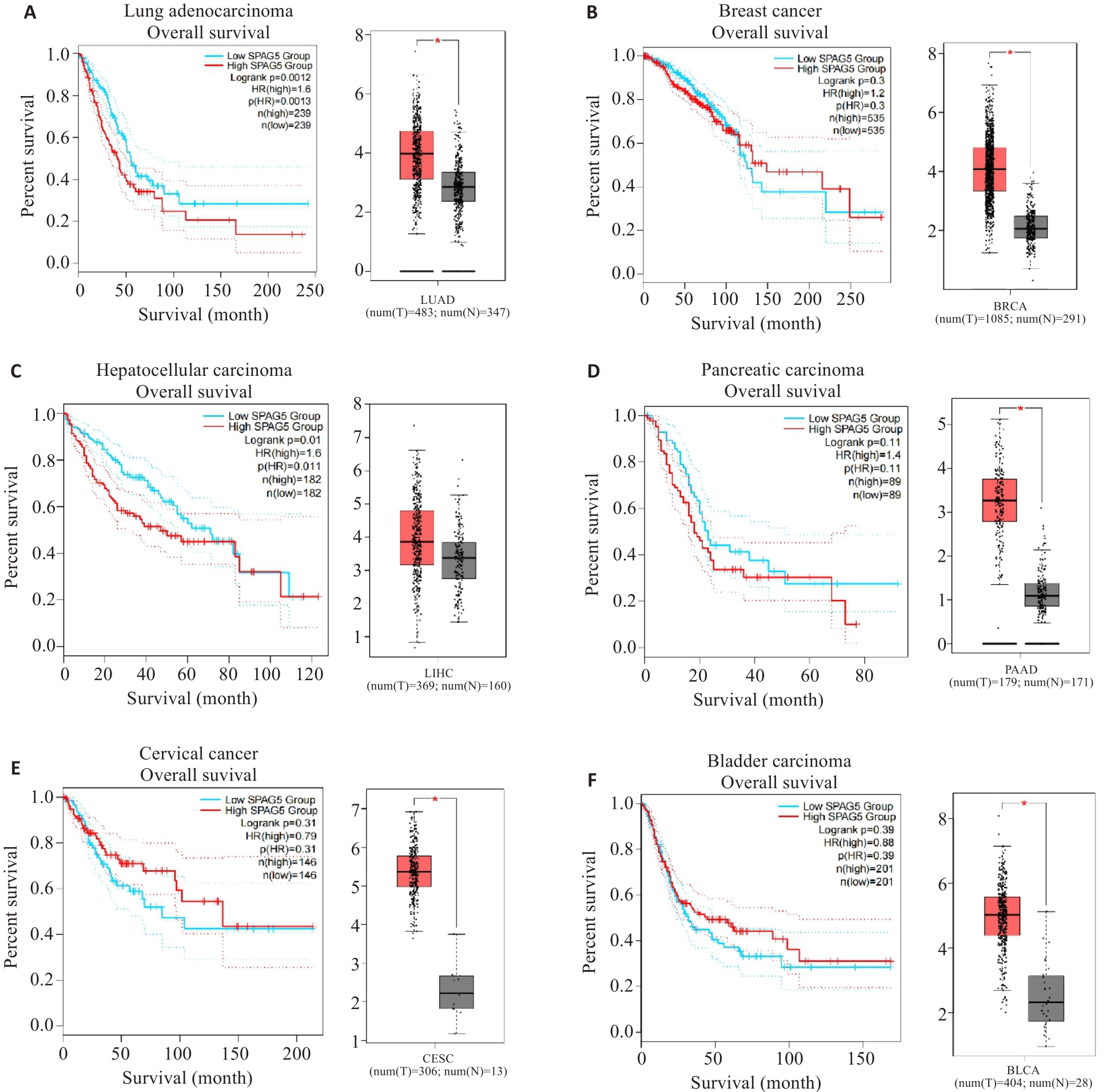
Fig.2 Kaplan-Meier plots and GEPIA database analysis of SPAG5 expression in lung adenocarcinoma (A), breast cancer (B), hepatocellular carcinoma (C), pancreatic cancer (D), cervical cancer (E), and bladder cancer (F) and its association with patient survival (*P<0.05).
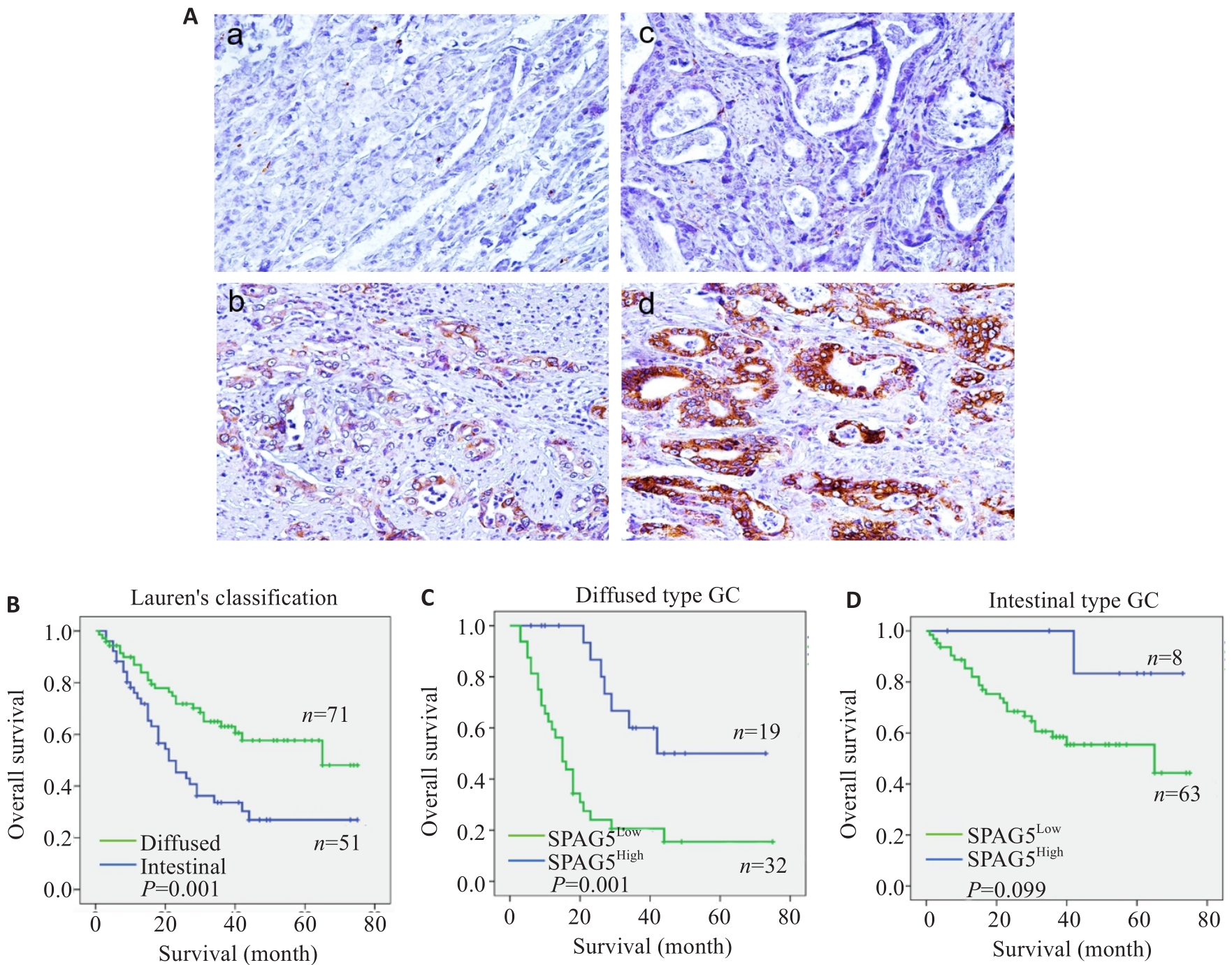
Fig.3 Immunohistochemistry for SPAG5 in gastric and adjacent tissues and survival analysis of SPAG5 expression. A: Immunohistochemistry for detecting expression of SPAG5 in gastric cancer and adjacent tissues (Original magnification: ×100). a: Normal gastric mucosal tissue Negative b: Normal gastric mucosal tissue Positive c: Gastric cancer tissue Negative d: Gastric cancer tissue Positive. B-D: Kaplan-Meier survival analysis of gastric cancer patients with high and low SPAG5 expression using Log-rank test and Breslow test.
| Type | Cases | SPAG5 expression [n (%)] | P | |
|---|---|---|---|---|
| High | Low | |||
| Nomal | 50 | 11 (22.0) | 39 (78.0) | <0.01 |
| Tumor | 122 | 95 (78.0) | 27 (22.0) | |
Tab.2-1 Expression of SPAG5 in gastric cancer and adjacent tissues
| Type | Cases | SPAG5 expression [n (%)] | P | |
|---|---|---|---|---|
| High | Low | |||
| Nomal | 50 | 11 (22.0) | 39 (78.0) | <0.01 |
| Tumor | 122 | 95 (78.0) | 27 (22.0) | |
| Type | Cases | MKi67 expression [n (%)] | P | |
|---|---|---|---|---|
| High | Low | |||
| Nomal | 50 | 27 (54.0) | 23 (46.0) | <0.01 |
| Tumor | 122 | 75 (61.5) | 47 (38.5) | |
Tab.2-2 Expression of MKi67 in gastric cancer and adjacent tissues
| Type | Cases | MKi67 expression [n (%)] | P | |
|---|---|---|---|---|
| High | Low | |||
| Nomal | 50 | 27 (54.0) | 23 (46.0) | <0.01 |
| Tumor | 122 | 75 (61.5) | 47 (38.5) | |
| Characteristics | Nomal-SPAG5 expression | Characteristics | Tumor-SPAG5 expression | ||
|---|---|---|---|---|---|
| Low | High | Low | High | ||
| Nomal-MKi67[High] | 17 | 10 | Tumor-MKi67[High] | 6 | 109 |
| Nomal-MKi67[Low] | 22 | 1 | Tumor-MKi67[Low] | 27 | 30 |
| P | 0.006 | P | <0.01 | ||
| Pearson's R | 0.393 | Pearson's R | 0.504 | ||
Tab.3 Analysis of the correlation between SPAG5 and MKi67 expressions in gastric cancer tissues
| Characteristics | Nomal-SPAG5 expression | Characteristics | Tumor-SPAG5 expression | ||
|---|---|---|---|---|---|
| Low | High | Low | High | ||
| Nomal-MKi67[High] | 17 | 10 | Tumor-MKi67[High] | 6 | 109 |
| Nomal-MKi67[Low] | 22 | 1 | Tumor-MKi67[Low] | 27 | 30 |
| P | 0.006 | P | <0.01 | ||
| Pearson's R | 0.393 | Pearson's R | 0.504 | ||
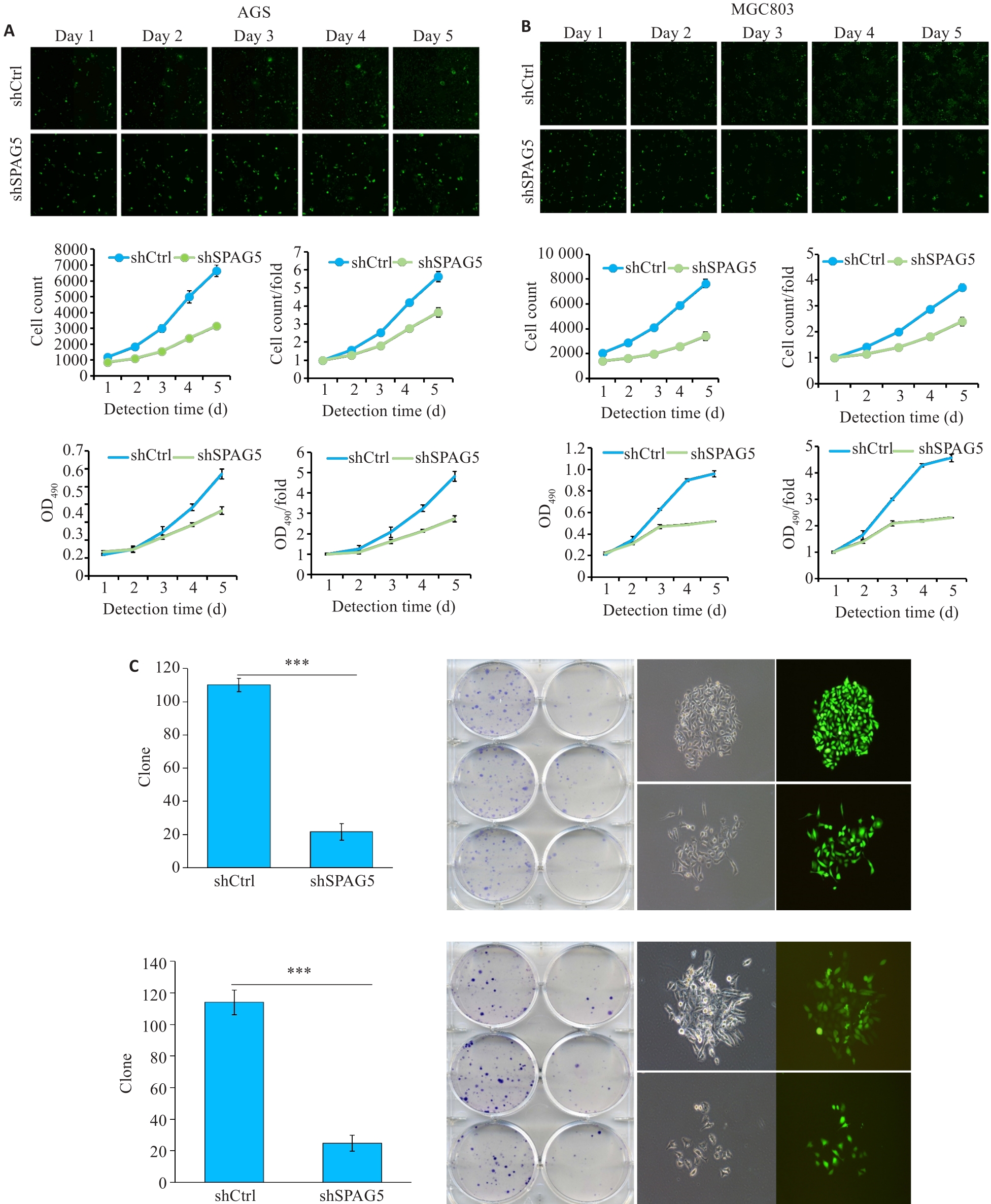
Fig.7 Lentivirus-mediated SPAG5 interference inhibits proliferation of AGS and MGC803 cells. A, B: Celigo assay and MTT assays showing inhibition of proliferation of AGS and MGC-803 cells 3 days after lentivirus infection in the experimental group was significantly inhibited. C: Number of clones formed by AGS and MGC-803 cells with SPAG5 knockdown (×100). ***P<0.001.
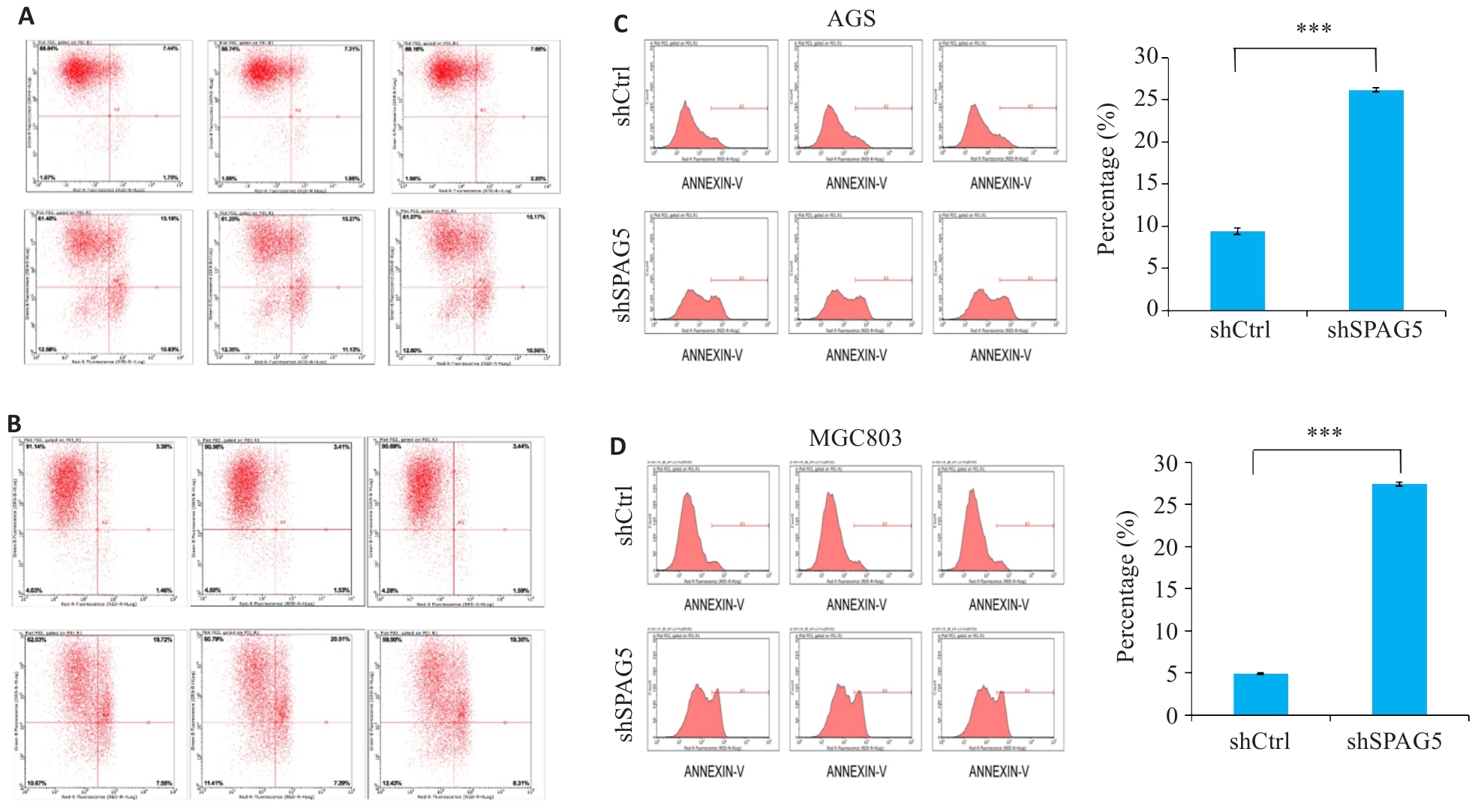
Fig.8 SPAG5 interference promotes apoptosis in AGS and MGC803 cells. A, B: Scatter plot of flow cytometry after SPAG5 interference in AGS cells and MGC803 cells. C, D: Apoptosis rates in AGS cells and MGC803 cells 5 days after SPAG5 interference (***P<0.001).
| 1 | Chen WQ, Zheng RS, Baade PD, et al. Cancer statistics in China, 2015[J]. CA A Cancer J Clin, 2016, 66(2): 115-32. |
| 2 | Smyth EC, Nilsson M, Grabsch HI, et al. Gastric cancer[J]. Lancet, 2020, 396(10251): 635-48. |
| 3 | Guan WL, He Y, Xu RH. Gastric cancer treatment: recent progress and future perspectives[J]. J Hematol Oncol, 2023, 16(1): 57. |
| 4 | Zeng XY, Xu WB, Tong JJ, et al. SPAG5 as a novel biomarker and potential therapeutic target via regulating AKT pathway in multiple myeloma[J]. Leuk Lymphoma, 2022, 63(11): 2565-72. |
| 5 | Xiao G, Xu X, Chen ZB, et al. SPAG5 expression predicts poor prognosis and is associated with adverse immune infiltration in lung adenocarcinomas[J]. Clin Med Insights Oncol, 2023, 17: 11795549231199915. |
| 6 | Gao XF, Bu HT, Gao XZ, et al. Pan-cancer analysis: SPAG5 is an immunological and prognostic biomarker for multiple cancers[J]. FASEB J, 2023, 37(10): e23159. |
| 7 | 葛柯乐, 方 成, 燕海娇, 等. 胃癌组织中SPAG5的表达及意义[J]. 山东医药, 2020, 60(10): 55-9. |
| 8 | Liu GD, Liu S, Cao GY, et al. SPAG5 contributes to the progression of gastric cancer by upregulation of Survivin depend on activating the Wnt/β-catenin pathway[J]. Exp Cell Res, 2019, 379(1): 83-91. |
| 9 | Chang MS, Huang CJ, Chen ML, et al. Cloning and characterization of hMAP126, a new member of mitotic spindle-associated proteins[J]. Biochem Biophys Res Commun, 2001, 287(1): 116-21. |
| 10 | Yang YC, Hsu YT, Wu CC, et al. Silencing of astrin induces the p53-dependent apoptosis by suppression of HPV18 E6 expression and sensitizes cells to paclitaxel treatment in HeLa cells[J]. Biochem Biophys Res Commun, 2006, 343(2): 428-34. |
| 11 | Kodani A, Yu TW, Johnson JR, et al. Centriolar satellites assemble centrosomal microcephaly proteins to recruit CDK2 and promote centriole duplication[J]. Elife, 2015, 4: e07519. |
| 12 | Gruber J, Harborth J, Schnabel J, et al. The mitotic-spindle-associated protein astrin is essential for progression through mitosis[J]. J Cell Sci, 2002, 115(Pt 21): 4053-9. |
| 13 | Chen W, Chen X, Li SJ, et al. Expression, immune infiltration and clinical significance of SPAG5 in hepatocellular carcinoma: a gene expression-based study[J]. J Gene Med, 2020, 22(4): e3155. |
| 14 | Wang CH, Su HY, Cheng R, et al. SPAG5 is involved in human gliomagenesis through the regulation of cell proliferation and apoptosis[J]. Front Oncol, 2021, 11: 673780. |
| 15 | He J, Green AR, Li Y, et al. SPAG5: an emerging oncogene[J]. Trends Cancer, 2020, 6(7): 543-7. |
| 16 | Liu P, Li H, Liao CF, et al. Identification of key genes and biological pathways in Chinese lung cancer population using bioinformatics analysis[J]. PeerJ, 2022, 10: e12731. |
| 17 | Xu J, Deng YJ, Wang Y, et al. SPAG5-AS1 inhibited autophagy and aggravated apoptosis of podocytes via SPAG5/AKT/mTOR pathway[J]. Cell Prolif, 2020, 53(2): e12738. |
| 18 | Liu HL, Hu JW, Wei R, et al. SPAG5 promotes hepatocellular carcinoma progression by downregulating SCARA5 through modifying β-catenin degradation[J]. J Exp Clin Cancer Res, 2018, 37(1): 229. |
| 19 | Li M, Li AQ, Zhou SL, et al. SPAG5 upregulation contributes to enhanced c-MYC transcriptional activity via interaction with c-MYC binding protein in triple-negative breast cancer[J]. J Hematol Oncol, 2019, 12(1): 14. |
| 20 | Li YW, Diao YC, Wang ZX, et al. The splicing factor SF3B4 drives proliferation and invasion in cervical cancer by regulating SPAG5[J]. Cell Death Discov, 2022, 8(1): 326. |
| 21 | Dang L, Shi CP, Zhang QQ, et al. Downregulation of sperm-associated antigen 5 inhibits melanoma progression by regulating forkhead box protein M1/a disintegrin and metalloproteinase 17/NOTCH1 signaling[J]. Bioengineered, 2022, 13(3): 4744-56. |
| 22 | Buechler S. Low expression of a few genes indicates good prognosis in estrogen receptor positive breast cancer[J]. BMC Cancer, 2009, 9: 243. |
| 23 | Abdel-Fatah TMA, Agarwal D, Liu DX, et al. SPAG5 as a prognostic biomarker and chemotherapy sensitivity predictor in breast cancer: a retrospective, integrated genomic, transcriptomic, and protein analysis[J]. Lancet Oncol, 2016, 17(7): 1004-18. |
| 24 | Du Q, Xiao RD, Luo RG, et al. Construction of long non-coding RNA- and microRNA-mediated competing endogenous RNA networks in alcohol-related esophageal cancer[J]. PLoS One, 2022, 17(6): e0269742. |
| 25 | Huang RJ, Li AL. SPAG5 is associated with unfavorable prognosis in patients with lung adenocarcinoma and promotes proliferation, motility and autophagy in A549 cells[J]. Exp Ther Med, 2020, 20(5): 77. |
| 26 | Li HY, Qin YF, Huang YR, et al. SPAG5, the upstream protein of Wnt and the target of curcumin, inhibits hepatocellular carcinoma[J]. Oncol Rep, 2023, 50(3): 172. |
| 27 | Zhang WW, Xiao YH, Zhou Q, et al. KNSTRN is a prognostic biomarker that is correlated with immune infiltration in breast cancer and promotes cell cycle and proliferation[J]. Biochem Genet, 2024: Online ahead of print.. |
| 28 | An J, Yang L, Pan YM, et al. SPAG5 activates PI3K/AKT pathway and promotes the tumor progression and chemo-resistance in gastric cancer[J]. DNA Cell Biol, 2022, 41(10): 893-902. |
| 29 | Hashmi AA, Hashmi KA, Irfan M, et al. Ki67 index in intrinsic breast cancer subtypes and its association with prognostic parameters[J]. BMC Res Notes, 2019, 12(1): 605. |
| 30 | Uxa S, Castillo-Binder P, Kohler R, et al. Ki-67 gene expression[J]. Cell Death Differ, 2021, 28(12): 3357-70. |
| 31 | Luo ZW, Zhu MG, Zhang ZQ, et al. Increased expression of Ki-67 is a poor prognostic marker for colorectal cancer patients: a meta analysis[J]. BMC Cancer, 2019, 19(1): 123. |
| 32 | Faragalla H, Plotkin A, Barnes P, et al. Ki67 in breast cancer assay: an ad hoc testing recommendation from the Canadian association of pathologists task force[J]. Curr Oncol, 2023, 30(3): 3079-90. |
| 33 | Gerdes J, Li L, Schlueter C, et al. Immunobiochemical and molecular biologic characterization of the cell proliferation-associated nuclear antigen that is defined by monoclonal antibody Ki-67[J]. Am J Pathol, 1991, 138(4): 867-73. |
| 34 | van Ree JH, Jeganathan KB, Malureanu L, et al. Overexpression of the E2 ubiquitin-conjugating enzyme UbcH10 causes chromosome missegregation and tumor formation[J]. J Cell Biol, 2010, 188(1): 83-100. |
| 35 | Chen Z, Zhang CP, Wu DY, et al. Phospho-MED1-enhanced UBE2C locus looping drives castration-resistant prostate cancer growth[J]. EMBO J, 2011, 30(12): 2405-19. |
| 36 | Ramos-Santillan V, Oshi M, Nelson E, et al. High Ki67 gene expression is associated with aggressive phenotype in hepatocellular carcinoma[J]. World J Oncol, 2024, 15(2): 257-67. |
| 37 | Li B, Severson E, Pignon JC, et al. Comprehensive analyses of tumor immunity: implications for cancer immunotherapy[J]. Genome Biol, 2016, 17(1): 174. |
| [1] | Xinyuan CHEN, Chengting WU, Ruidi LI, Xueqin PAN, Yaodan ZHANG, Junyu TAO, Caizhi LIN. Shuangshu Decoction inhibits growth of gastric cancer cell xenografts by promoting cell ferroptosis via the P53/SLC7A11/GPX4 axis [J]. Journal of Southern Medical University, 2025, 45(7): 1363-1371. |
| [2] | Xuan WU, Jiamin FANG, Weiwei HAN, Lin CHEN, Jing SUN, Qili JIN. High PRELID1 expression promotes epithelial-mesenchymal transition in gastric cancer cells and is associated with poor prognosis [J]. Journal of Southern Medical University, 2025, 45(7): 1535-1542. |
| [3] | Xinrui HOU, Zhendong ZHANG, Mingyuan CAO, Yuxin DU, Xiaoping WANG. Salidroside inhibits proliferation of gastric cancer cells by regulating the miR-1343-3p-OGDHL/PDHB glucose metabolic axis [J]. Journal of Southern Medical University, 2025, 45(6): 1226-1239. |
| [4] | Yumei ZENG, Jike LI, Zhongxi HUANG, Yibo ZHOU. Villin-like protein VILL suppresses proliferation of nasopharyngeal carcinoma cells by interacting with LMO7 protein [J]. Journal of Southern Medical University, 2025, 45(5): 954-961. |
| [5] | Yi ZHANG, Yu SHEN, Zhiqiang WAN, Song TAO, Yakui LIU, Shuanhu WANG. High expression of CDKN3 promotes migration and invasion of gastric cancer cells by regulating the p53/NF-κB signaling pathway and inhibiting cell apoptosis [J]. Journal of Southern Medical University, 2025, 45(4): 853-861. |
| [6] | Qingqing HUANG, Wenjing ZHANG, Xiaofeng ZHANG, Lian WANG, Xue SONG, Zhijun GENG, Lugen ZUO, Yueyue WANG, Jing LI, Jianguo HU. High MYO1B expression promotes proliferation, migration and invasion of gastric cancer cells and is associated with poor patient prognosis [J]. Journal of Southern Medical University, 2025, 45(3): 622-631. |
| [7] | Xue SONG, Yue CHEN, Min ZHANG, Nuo ZHANG, Lugen ZUO, Jing LI, Zhijun GENG, Xiaofeng ZHANG, Yueyue WANG, Lian WANG, Jianguo HU. GPSM2 is highly expressed in gastric cancer to affect patient prognosis by promoting tumor cell proliferation [J]. Journal of Southern Medical University, 2025, 45(2): 229-238. |
| [8] | Xiaohua CHEN, Hui LU, Ziliang WANG, Lian WANG, Yongsheng XIA, Zhijun GENG, Xiaofeng ZHANG, Xue SONG, Yueyue WANG, Jing LI, Jianguo HU, Lugen ZUO. Role of Abelson interactor 2 in progression and prognosis of gastric cancer and its regulatory mechanisms [J]. Journal of Southern Medical University, 2024, 44(9): 1653-1661. |
| [9] | Liangjun XUE, Qiuyu TAN, Jingwen XU, Lu FENG, Wenjin LI, Liang YAN, Yulei LI. MiR-6838-5p overexpression inhibits proliferation of breast cancer MCF-7 cells by downregulating DDR1 expression [J]. Journal of Southern Medical University, 2024, 44(9): 1677-1684. |
| [10] | Mengnan YE, Hongmei WU, Yan MEI, Qingling ZHANG. High expression of CREM is associated with poor prognosis in gastric cancer patients [J]. Journal of Southern Medical University, 2024, 44(9): 1776-1782. |
| [11] | Zhijun GENG, Jingjing YANG, Minzhu NIU, Xinyue LIU, Jinran SHI, Yike LIU, Xinyu YAO, Yulu ZHANG, Xiaofeng ZHANG, Jianguo HU. Kuwanon G inhibits growth, migration and invasion of gastric cancer cells by regulating the PI3K/AKT/mTOR pathway [J]. Journal of Southern Medical University, 2024, 44(8): 1476-1484. |
| [12] | Yongsheng XIA, Lian WANG, Xiaohua CHEN, Yulu ZHANG, Aofei SUN, Deli CHEN. TSR2 overexpression inhibits proliferation and invasion of gastric cancer cells by downregulating the PI3K/AKT signaling pathway [J]. Journal of Southern Medical University, 2024, 44(5): 913-919. |
| [13] | LIANG Yihao, LAI Yingjun, YUAN Yanwen, YUAN Wei, ZHANG Xibo, ZHANG Bashan, LU Zhifeng. Screening of differentially expressed genes in gastric cancer based on GEO database and function and pathway enrichment analysis [J]. Journal of Southern Medical University, 2024, 44(3): 605-616. |
| [14] | SHEN Mengdi, ZHAO Na, DENG Xiaojing, DENG Min. High expression of COX6B2 in gastric cancer is associated with poor long-term prognosis and promotes cell proliferation and cell cycle progression by inhibiting p53 signaling [J]. Journal of Southern Medical University, 2024, 44(2): 289-297. |
| [15] | ZHANG Nuo, ZHANG Zhen, ZHANG Yulu, SONG Xue, ZHANG Xiaofeng, LI Jing, ZUO Lugen, HU Jianguo. PCID2 is highly expressed in gastric cancer and affects the prognosis by regulating cancer cell cycle and proliferation [J]. Journal of Southern Medical University, 2024, 44(2): 324-332. |
| Viewed | ||||||
|
Full text |
|
|||||
|
Abstract |
|
|||||