Journal of Southern Medical University ›› 2025, Vol. 45 ›› Issue (1): 10-17.doi: 10.12122/j.issn.1673-4254.2025.01.02
Previous Articles Next Articles
Shenyao ZHANG1,3( ), Min LU1,2(), Gaoyan KUANG1,2, Xiaotong XU1,2, Jun FU1, Churan ZENG1
Received:2024-08-21
Online:2025-01-20
Published:2025-01-20
Contact:
Min LU
E-mail:dirkzhang8627@hnucm.edu.cn;lumin6563@163.com
Shenyao ZHANG, Min LU, Gaoyan KUANG, Xiaotong XU, Jun FU, Churan ZENG. HDAC1 overexpression inhibits steroid-induced apoptosis of mouse osteocyte-like MLO-Y4 cells by inducing SP1 deacetylation[J]. Journal of Southern Medical University, 2025, 45(1): 10-17.
Add to citation manager EndNote|Ris|BibTeX
URL: https://www.j-smu.com/EN/10.12122/j.issn.1673-4254.2025.01.02
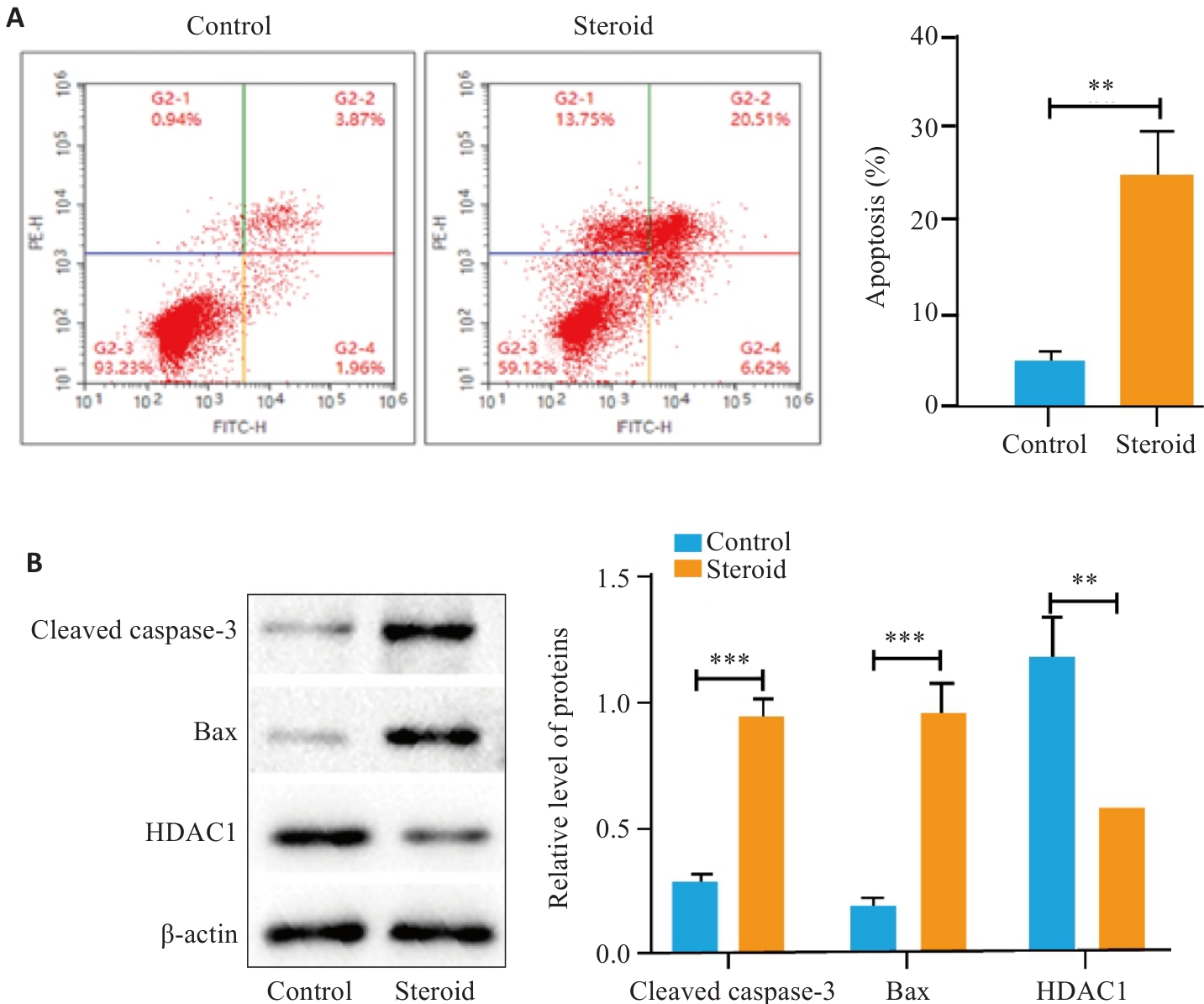
Fig.1 Cell apoptosis and HDAC1 expression in control and dexamethasone-treated mouse osteocyte-like MLO-Y4 cells. A: Cell apoptosis detected by flow cytometry. B: Expression levels of cleaved caspase-3, Bax and HDAC1 in the cells detected by Western blotting. **P<0.01, ***P<0.001.
| Group | Treatment time (h) | F P | |||
|---|---|---|---|---|---|
| 24 | 48 | 72 | |||
| Control | 0.59±0.07 | 1.12±0.09 | 1.25±0.09 | 52.89 | 0.009 |
| Steroid | 0.55±0.09 | 0.76±0.08 | 0.95±0.09 | 27.32 | 0.028 |
| t | 0.501 | 4.299 | 3.308 | ||
| P | 0.643 | 0.013 | 0.03 | ||
Tab.1 A450 nm in MLO-Y4 cells in CCK-8 assay 24, 48 and 72 h after dexamethasone treatment (n=3, Mean±SD)
| Group | Treatment time (h) | F P | |||
|---|---|---|---|---|---|
| 24 | 48 | 72 | |||
| Control | 0.59±0.07 | 1.12±0.09 | 1.25±0.09 | 52.89 | 0.009 |
| Steroid | 0.55±0.09 | 0.76±0.08 | 0.95±0.09 | 27.32 | 0.028 |
| t | 0.501 | 4.299 | 3.308 | ||
| P | 0.643 | 0.013 | 0.03 | ||
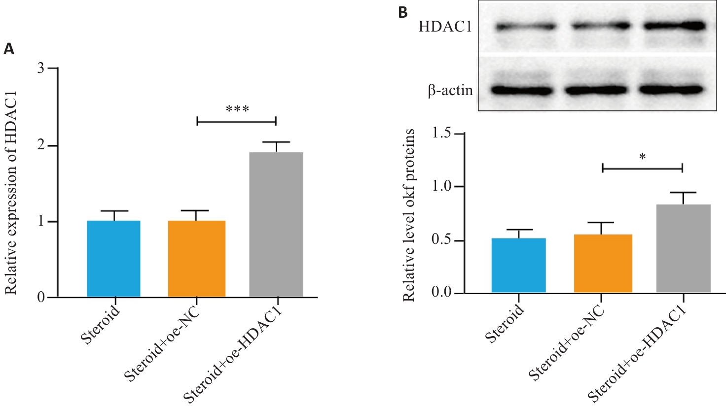
Fig.2 Identification of transfection efficiency of HDAC1 overexpression vector. A: qPCR for HDAC1 expression; B: western blot for HDAC1 protein. *P < 0.05, ***P<0.001.
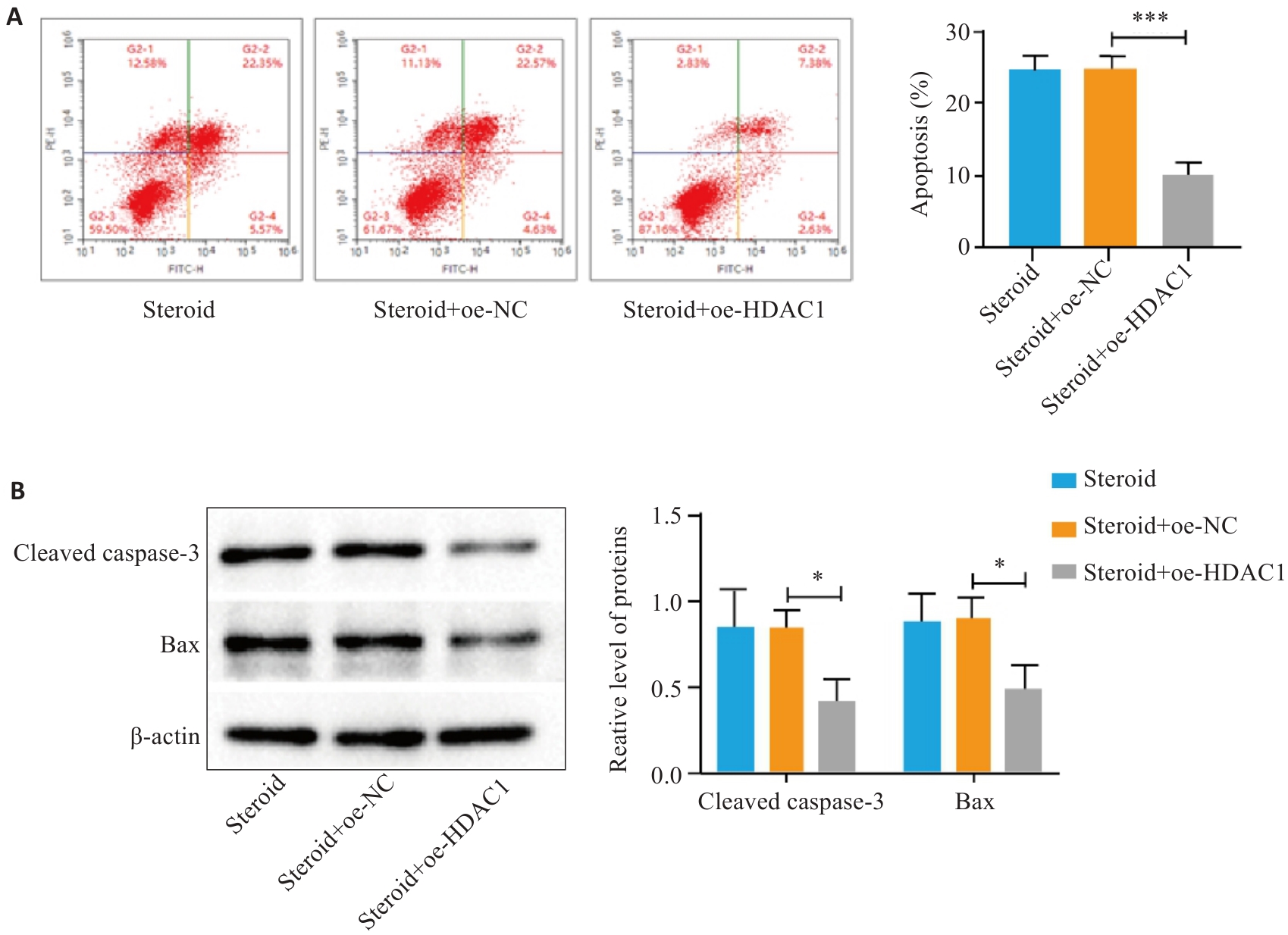
Fig.3 Up-regulation of HDAC1 inhibits apoptosis of dexamethasone-treated MLO-Y4 cells. A: Cell apoptosis detected by flow cytometry. B: Expressions of cleaved caspase-3 and Bax detected by Western blotting. *P<0.05, ***P<0.001.
| Group | Treatment time (h) | F | P | ||
|---|---|---|---|---|---|
| 24 | 48 | 72 | |||
| Steroid | 0.56±0.07 | 0.79±0.09 | 0.96±0.11 | 47.72 | 0.016 |
| Steroid+oe-NC | 0.45±0.04 | 0.80±0.09 | 0.97±0.12 | 15.12 | 0.048 |
| Steroid+oe-HDAC1 | 0.59±0.08* | 1.15±0.11* | 1.32±0.10* | 30.63 | 0.031 |
| F | 2.177 | 8.92 | 7.279 | ||
| P | 0.195 | 0.016 | 0.025 | ||
Tab.2 A450 nm at 24, 48 and 72 h after the transfection of HDAC1 overexpressing vector
| Group | Treatment time (h) | F | P | ||
|---|---|---|---|---|---|
| 24 | 48 | 72 | |||
| Steroid | 0.56±0.07 | 0.79±0.09 | 0.96±0.11 | 47.72 | 0.016 |
| Steroid+oe-NC | 0.45±0.04 | 0.80±0.09 | 0.97±0.12 | 15.12 | 0.048 |
| Steroid+oe-HDAC1 | 0.59±0.08* | 1.15±0.11* | 1.32±0.10* | 30.63 | 0.031 |
| F | 2.177 | 8.92 | 7.279 | ||
| P | 0.195 | 0.016 | 0.025 | ||
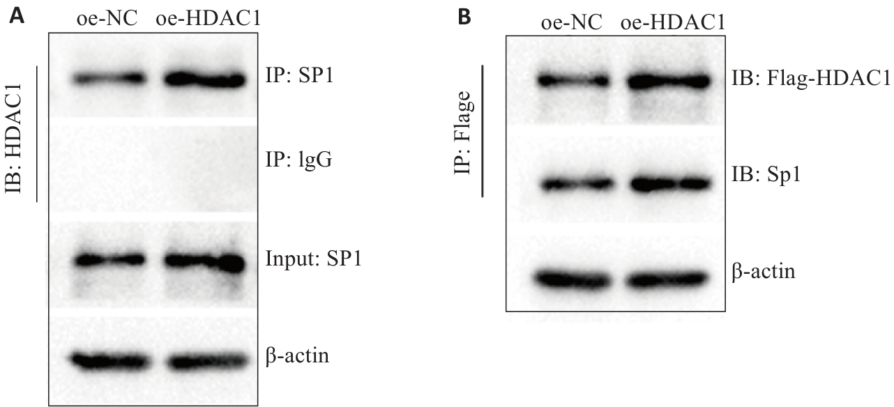
Fig.4 Identification of the interaction between HDAC1 and SP1 by immunoprecipitation.
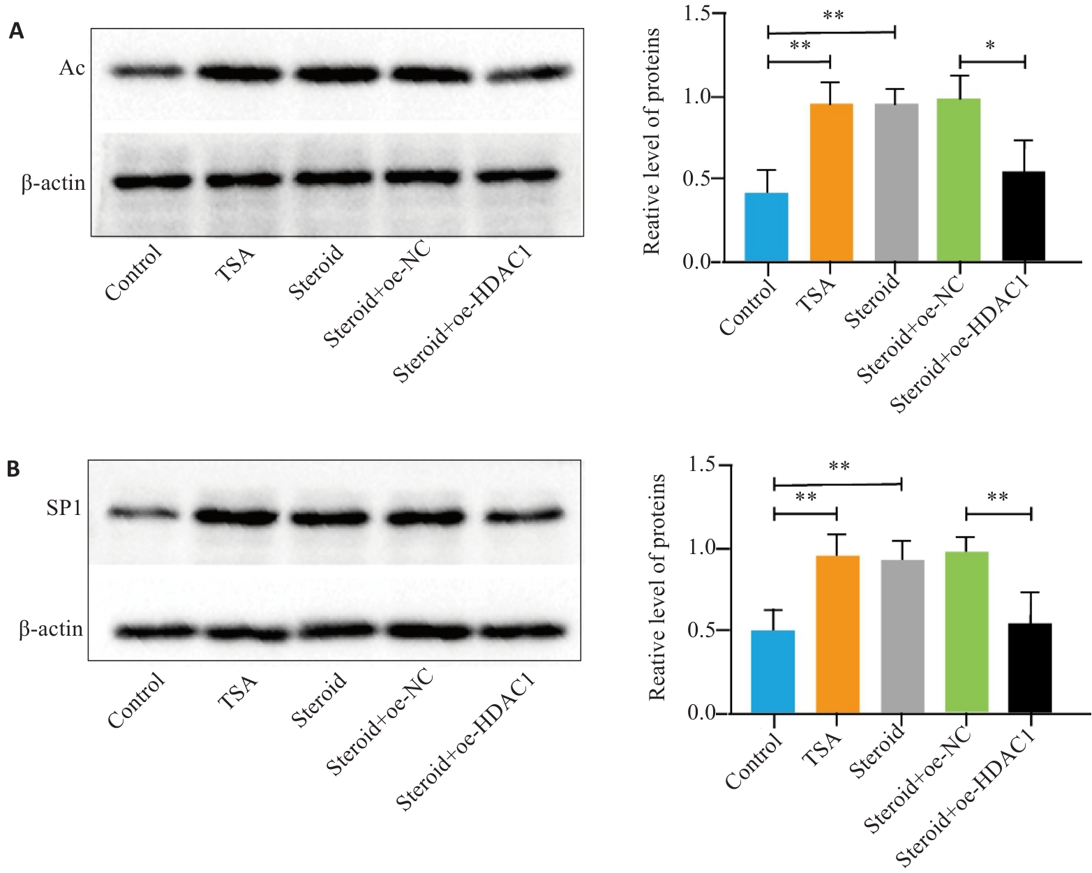
Fig.5 Regulation of protein acetylation levels by the HDAC1 axis. A: Cellular acetylation levels detected by immunoprecipitation assay and Western blotting. B: SP1 protein detected by Western blotting. *P<0.05, **P<0.01.
| 1 | Chang C, Greenspan A, Gershwin ME. The pathogenesis, diagnosis and clinical manifestations of steroid-induced osteonecrosis[J]. J Autoimmun, 2020, 110: 102460. |
| 2 | Motta F, Timilsina S, Gershwin ME, et al. Steroid-induced osteonecrosis[J]. J Transl Autoimmun, 2022, 5: 100168. |
| 3 | Huang C, Wen ZQ, Niu JJ, et al. Steroid-induced osteonecrosis of the femoral head: novel insight into the roles of bone endothelial cells in pathogenesis and treatment[J]. Front Cell Dev Biol, 2021, 9: 777697. |
| 4 | Zhang SQ, Wang CB, Shi L, et al. Beware of steroid-induced avascular necrosis of the femoral head in the treatment of COVID-19-experience and lessons from the SARS epidemic[J]. Drug Des Devel Ther, 2021, 15: 983-95. |
| 5 | Kimura K, Jackson TLB, Huang RCC. Interaction and collaboration of SP1, HIF-1, and MYC in regulating the expression of cancer-related genes to further enhance anticancer drug development[J]. Curr Issues Mol Biol, 2023, 45(11): 9262-83. |
| 6 | Yu X, Rong PZ, Song MS, et al. lncRNA SNHG1 induced by SP1 regulates bone remodeling and angiogenesis via sponging miR-181c-5p and modulating SFRP1/Wnt signaling pathway[J]. Mol Med, 2021, 27(1): 141. |
| 7 | 卢 敏, 许晓彤, 申 楠, 等. 黄芪多糖对激素性股骨头坏死模型骨细胞凋亡和SP1/MEK/ERK轴的影响[J]. 中国现代医学杂志, 2023, 33(12): 33-40. |
| 8 | Parveen R, Harihar D, Chatterji BP. Recent histone deacetylase inhibitors in cancer therapy[J]. Cancer, 2023, 129(21): 3372-80. |
| 9 | Banerjee A, Mahata B, Dhir A, et al. Elevated histone H3 acetylation and loss of the Sp1-HDAC1 complex de-repress the GM2-synthase gene in renal cell carcinoma[J]. J Biol Chem, 2019, 294(3): 1005-18. |
| 10 | Duan P, Wang HY, Yi X, et al. C/EBPα regulates the fate of bone marrow mesenchymal stem cells and steroid-induced avascular necrosis of the femoral head by targeting the PPARγ signalling pathway[J]. Stem Cell Res Ther, 2022, 13(1): 342. |
| 11 | Al-Omari AA, Aleshawi AJ, Marei OA, et al. Avascular necrosis of the femoral head after single steroid intra-articular injection[J]. Eur J Orthop Surg Traumatol, 2020, 30(2): 193-7. |
| 12 | Fan ZQ, Bai SC, Xu Q, et al. Oxidative stress induced osteocyte apoptosis in steroid-induced femoral head necrosis[J]. Orthop Surg, 2021, 13(7): 2145-52. |
| 13 | Li GQ, Wang ZY. MiR-20b promotes osteocyte apoptosis in rats with steroid-induced necrosis of the femoral head through BMP signaling pathway[J]. Eur Rev Med Pharmacol Sci, 2019, 23(11): 4599-608. |
| 14 | 李昊然, 高 璐, 迟湘胤, 等. 糖皮质激素性骨质疏松症的药物作用机制研究进展[J]. 中国骨质疏松杂志, 2021, 27(6): 910-3, 921. |
| 15 | Xu K, Lu C, Ren XY, et al. Overexpression of HIF-1α enhances the protective effect of mitophagy on steroid-induced osteocytes apoptosis[J]. Environ Toxicol, 2021, 36(11): 2123-37. |
| 16 | Jiang JF, Zhou ZY, Liu YZ, et al. Role of Sp1 in atherosclerosis[J]. Mol Biol Rep, 2022, 49(10): 9893-902. |
| 17 | Safe S. Specificity proteins (sp) and cancer[J]. Int J Mol Sci, 2023, 24(6): 5164. |
| 18 | Cai Y, Zhang MM, Wang M, et al. Bone marrow-derived mesenchymal stem cell-derived exosomes containing Gli1 alleviate microglial activation and neuronal apoptosis in vitro and in a mouse parkinson disease model by direct inhibition of Sp1 signaling[J]. J Neuropathol Exp Neurol, 2022, 81(7): 522-34. |
| 19 | Zhang P, Li YN, Tu S, et al. SP1-induced lncRNA TUG1 regulates proliferation and apoptosis in islet cells of type 2 diabetes mellitus via the miR-188-3p/FGF5 axis[J]. Eur Rev Med Pharmacol Sci, 2021, 25(4): 1959-66. |
| 20 | Guo L, Fang L, Liu Y. SP1-regulated LINC01638 promotes proliferation and inhibits apoptosis in non-small cell lung cancer[J]. Eur Rev Med Pharmacol Sci, 2019, 23(20): 8913-20. |
| 21 | Liu D, Wang YQ, Pan ZY, et al. cAMP regulates 11β-hydroxysteroid dehydrogenase-2 and Sp1 expression in MLO-Y4/MC3T3-E1 cells[J]. Exp Ther Med, 2020, 20(3): 2166-72. |
| 22 | Zhang SY, Dong KF, Zeng XJ, et al. Astragalus polysaccharide ameliorates steroid-induced osteonecrosis of the femoral head by regulating miR-200b-3p-mediated Wnt/β-catenin signaling pathway via inhibiting SP1 expression: Astragalus polysaccharide regulates SONFH via SP1[J]. BMC Musculoskelet Disord, 2023, 24(1): 369. |
| 23 | Dunaway LS, Pollock JS. HDAC1: an environmental sensor regulating endothelial function[J]. Cardiovasc Res, 2022, 118(8): 1885-903. |
| 24 | Wang YF, Wang HY. The emerging role of histone deacetylase 1 in allergic diseases[J]. Front Immunol, 2022, 13: 1027403. |
| 25 | Jain R, Epstein JA. Epigenetics[J]. Adv Exp Med Biol, 2024, 1441: 341-64. |
| 26 | Sun ZH, Ma YF, Liu Y, et al. The acetylation modification of SP1 regulates the protein stability in silkworm[J]. Appl Biochem Biotechnol, 2022, 194(4): 1621-35. |
| 27 | Yang WB, Hsu CC, Hsu TI, et al. Increased activation of HDAC1/2/6 and Sp1 underlies therapeutic resistance and tumor growth in glioblastoma[J]. Neuro Oncol, 2020, 22(10): 1439-51. |
| 28 | Ran XH, Zhu JW, Ni RZ, et al. TRIM5α recruits HDAC1 to p50 and Sp1 and promotes H3K9 deacetylation at the HIV-1 LTR[J]. Nat Commun, 2023, 14(1): 3343. |
| 29 | Qiu Y, Zhao YM, Becker M, et al. HDAC1 acetylation is linked to progressive modulation of steroid receptor-induced gene transcription[J]. Mol Cell, 2006, 22(5): 669-79. |
| 30 | Kuzmochka C, Abdou HS, Haché RJG, et al. Inactivation of histone deacetylase 1 (HDAC1) but not HDAC2 is required for the gluco-corticoid-dependent CCAAT/enhancer-binding protein α (C/EBPα) expression and preadipocyte differentiation[J]. Endocrinology, 2014, 155(12): 4762-73. |
| 31 | Wen YX, Shi HS, Wu ZX, et al. GR/Sp3/HDAC1/UGDH signaling participated in the maternal dexamethasone-induced dysplasia of the rat fetal growth plate[J]. FASEB J, 2020, 34(9): 12834-46. |
| Viewed | ||||||
|
Full text |
|
|||||
|
Abstract |
|
|||||