
Journal of Southern Medical University ›› 2026, Vol. 46 ›› Issue (10): 2325-2337.doi: 10.12122/j.issn.1673-4254.2026.10.04
Qianqian REN1( ), Baowen ZHANG1, Xingran LIU1,2, Weili DING3,4, Dandan CHEN1,5, Xianjuan KOU1()
Received:2025-03-16
Online:2026-10-20
Published:2026-10-26
Contact:
Xianjuan KOU
E-mail:906371632@qq.com;kouxianjuan@126.com
Qianqian REN, Baowen ZHANG, Xingran LIU, Weili DING, Dandan CHEN, Xianjuan KOU. 40 Hz flicker light stimulation suppresses STING signaling to improve cognitive dysfunction in mice with Parkinson disease[J]. Journal of Southern Medical University, 2026, 46(10): 2325-2337.
Add to citation manager EndNote|Ris|BibTeX
URL: https://www.j-smu.com/EN/10.12122/j.issn.1673-4254.2026.10.04
| Gene | Forward primer (5'-3') | Reverse primer (5'-3') |
|---|---|---|
| BDNF | TCATACTTCGGTTGCATGAAGG | AGACCTCTCGAACCTGCCC |
| SYN | AGCTCAACAAATCCCAGTCTCT | CGGATGGTCTCAGCTTTCAC |
| PSD93 | ATCATTCCTTACCTCGGCTAACT | TCAGAGGAGAGATGTGAGACTG |
| PSD95 | TGAGATCAGTCATAGCAGCTACT | CTTCCTCCCCTAGCAGGTCC |
| C1q | TGGAGACCTCTCAGGGATGG | CTTTCACGCCCTTCAGTCCT |
| Arg-1 | CTCCAAGCCAAAGTCCTTAGAG | AGGAGCTGTCATTAGGGACATC |
| C3 | TAGAGGCCCATGATGCTCAG | AAGTTTGCCACCACTGTCAC |
| S100A10 | TGGAAACCATGATGCTTACGTT | GAAGCCCACTTTGCCATCTC |
| IL-6 | TAGTCCTTCCTACCCCAATTTCC | TTGGTCCTTAGCCACTCCTTC |
| TNF-ɑ | GACGTGGAACTGGCAGAAGAG | TTGGTGGTTTGTGAGTGTGAG |
| GAPDH | GGTTGTCTCCTGCGACTTCA | TGGTCCAGGGTTTCTTACTCC |
Tab.1 Primer sequence for RT-qPCR
| Gene | Forward primer (5'-3') | Reverse primer (5'-3') |
|---|---|---|
| BDNF | TCATACTTCGGTTGCATGAAGG | AGACCTCTCGAACCTGCCC |
| SYN | AGCTCAACAAATCCCAGTCTCT | CGGATGGTCTCAGCTTTCAC |
| PSD93 | ATCATTCCTTACCTCGGCTAACT | TCAGAGGAGAGATGTGAGACTG |
| PSD95 | TGAGATCAGTCATAGCAGCTACT | CTTCCTCCCCTAGCAGGTCC |
| C1q | TGGAGACCTCTCAGGGATGG | CTTTCACGCCCTTCAGTCCT |
| Arg-1 | CTCCAAGCCAAAGTCCTTAGAG | AGGAGCTGTCATTAGGGACATC |
| C3 | TAGAGGCCCATGATGCTCAG | AAGTTTGCCACCACTGTCAC |
| S100A10 | TGGAAACCATGATGCTTACGTT | GAAGCCCACTTTGCCATCTC |
| IL-6 | TAGTCCTTCCTACCCCAATTTCC | TTGGTCCTTAGCCACTCCTTC |
| TNF-ɑ | GACGTGGAACTGGCAGAAGAG | TTGGTGGTTTGTGAGTGTGAG |
| GAPDH | GGTTGTCTCCTGCGACTTCA | TGGTCCAGGGTTTCTTACTCC |
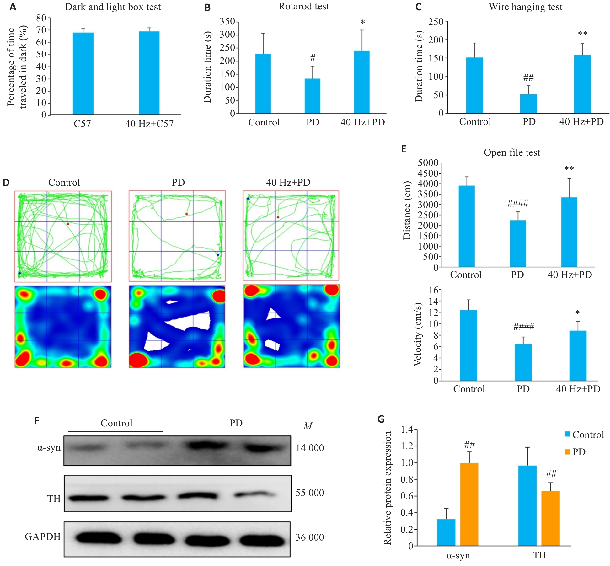
Fig.1 Modeling of Parkinson disease (PD) mice and motor function test results. A: Dark box residence time in the black-and-white box test. B: Duration time in the rotating rod test. C: Duration time in the hanging test. D: Pathway map and heat map of open field test. E: Total distance and movement speed in the open field test. F: Western blotting detection results. G: Expression levels of α-syn and TH proteins. Data are presented as Mean±SD and analyzed using one-way ANOVA. #P<0.05, ##P<0.01, ####P<0.0001 vs Control. *P<0.05, **P<0.01 vs PD.
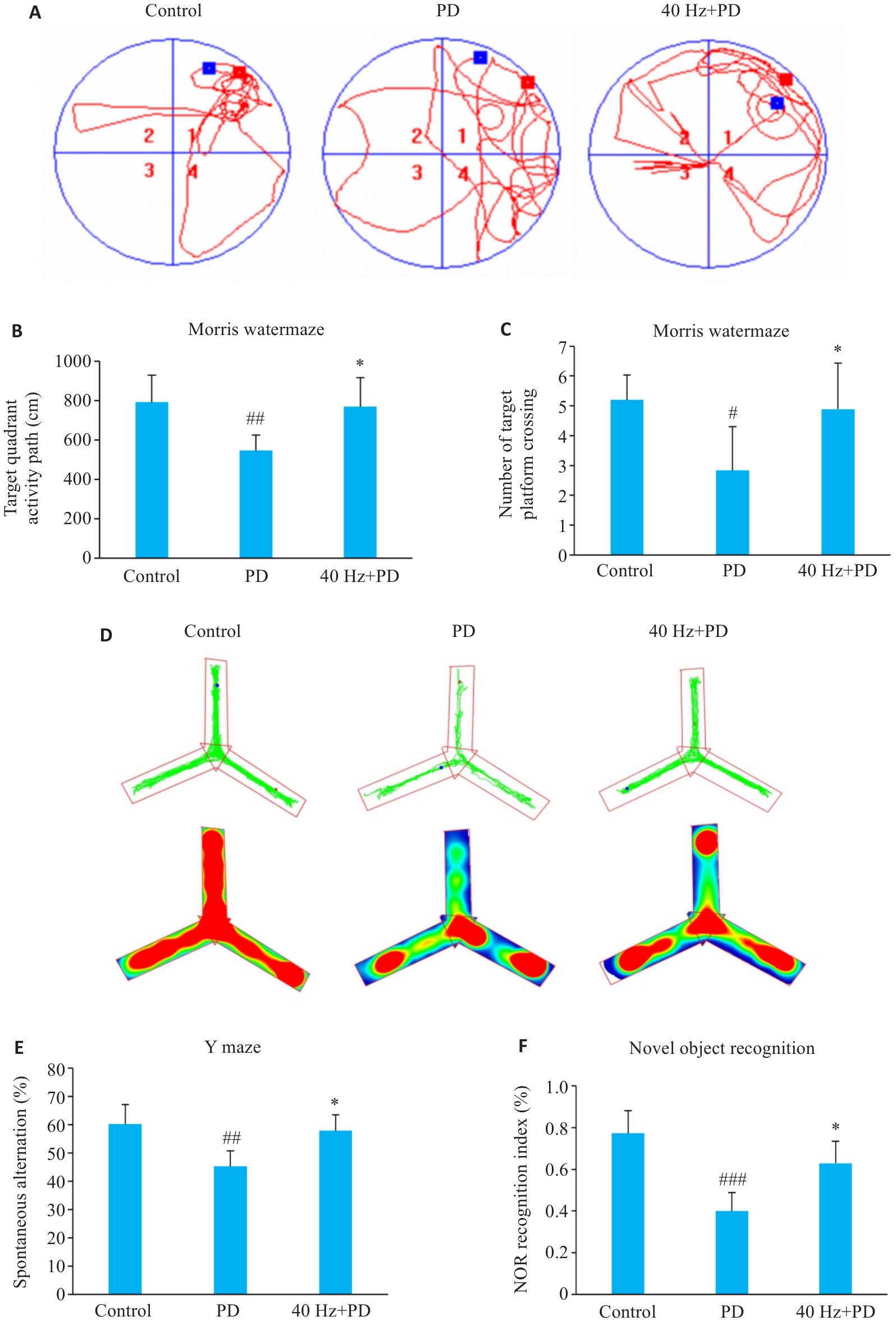
Fig.2 Cognitive function test results in each group. A: Water maze test trajectory. B: Target quadrant activity distance within the water maze. C: Number of target platform crossing within the water maze. D: Y maze test trajectory and heat map. E: Y maze spontaneous response alternation rate. F: Novel object recognition index. Data are presented as Mean±SD. #P<0.05,##P<0.01,###P<0.001 vs Control. *P<0.05 vs PD.
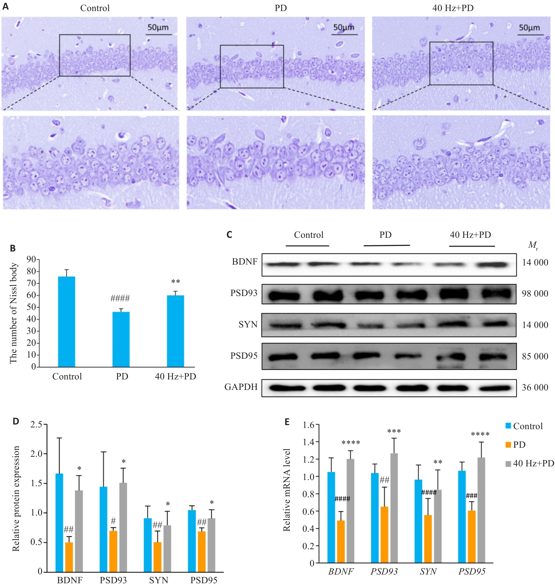
Fig.3 Detection results of hippocampal neurons and synapses in each group. A: Nissl staining of the hippocampus in each group (Original magnification: × 50). B: Nissl neuronal count statistics in the hippocampus in each group. C: Western blotting detection results. D: Analysis of Western blotting detection results. E: Analysis of RT-qPCR detection results. Data are presented as Mean±SD and analyzed using one-way ANOVA. #P<0.05,##P<0.01, ###P<0.001,####P<0.0001 vs Control. *P<0.05, **P<0.01, ***P<0.001, ****P<0.0001 vs PD.
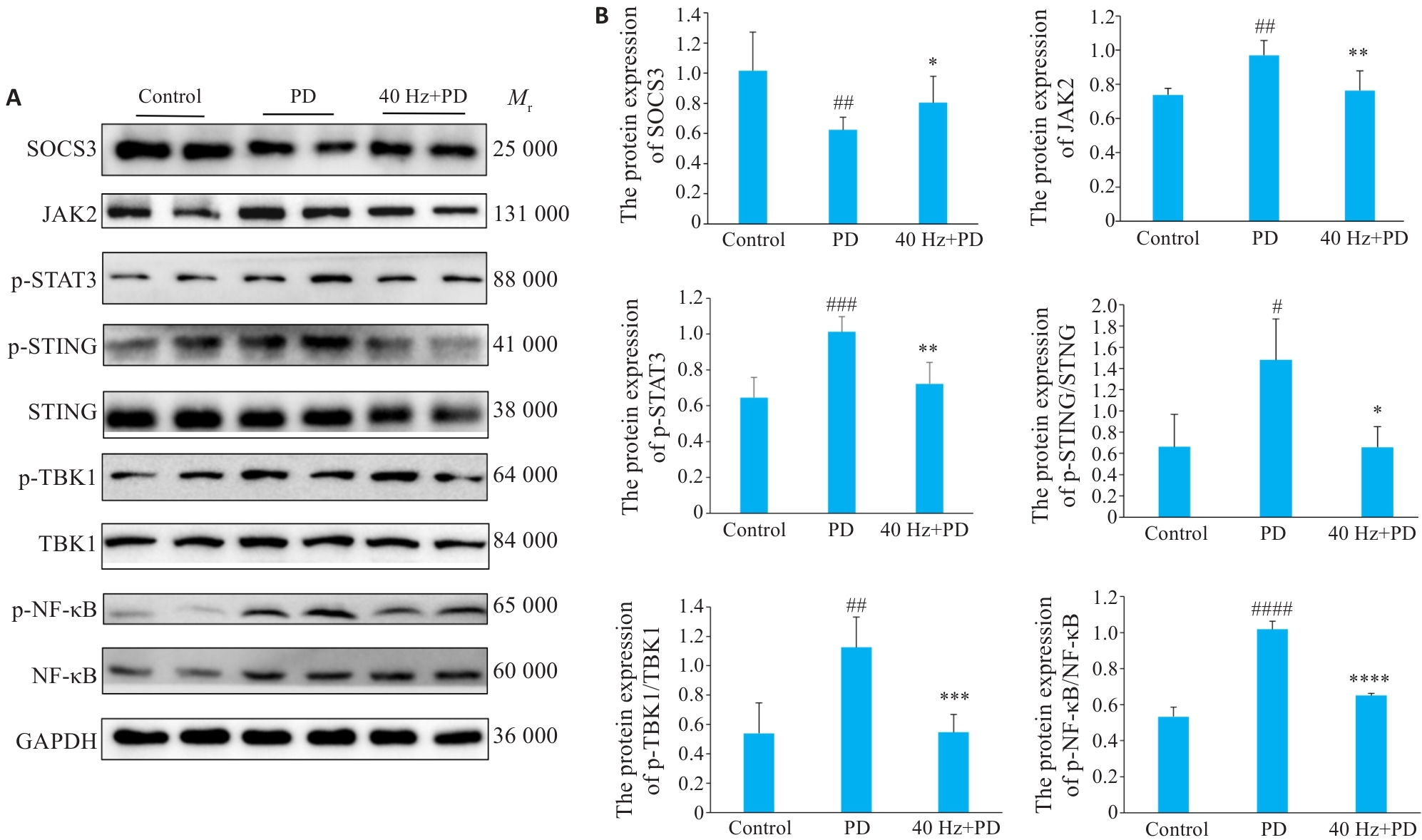
Fig.4 Detection results of SOCS3 and the JAK2-STAT3-STING pathway in mouse hippocampus in each group. A: Western blotting detection results. B: Analysis of the results of Western blotting. Data are presented as Mean±SD and analyzed using one-way ANOVA. #P<0.05,##P<0.01, ###P<0.001,####P<0.0001 vs Control. *P<0.05, **P<0.01, ***P<0.001, ****P<0.0001 vs PD.
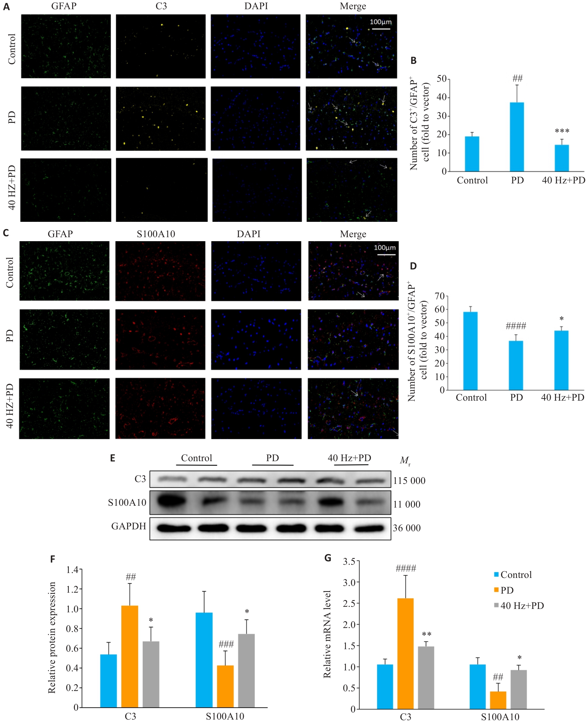
Fig.5 Detection results of hippocampal astrocyte polarization phenotypes in the mice in each group. A: Immunofluorescence staining of detecting hippocampal GFAP (green) and C3 (yellow) in each group (×100; arrows indicate co-staining regions of GFAP and C3). B: Quantitative analysis of immunofluorescence staining results. C: Immunofluorescence staining for detecting hippocampal GFAP (green) and S100A10 (red) in each group (×100; arrows indicate co-staining regions of GFAP and S100A10). D: Quantitative analysis of immunofluorescence staining results. E: Western blotting detection results. F: Quantitative analysis of Western blotting results. G: RT-qPCR detection analysis. Data are presented as Mean±SD and analyzed using one-way ANOVA. ##P<0.01, ###P<0.001,####P<0.0001 vs Control. *P<0.05, **P<0.01, ***P<0.001 vs PD.
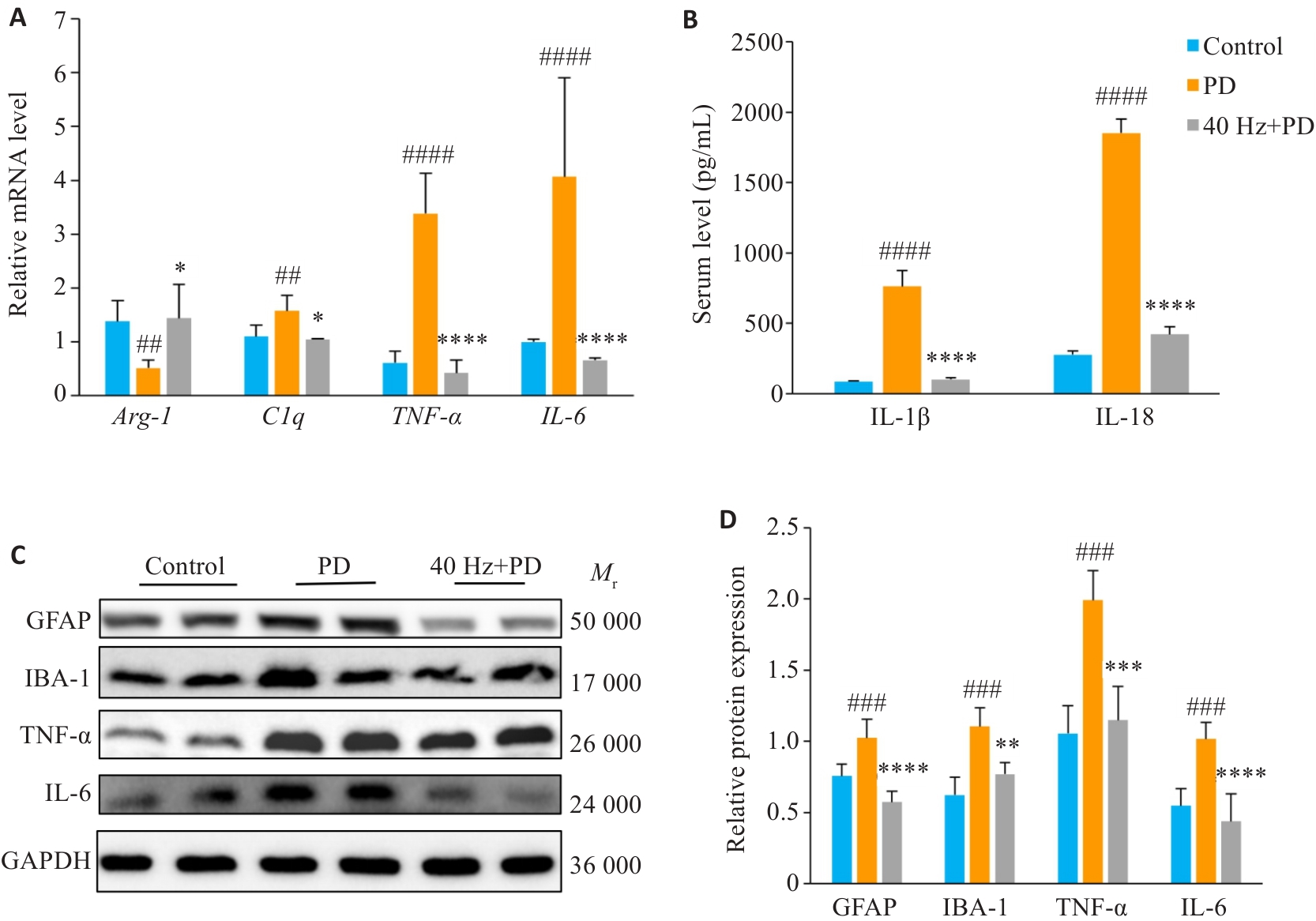
Fig.6 Detection results of hippocampal neuroinflammation and systemic inflammation in each group. A: Analysis of RT-qPCR detection results. B: Serum ELISA detection results. C: Western blotting detection results. D: Quantitative analysis of Western blotting detection results. Data are presented as Mean±SD and analyzed using one-way ANOVA. ##P<0.01, ###P<0.001, ####P<0.0001 vs Control. *P<0.05, **P<0.01, ***P<0.001, ****P<0.0001 vs PD.
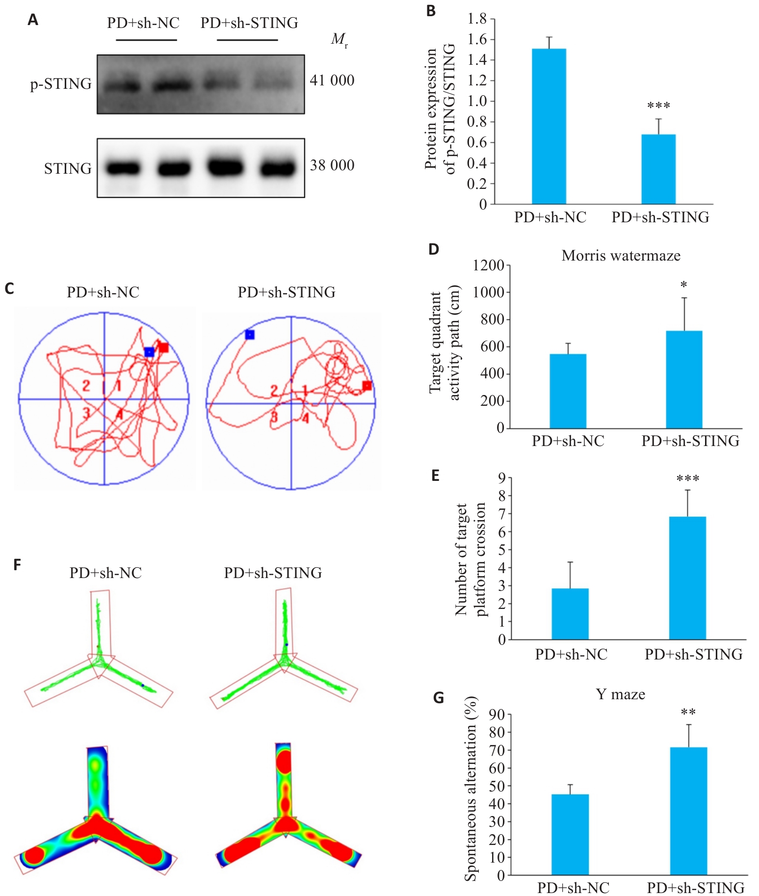
Fig.7 Effect of STING knockdown on cognitive function of PD mice. A: Western blotting detection results. B: Quantitative analysis of Western blotting results. C: Water maze test trajectory. D: Target quadrant activity path within the water maze. E: Number of target platform crossings within the water maze. F: Y maze test trajectory and heat map. G: Y maze spontaneous response alternation rate. Data are presented as Mean±SD and analyzed using one-way ANOVA. *P<0.05, **P<0.01, ***P<0.001 vs PD+sh-NC.
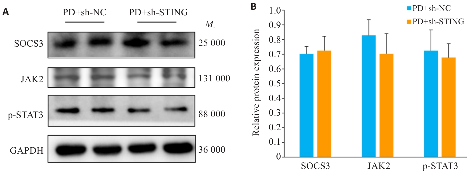
Fig.8 Detection results of the SOCS3-JAK2-STAT3 signaling pathway in the hippocampus of PD mice with STING knockdown. A: Western blotting detection results. B: Quantitative analysis of Western blotting results.
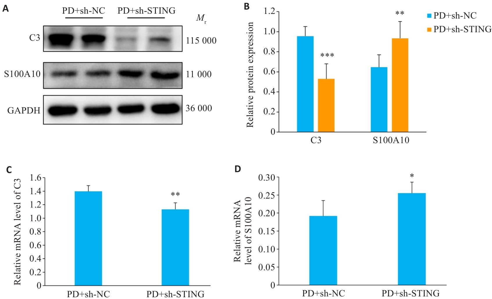
Fig. 9 STING knockdown affects detection results of hippocampal astrocyte polarization in mice. A: Western blotting detection results. B: Quantitative analysis of Western blotting results. C: RT-qPCR for detection of C3. D: RT-qPCR for detection of S100A10. Data are presented as Mean±SD and analyzed using one-way ANOVA. *P<0.05, **P<0.01, ***P<0.001 vs sh-NC.
| [1] | Wallace ER, Segerstrom SC, van Horne CG, et al. Meta-analysis of cognition in Parkinson's disease mild cognitive impairment and dementia progression[J]. Neuropsychol Rev, 2022, 32(1): 149-60. doi:10.1007/s11065-021-09502-7 |
| [2] | Gibson LL, Weintraub D, Lemmen R, et al. Risk of dementia in Parkinson's disease: a systematic review and meta-analysis[J]. Mov Disord, 2024, 39(10): 1697-709. doi:10.1002/mds.29918 |
| [3] | Gallagher J, Gochanour C, Caspell-Garcia C, et al. Long-Term Dementia Risk in Parkinson Disease[J]. Neurology, 2024,103(5): e209699. |
| [4] | Ye H, Robak LA, Yu MG, et al. Genetics and pathogenesis of Parkinson's syndrome[J]. Annu Rev Pathol, 2023, 18: 95-121. doi:10.1146/annurev-pathmechdis-031521-034145 |
| [5] | Kouli A, Spindler LRB, Fryer TD, et al. Neuroinflammation is linked to dementia risk in Parkinson's disease[J]. Brain, 2024, 147(3): 923-35. doi:10.1093/brain/awad322 |
| [6] | Smajić S, Prada-Medina CA, Landoulsi Z, et al. Single-cell sequencing of human midbrain reveals glial activation and a Parkinson-specific neuronal state[J]. Brain, 2022, 145(3): 964-78. doi:10.1093/brain/awab446 |
| [7] | Jiang SY, Tian T, Yao H, et al. The cGAS-STING-YY1 axis accelerates progression of neurodegeneration in a mouse model of Parkinson's disease via LCN2-dependent astrocyte senescence[J]. Cell Death Differ, 2023, 30(10): 2280-92. doi:10.1038/s41418-023-01216-y |
| [8] | Chakrabarti S, Roy A, Prorok T, et al. Aspirin up-regulates suppressor of cytokine signaling 3 in glial cells via PPARα[J]. J Neurochem, 2019, 151(1): 50-63. doi:10.1111/jnc.14813 |
| [9] | Zhang H, Meng FB, Tang JX, et al. Lactate inhibits T-cell activation in sepsis through CD40LG downregulation and SOCS3-mediated JAK1/STAT3 pathway suppression[J]. Biochim Biophys Acta BBA Mol Basis Dis, 2025, 1871(7): 167923. doi:10.1016/j.bbadis.2025.167923 |
| [10] | Gao Y, Liu WN, Shi L, et al. Narirutin reduces microglia-mediated neuroinflammation by inhibiting the JAK2/STAT3 pathway in MPP+/MPTP-induced Parkinson's disease models[J]. Exp Neurol, 2025, 389: 115232. doi:10.1016/j.expneurol.2025.115232 |
| [11] | Meissner WG, Remy P, Giordana C, et al. Trial of lixisenatide in early Parkinson's disease[J]. N Engl J Med, 2024, 390(13): 1176-85. doi:10.1056/nejmoa2312323 |
| [12] | Adaikkan C, Tsai LH. Gamma entrainment: impact on neurocircuits, Glia, and therapeutic opportunities[J]. Trends Neurosci, 2020, 43(1): 24-41. doi:10.1016/j.tins.2019.11.001 |
| [13] | Zhang QS, Heng Y, Mou Z, et al. Reassessment of subacute MPTP-treated mice as animal model of Parkinson's disease[J]. Acta Pharmacol Sin, 2017, 38(10): 1317-28. doi:10.1038/aps.2017.49 |
| [14] | 韦雅露, 张玉雪, 卿天钰, 等. rAAV-PHP.eB病毒包装条件优化及其体外感染能力的研究[J]. 基因组学与应用生物学, 2025, 44(11): 1157-70. |
| [15] | 张 蒙, 孟祎静, 聂碧徽, 等. 自噬-STING信号在跑台运动抑制细胞焦亡改善PD小鼠认知功能障碍中的机制研究[J]. 体育科学, 2024, 44(10): 58-76. |
| [16] | Guillemaud O, Ceyzériat K, Saint-Georges T, et al. Complex roles for reactive astrocytes in the triple transgenic mouse model of Alzheimer disease[J]. Neurobiol Aging, 2020, 90: 135-46. doi:10.1016/j.neurobiolaging.2020.02.010 |
| [17] | Leite Silva ABR, Gonçalves de Oliveira RW, Diógenes GP, et al. Premotor, nonmotor and motor symptoms of Parkinson's Disease: a new clinical state of the art[J]. Ageing Res Rev, 2023, 84: 101834. doi:10.1016/j.arr.2022.101834 |
| [18] | Rau A, Philipsen L, Frings L, et al. Hippocampus and basal forebrain degeneration differentially impact cognition in Lewy body spectrum disorders[J]. Brain, 2025, 148(8): 2772-84. doi:10.1093/brain/awaf070 |
| [19] | 杨佳瑶, 何玉莲, 郭延垒, 等. 党参-茯苓配伍调控ERα/PI3K/Akt信号通路改善单侧颈总动脉永久结扎痴呆大鼠的认知障碍[J]. 南方医科大学学报, 2026, 46(2): 247-58. |
| [20] | Huang SY, Su ZY, Han YY, et al. Cordycepin improved the cognitive function through regulating adenosine A2A receptors in MPTP induced Parkinson's disease mice model[J]. Phytomedicine, 2023, 110: 154649. doi:10.1016/j.phymed.2023.154649 |
| [21] | Manippa V, Palmisano A, Nitsche MA, et al. Cognitive and neuropathophysiological outcomes of gamma-tACS in dementia: a systematic review[J]. Neuropsychol Rev, 2024, 34(1): 338-61. doi:10.1007/s11065-023-09589-0 |
| [22] | Huang FK, Huang QK, Zheng LY, et al. Effect of 40 Hz light flicker on behaviors of adult C57BL/6J mice[J]. Brain Res, 2023, 1814: 148441. doi:10.1016/j.brainres.2023.148441 |
| [23] | Rodríguez-Cruz A, Romo-Mancillas A, Mendiola-Precoma J, et al. "Effect of valerenic acid on neuroinflammation in a MPTP-induced mouse model of Parkinson's disease"[J]. IBRO Rep, 2020, 8: 28-35. doi:10.1016/j.ibror.2019.12.002 |
| [24] | Wolf D, Ayon-Olivas M, Sendtner M. BDNF-regulated modulation of striatal circuits and implications for Parkinson's disease and dystonia[J]. Biomedicines, 2024, 12(8): 1761. doi:10.3390/biomedicines12081761 |
| [25] | Soman SK, Swain M, Dagda RK. BDNF-TrkB signaling in mitochondria: implications for neurodegenerative diseases[J]. Mol Neurobiol, 2025, 62(2): 1756-69. doi:10.1007/s12035-024-04357-4 |
| [26] | Park SS, Park HS, Kim CJ, et al. Combined effects of aerobic exercise and 40-Hz light flicker exposure on early cognitive impairments in Alzheimer's disease of 3×Tg mice[J]. J Appl Physiol, 2022, 132(4): 1054-68. doi:10.1152/japplphysiol.00751.2021 |
| [27] | Lin ZL, Hou GQ, Yao YL, et al. 40-hz blue light changes hippocampal activation and functional connectivity underlying recognition memory[J]. Front Hum Neurosci, 2021, 15: 739333. doi:10.3389/fnhum.2021.739333 |
| [28] | 张宝文, 吕梦林, 陈丹丹, 等. 40 Hz光闪烁刺激调节NAD+/Sirt1/PGC-1α信号通路对帕金森病伴抑郁小鼠的影响[J]. 中国激光, 2025, 52(15): 113-25. |
| [29] | Zeng JL, Indajang J, Pitt D, et al. Lysosomal acidification impairment in astrocyte-mediated neuroinflammation[J]. J Neuroinflammation, 2025, 22(1): 72. doi:10.1186/s12974-025-03410-w |
| [30] | 黄鹏伟, 陈 洁, 邹金虎, 等. 槲皮素促进应激颗粒G3BP1解聚改善HIV-1 gp120诱导的星形胶质细胞神经毒性[J]. 南方医科大学学报, 2025, 45(2): 304-12. |
| [31] | Guo M, Guan A, Zhang MJ, et al. Exosome-mediated microglia-astrocyte interactions drive neuroinflammation in Parkinson's disease with Peli1 as a potential therapeutic target[J]. Pharmacol Res, 2025, 219: 107908. doi:10.1016/j.phrs.2025.107908 |
| [32] | Franklin TC, Bitarafan S, King AT, et al. Sensory neurostimulation promotes stress resilience with frequency-specificity[J]. bioRxiv. 2025, 2025:664223. |
| [33] | Xia XY, He XM, Zhao TM, et al. Inhibiting mtDNA-STING-NLRP3/IL-1β axis-mediated neutrophil infiltration protects neurons in Alzheimer's disease[J]. Cell Prolif, 2024, 57(1): e13529. doi:10.1111/cpr.13529 |
| [34] | Ferecskó AS, Smallwood MJ, Moore A, et al. STING-triggered CNS inflammation in human neurodegenerative diseases[J]. Biomedicines, 2023, 11(5): 1375. doi:10.3390/biomedicines11051375 |
| [35] | Huang XL, Hu Y, Jiang W, et al. Suppression of cGAS/STING pathway-triggered necroptosis in the hippocampus relates H2S to attenuate cognitive dysfunction of Parkinson's disease[J]. Exp Neurol, 2025, 385: 115093. doi:10.1016/j.expneurol.2024.115093 |
| [36] | Yue RZ, Guo X, Li WQ, et al. HDAC7 knockout mitigates astrocyte reactivity and neuroinflammation via the IRF3/cGAS/STING signaling pathway[J]. Front Cell Neurosci, 2025, 19: 1683595. doi:10.3389/fncel.2025.1683595 |
| [37] | He JQ, Qin ZJ, Cai YN, et al. USP11-PGAM5 axis promotes neurotoxic astrocyte reactivity by aggravating the mtDNA-cGAS-STING pathway after intracerebral hemorrhage[J]. Adv Sci, 2026, 13(1): e14283. doi:10.1002/advs.202514283 |
| [38] | Zhang W, Xu MJ, Chen FY, et al. Targeting the JAK2-STAT3 pathway to inhibit cGAS-STING activation improves neuronal senescence after ischemic stroke[J]. Exp Neurol, 2023, 368: 114474. doi:10.1016/j.expneurol.2023.114474 |
| [39] | Meng Y, Li XY, Bi YH, et al. Tregs promote astrocyte-neuron lactate shuttle via inhibiting STING pathway to improve neurological recovery after ischemic stroke[J]. CNS Neurosci Ther, 2026, 32(1): e70753. doi:10.1002/cns.70753 |
| Viewed | ||||||
|
Full text |
|
|||||
|
Abstract |
|
|||||
