Journal of Southern Medical University ›› 2025, Vol. 45 ›› Issue (11): 2365-2374.doi: 10.12122/j.issn.1673-4254.2025.11.09
Daiping HUA1( ), Qiaoyu XUAN1, Lanting SUN1, Qingsheng YU2, Qin WANG3, Tao WANG4, Qiyan MA4, Wenming YANG1,5, Han WANG1,5()
Received:2025-05-01
Online:2025-11-20
Published:2025-11-28
Contact:
Han WANG
E-mail:15655171331@163.com;neuwhah@126.com
Supported by:Daiping HUA, Qiaoyu XUAN, Lanting SUN, Qingsheng YU, Qin WANG, Tao WANG, Qiyan MA, Wenming YANG, Han WANG. LncRNA Meg3 expression level is negatively correlated with liver fibrosis severity in patients with Wilson disease[J]. Journal of Southern Medical University, 2025, 45(11): 2365-2374.
Add to citation manager EndNote|Ris|BibTeX
URL: https://www.j-smu.com/EN/10.12122/j.issn.1673-4254.2025.11.09
| Variable | WD group (n=100) | Control group (n=50) | χ2 /z/t | P |
|---|---|---|---|---|
| Male [n (%)] | 61 (61.00) | 29 (58.00) | 0.125 | 0.724 |
| Age (year) | 26.00 (22.00, 35.00) | 25.00 (23.00, 31.00) | -0.647 | 0.518 |
| BMI (kg/m2) | 21.62 (20.21, 22.69) | 21.05 (20.28, 22.64) | -0.451 | 0.652 |
| ALT (U/L) | 30.00 (20.45, 54.20) | 16.70 (14.40, 22.40) | -5.132 | <0.001 |
| AST (U/L) | 30.40 (22.15, 42.15) | 19.00 (16.40, 22.60) | -6.127 | <0.001 |
| APRI | 0.53 (0.33, 0.78) | 0.32 (0.28, 0.39) | -5.482 | <0.001 |
| FIB-4 | 0.98 (0.56, 1.44) | 0.52 (0.39, 0.58) | -5.753 | <0.001 |
| HA (ng/mL) | 108.07 (66.65, 175.39) | 67.47 (54.74, 83.28) | -5.116 | <0.001 |
| LN (ng/mL) | 112.12 (84.56, 160.35) | 80.77 (76.59, 90.46) | -4.956 | <0.001 |
| PⅢNP (ng/mL) | 16.20 (10.01, 26.36) | 9.62 (7.90, 10.33) | -5.759 | <0.001 |
| C-Ⅳ (ng/mL) | 72.31 (53.44, 100.65) | 48.70 (38.52, 59.83) | -5.512 | <0.001 |
| LSM (kPa) | 10.17 (7.93, 13.82) | 5.20 (3.81, 5.77) | -9.217 | <0.001 |
| SWV (m/s) | 1.85 (1.62, 2.11) | 1.26 (1.09, 1.32) | -9.603 | <0.001 |
| LncRNA Meg3 | 0.59 (0.11, 1.42) | 1.20 (0.56, 1.84) | 3.700 | <0.001 |
| Beclin-1 | 1.18 (0.72, 1.61) | 1.11 (0.95, 1.30) | -0.339 | 0.735 |
| LC3B | 1.35 (0.54, 2.02) | 1.17 (0.85, 1.45) | -0.797 | 0.425 |
Tab.1 Baseline data and observation indexes in WD and control groups
| Variable | WD group (n=100) | Control group (n=50) | χ2 /z/t | P |
|---|---|---|---|---|
| Male [n (%)] | 61 (61.00) | 29 (58.00) | 0.125 | 0.724 |
| Age (year) | 26.00 (22.00, 35.00) | 25.00 (23.00, 31.00) | -0.647 | 0.518 |
| BMI (kg/m2) | 21.62 (20.21, 22.69) | 21.05 (20.28, 22.64) | -0.451 | 0.652 |
| ALT (U/L) | 30.00 (20.45, 54.20) | 16.70 (14.40, 22.40) | -5.132 | <0.001 |
| AST (U/L) | 30.40 (22.15, 42.15) | 19.00 (16.40, 22.60) | -6.127 | <0.001 |
| APRI | 0.53 (0.33, 0.78) | 0.32 (0.28, 0.39) | -5.482 | <0.001 |
| FIB-4 | 0.98 (0.56, 1.44) | 0.52 (0.39, 0.58) | -5.753 | <0.001 |
| HA (ng/mL) | 108.07 (66.65, 175.39) | 67.47 (54.74, 83.28) | -5.116 | <0.001 |
| LN (ng/mL) | 112.12 (84.56, 160.35) | 80.77 (76.59, 90.46) | -4.956 | <0.001 |
| PⅢNP (ng/mL) | 16.20 (10.01, 26.36) | 9.62 (7.90, 10.33) | -5.759 | <0.001 |
| C-Ⅳ (ng/mL) | 72.31 (53.44, 100.65) | 48.70 (38.52, 59.83) | -5.512 | <0.001 |
| LSM (kPa) | 10.17 (7.93, 13.82) | 5.20 (3.81, 5.77) | -9.217 | <0.001 |
| SWV (m/s) | 1.85 (1.62, 2.11) | 1.26 (1.09, 1.32) | -9.603 | <0.001 |
| LncRNA Meg3 | 0.59 (0.11, 1.42) | 1.20 (0.56, 1.84) | 3.700 | <0.001 |
| Beclin-1 | 1.18 (0.72, 1.61) | 1.11 (0.95, 1.30) | -0.339 | 0.735 |
| LC3B | 1.35 (0.54, 2.02) | 1.17 (0.85, 1.45) | -0.797 | 0.425 |
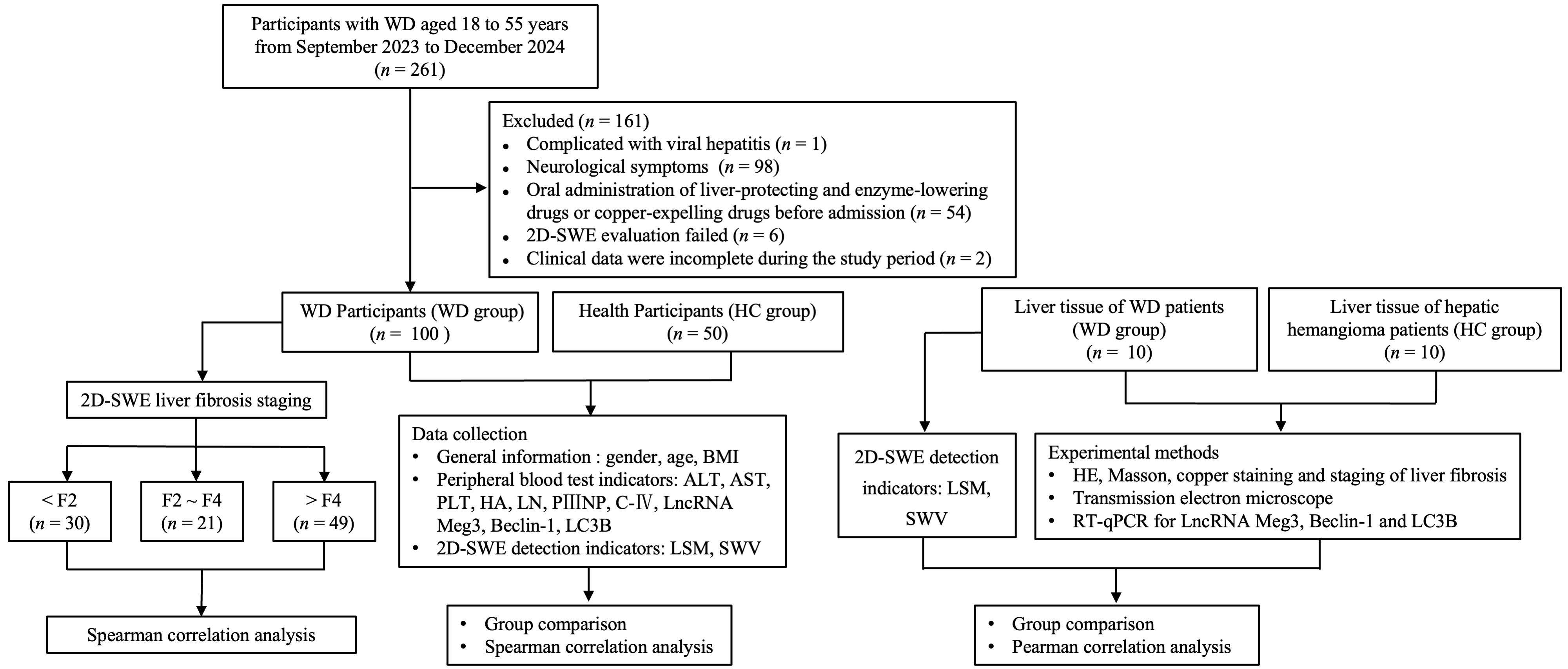
Fig.1 Flow chart of the study.
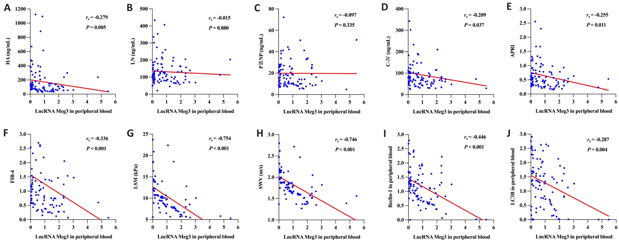
Fig.2 Scatter plot diagram showing the correlation of LncRNA Meg3 in peripheral blood of WD patients with liver fibrosis indexes and Beclin-1 and LC3B. LncRNA Meg3 in peripheral blood was negatively correlated with HA (A), LN (B), PⅢNP (C), C-Ⅳ (D), APRI (E), FIB-4 (F), LSM (G), SWV (H), Beclin-1 (I) and LC3B (J).
| Variable | β | t | P |
|---|---|---|---|
| HA | -0.140 | -1.376 | 0.172 |
| LN | -0.072 | -0.710 | 0.480 |
| PⅢNP | -0.032 | -0.308 | 0.758 |
| C-Ⅳ | -0.125 | -1.240 | 0.218 |
| APRI | -0.231 | -2.340 | 0.021 |
| FIB-4 | -0.276 | -2.566 | 0.012 |
| LSM | -0.569 | -6.441 | <0.001 |
| SWV | 0.572 | -6.500 | <0.001 |
| Beclin-1 | -0.402 | -4.321 | <0.001 |
| LC3B | -0.278 | -2.821 | 0.006 |
Tab.2 Multiple linear regression analysis of peripheral blood LncRNA Meg3 and the continuous variables in WD patients
| Variable | β | t | P |
|---|---|---|---|
| HA | -0.140 | -1.376 | 0.172 |
| LN | -0.072 | -0.710 | 0.480 |
| PⅢNP | -0.032 | -0.308 | 0.758 |
| C-Ⅳ | -0.125 | -1.240 | 0.218 |
| APRI | -0.231 | -2.340 | 0.021 |
| FIB-4 | -0.276 | -2.566 | 0.012 |
| LSM | -0.569 | -6.441 | <0.001 |
| SWV | 0.572 | -6.500 | <0.001 |
| Beclin-1 | -0.402 | -4.321 | <0.001 |
| LC3B | -0.278 | -2.821 | 0.006 |
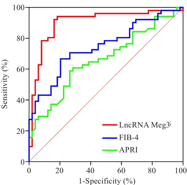
Fig.3 ROC curves for peripheral blood LncRNA Meg3, FIB-4, and APRI in WD patients. The AUC for LncRNA Meg3 was 0.902 (95% CI: 0.835-0.969), significantly higher than that of FIB-4 (AUC=0.661) and APRI (AUC=0.746).
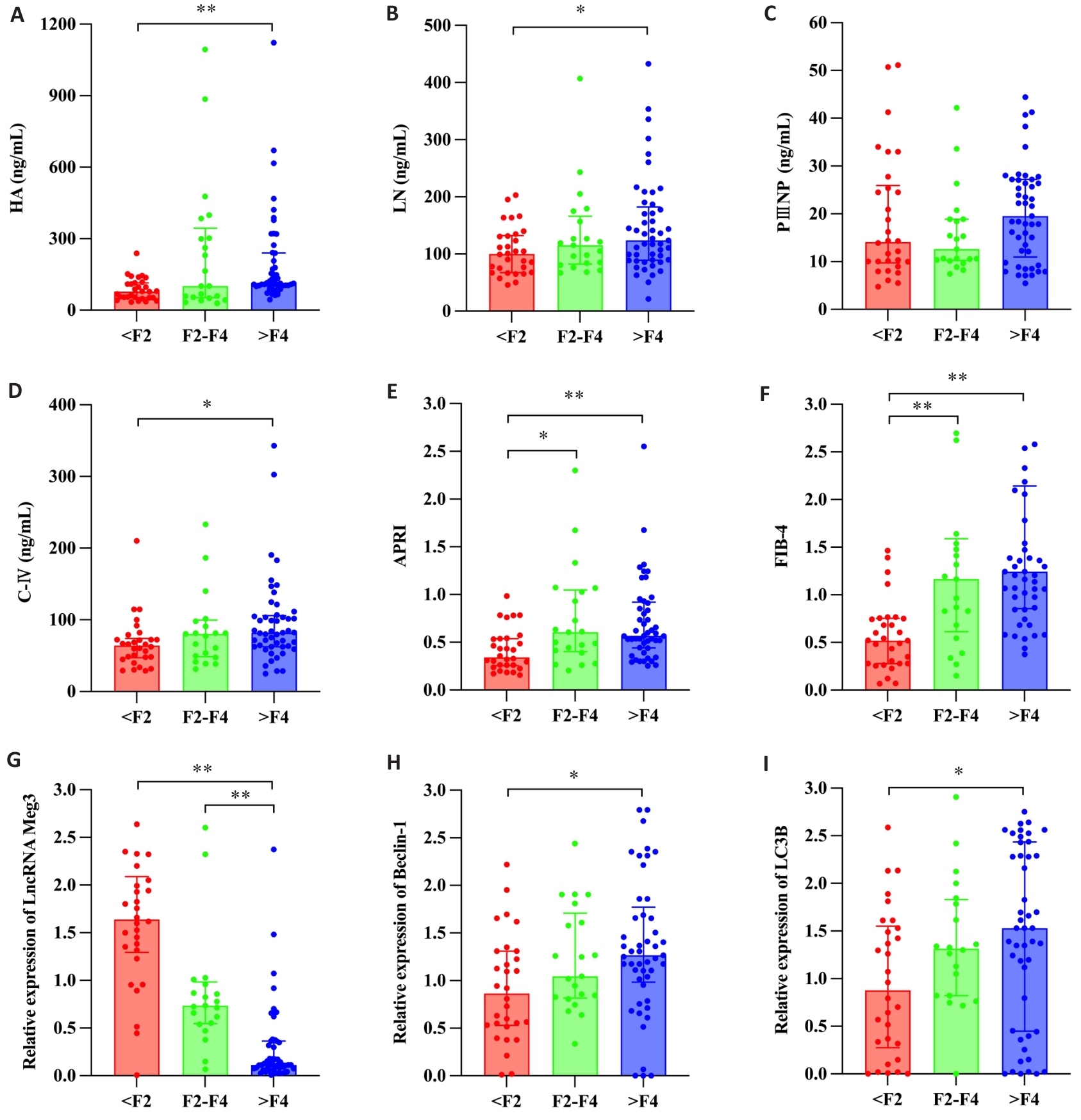
Fig.4 2D-SWE liver fibrosis stage and observation index analysis box plot of WD patients.Histograms are shown for HA (A), LN (B), PⅢNP (C), C-Ⅳ (D), APRI (E), FIB-4 (F), LncRNA Meg3 (G), Beclin-1 (H) and LC3B (I) for different 2D-SWE liver fibrosis stages. *P<0.05, **P<0.01.
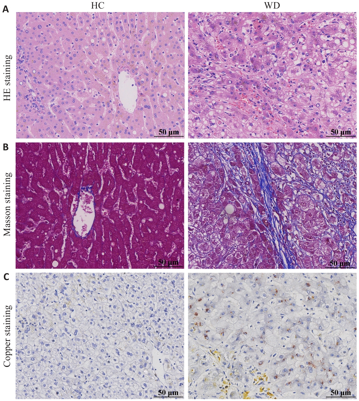
Fig.5 HE (A), Masson (B) and copper staining (C) of the liver tissues from WD and healthy control (HC) groups (scale bar=50 μm).
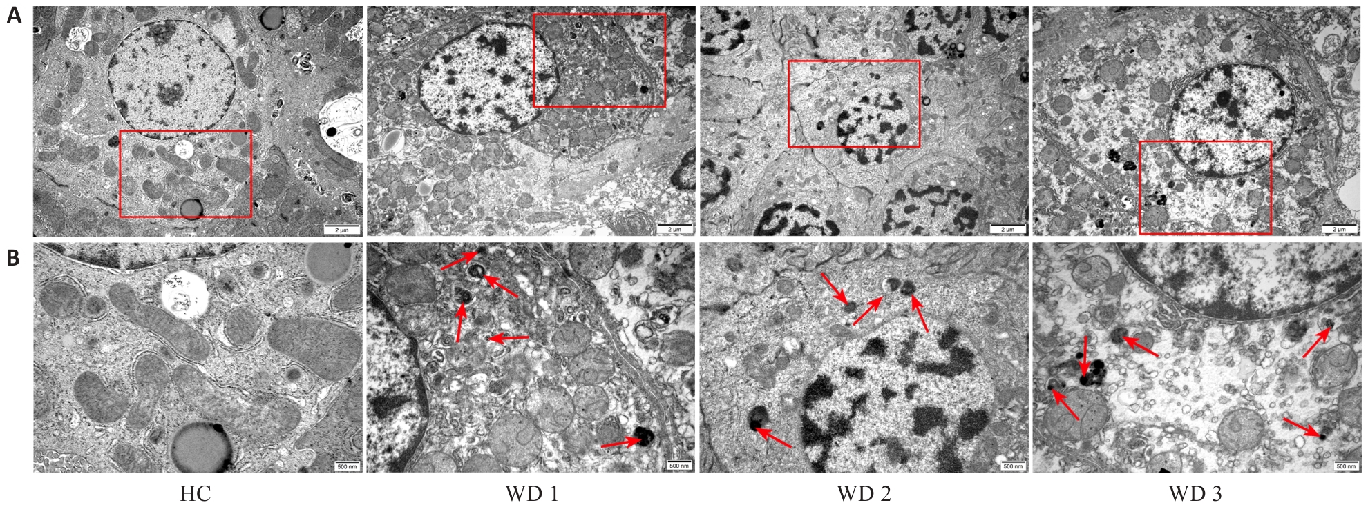
Fig.6 Transmission electron microscopy of the liver tissues in WD and HC groups (A: original magnification: ×10 000; B: ×25 000). Red arrows indicate the autophagosomes.
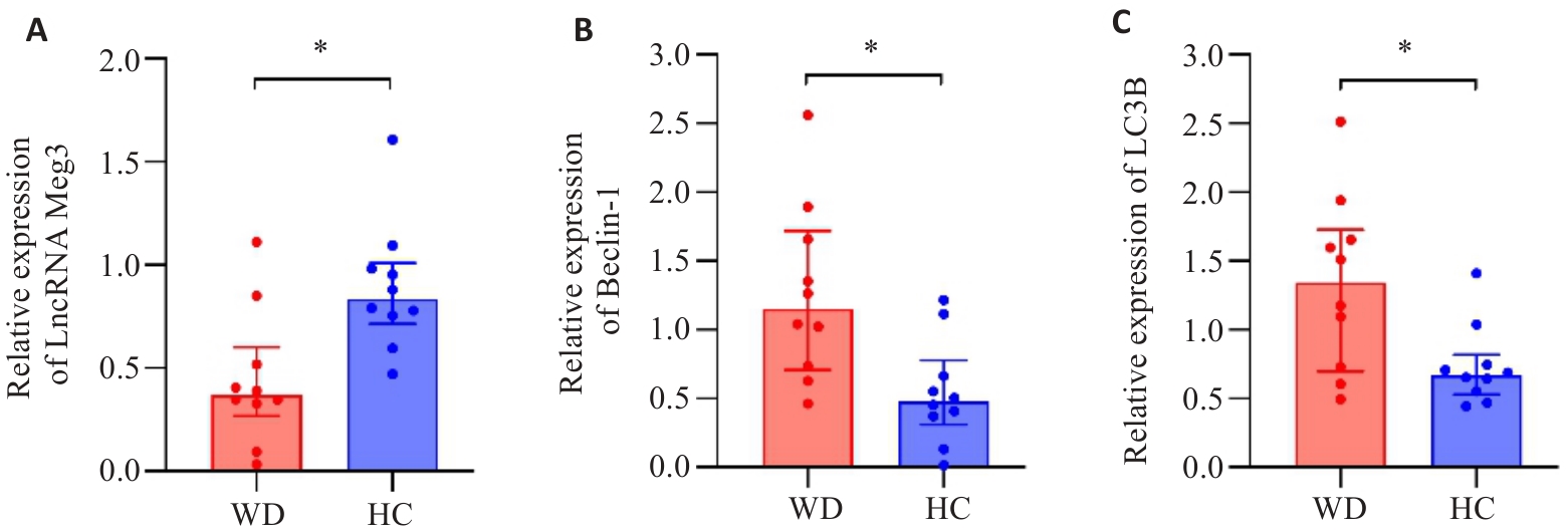
Fig.7 Box plots of the expression levels of LncRNA Meg3 (A), Beclin-1 (B) and LC3B (C) in the liver tissues in WD and HC groups. *P<0.05.
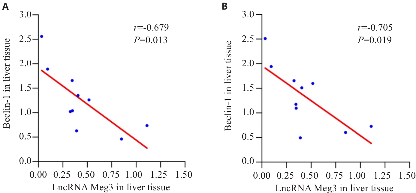
Fig.8 Scatter plots for correlation analysis of LncRNA Meg3 with Beclin-1 (A) and LC3B (B) in the liver tissue of WD group.
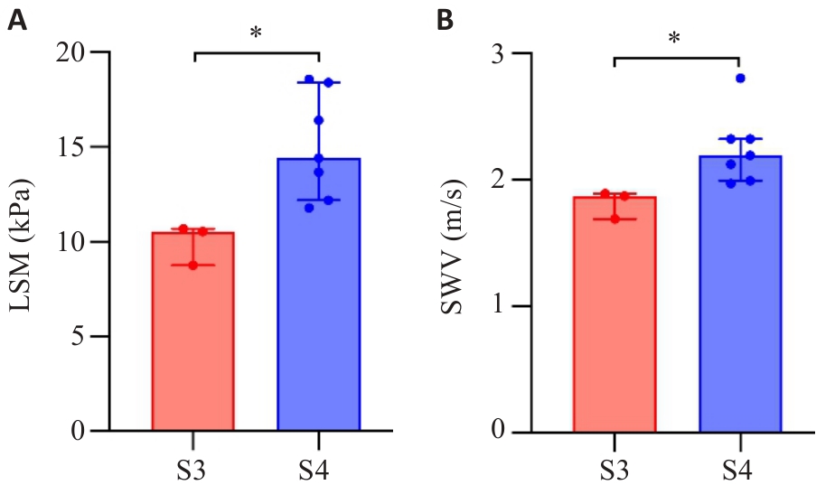
Fig.9 Box plot of LSM (A) and SWV (B) levels in WD patients with different liver fibrosis stages. *P<0.05
| [1] | Członkowska A, Litwin T, Dusek P, et al. Wilson disease[J]. Nat Rev Dis Primers, 2018, 4: 21. doi:10.1038/s41572-018-0018-3 |
| [2] | Bandmann O, Weiss KH, Kaler SG. Wilson's disease and other neurological copper disorders[J]. Lancet Neurol, 2015, 14(1): 103-13. doi:10.1016/s1474-4422(14)70190-5 |
| [3] | 中华医学会肝病学分会遗传代谢性肝病协作组. 肝豆状核变性诊疗指南(2022年版)[J].中华肝脏病杂志, 2022, 30(1): 9-20. |
| [4] | Kang ZL, Qiao N, Liu GY, et al. Copper-induced apoptosis and autophagy through oxidative stress-mediated mitochondrial dysfunction in male germ cells[J]. Toxicol Vitro, 2019, 61: 104639. doi:10.1016/j.tiv.2019.104639 |
| [5] | Kisseleva T, Brenner D. Molecular and cellular mechanisms of liver fibrosis and its regression[J]. Nat Rev Gastroenterol Hepatol, 2021, 18(3): 151-66. doi:10.1038/s41575-020-00372-7 |
| [6] | 中华医学会肝病学分会, 中华医学会消化病学分会, 中华医学会感染病学分会.肝纤维化诊断及治疗共识(2019年)[J]. 中华肝脏病杂志, 2019, 27(9): 657-67. |
| [7] | Gao R, Mao J. Noncoding RNA-mediated epigenetic regulation in hepatic stellate cells of liver fibrosis[J]. Noncoding RNA, 2024, 10(4): 44. doi:10.3390/ncrna10040044 |
| [8] | Chen MJ, Wang XG, Sun ZX, et al. Diagnostic value of LncRNA-MEG3 as a serum biomarker in patients with hepatitis B complicated with liver fibrosis[J]. Eur Rev Med Pharmacol Sci, 2019, 23(10): 4360-7. |
| [9] | 赵红英, 王文浩, 孙宁宁.长链非编码RNA MEG3对老年乙型肝炎并发肝硬化和肝纤维化的诊断价值[J].国际消化病杂志, 2021, 41(3): 221-4. |
| [10] | Zhang J, Ma Y, Xie D, et al. Differentially expressed lncRNAs in liver tissues of TX mice with hepatolenticular degeneration[J]. Sci Rep, 2021, 11(1): 1377. doi:10.1038/s41598-020-80635-0 |
| [11] | Ferenci P, Caca K, Loudianos G, et al. Diagnosis and phenotypic classification of Wilson disease[J]. Liver Int, 2003, 23(3): 139-42. doi:10.1034/j.1600-0676.2003.00824.x |
| [12] | Wai CT, Greenson JK, Fontana RJ, et al. A simple noninvasive index can predict both significant fibrosis and cirrhosis in patients with chronic hepatitis C[J]. Hepatology, 2003, 38(2): 518-26. doi:10.1053/jhep.2003.50346 |
| [13] | Sterling RK, Lissen E, Clumeck N, et al. Development of a simple noninvasive index to predict significant fibrosis in patients with HIV/HCV coinfection[J]. Hepatology, 2006, 43(6): 1317-25. doi:10.1002/hep.21178 |
| [14] | Wang JF, Wu ML, Linghu RZ, et al. Usefulness of new shear wave elastography technique for noninvasive assessment of liver fibrosis in patients with chronic hepatitis B: a prospective multicenter study[J]. Ultraschall Med, 2022, 43(2): e1-e10. doi:10.1055/a-1376-6734 |
| [15] | Desmet VJ, Gerber M, Hoofnagle JH, et al. Classification of chronic hepatitis: diagnosis, grading and staging[J]. Hepatology, 1994, 19(6): 1513-20. doi:10.1002/hep.1840190629 |
| [16] | Wu YY, Wu S, Li XF, et al. LncRNA MEG3 reverses CCl4-induced liver fibrosis by targeting NLRC5[J]. Eur J Pharmacol, 2021, 911: 174462. doi:10.1016/j.ejphar.2021.174462 |
| [17] | Chen T, Lin HJ, Chen X, et al. LncRNA Meg8 suppresses activation of hepatic stellate cells and epithelial-mesenchymal transition of hepatocytes via the Notch pathway[J]. Biochem Biophys Res Commun, 2020, 521(4): 921-7. doi:10.1016/j.bbrc.2019.11.015 |
| [18] | Qin R, Huang W, Huang Y, et al. lncRNA MEG3 modulates hepatic stellate cell activation by sponging miR-145 to regulate PPARγ[J]. Mol Med Rep, 2022, 25(1): 3. doi:10.3892/mmr.2021.12519 |
| [19] | Hussain MS, Majami AA, Ali H, et al. The complex role of MEG3: an emerging long non-coding RNA in breast cancer[J]. Pathol Res Pract, 2023, 251: 154850. doi:10.1016/j.prp.2023.154850 |
| [20] | 朱 权, 黄柏胜, 位磊艳, 等. 过表达LncRNAMEG3通过促进铁死亡增强肝癌细胞对顺铂的化疗敏感性[J]. 南方医科大学学报, 2024, 44(1): 17-24. |
| [21] | 李元香, 郭艳艳, 莫代芬, 等.血清LncRNA MEG3、miR-23b-3p与艾滋病患者特应性皮炎严重程度的相关性研究[J].湖南师范大学学报(医学版), 2023, 20(2): 104-8. |
| [22] | 骆欣敏, 何 玉, 吴凤娇, 等.外泌体LncRNA MEG3在子宫内膜癌中表达及其临床意义[J]. 蚌埠医学院学报, 2022, 47(5): 627-30. |
| [23] | Jiang XY. Long noncoding RNA MEG3: an active player in fibrosis[J]. Pharmacol Rep, 2025, 77(1): 21-30. doi:10.1007/s43440-024-00661-x |
| [24] | Moradi M, Mard SA, Farbood Y, et al. The protective effect of p-Coumaric acid on hepatic injury caused by particulate matter in the rat and determining the role of long noncoding RNAs MEG3 and HOTAIR[J]. J Biochem Mol Toxicol, 2023, 37(7): e23364. doi:10.1002/jbt.23364 |
| [25] | 汪 瀚, 花代平, 殷 馨, 等.从LncRNA Meg3调控细胞自噬途径探讨肝豆灵治疗Wilson病肝纤维化的作用机制[J].中华中医药杂志, 2025, 40(5): 2574-9. |
| [26] | 殷 馨, 汪 瀚, 孙兰婷, 等.肝豆灵通过LncRNA Meg3调控自噬减轻铜负荷Wilson病大鼠肝纤维化[J].医学研究杂志,2025,54(01):37-42. |
| [27] | 中华医学会超声医学分会, 中国研究型医院学会肿瘤介入专业委员会, 国家卫生和健康委员会能力建设和继续教育中心超声医学专家委员会, 等. 肝病超声诊断指南[J]. 中华肝脏病杂志, 2021, 29(5): 385-402. |
| [28] | Osman AM, El Shimy A, Abd El Aziz MM. 2D shear wave elastography (SWE) performance versus vibration-controlled transient elastography (VCTE/fibroscan) in the assessment of liver stiffness in chronic hepatitis[J]. Insights Imaging, 2020, 11(1): 38. doi:10.1186/s13244-020-0839-y |
| [29] | Kavak S, Kaya S, Senol A, et al. Evaluation of liver fibrosis in chronic hepatitis B patients with 2D shear wave elastography with propagation map guidance: a single-centre study[J]. BMC Med Imaging, 2022, 22(1): 50. doi:10.1186/s12880-022-00777-7 |
| [30] | 刘安生, 李 燕, 伍宏兵.实时二维剪切波弹性成像与血清肝纤维化四项指标评估肝豆状核变性肝硬化[J].影像研究与医学应用, 2024,8(10): 84-6. |
| [31] | Wang J, Hu M, Zhu Q, et al. Liver stiffness assessed by real-time two-dimensional shear wave elastography predicts hypersplenism in patients with Wilson's disease: a prospective study[J]. BMC Med Imaging, 2022, 22(1): 25. doi:10.1186/s12880-022-00749-x |
| [32] | Hwang J, Yoon HM, Jung AY, et al. Diagnostic performance of ultrasound elastography and serologic fibrosis indices for evaluation of hepatic involvement in Wilson disease[J]. J Ultrasound Med, 2020, 39(11): 2231-42. doi:10.1002/jum.15334 |
| [33] | 滕 阅. 应用2D-SWE技术评估儿童肝豆状核变性及糖原贮积症肝脏硬度的价值 [D]. 广州: 广州医科大学, 2022. |
| [34] | Dikic I, Elazar Z. Mechanism and medical implications of mammalian autophagy[J]. Nat Rev Mol Cell Biol, 2018, 19(6): 349-64. doi:10.1038/s41580-018-0003-4 |
| [35] | Polishchuk EV, Merolla A, Lichtmannegger J, et al. Activation of autophagy, observed in liver tissues from patients with Wilson disease and from ATP7B-deficient animals, protects hepatocytes from copper-induced apoptosis[J]. Gastroenterology, 2019, 156(4): 1173-89. e5. doi:10.1053/j.gastro.2018.11.032 |
| [36] | Zhang Y, Wang M, Tang L, et al. FoxO1 silencing in Atp7b-/- neural stem cells attenuates high copper-induced apoptosis via regulation of autophagy[J]. J Neurochem, 2024, 168(9): 2762-74. doi:10.1111/jnc.16136 |
| [37] | Pantoom S, Pomorski A, Huth K, et al. Direct interaction of ATP7B and LC3B proteins suggests a cooperative role of copper transportation and autophagy[J]. Cells, 2021, 10(11): 3118. doi:10.3390/cells10113118 |
| [38] | Li X, Yang KB, Chen W, et al. CUL3 (cullin 3)‑mediated ubiquitination and degradation of BECN1 (beclin 1) inhibit autophagy and promote tumor progression[J]. Autophagy, 2021, 17(12): 4323-40. doi:10.1080/15548627.2021.1912270 |
| [1] | Weiyi LI, Lu JIANG, Zongxing ZHANG, Dan CHEN, Zhuoma BAO, Li HUANG, Lin YUAN. Qianggu Kangshu Formula attenuates osteoclast differentiation in rheumatoid arthritis by inhibiting the HIF-1α/BNIP3 autophagy signaling pathway [J]. Journal of Southern Medical University, 2025, 45(7): 1389-1396. |
| [2] | Tingting YANG, Li ZHAO. A stable mouse model of chronic liver fibrosis induced by vitamin A deficiency and intraperitoneal CCl4 injection [J]. Journal of Southern Medical University, 2025, 45(7): 1527-1534. |
| [3] | Xinheng WANG, Xiaohan SHAO, Tongtong LI, Lu ZHANG, Qinjun YANG, Weidong YE, Jiabing TONG, Zegeng LI, Xiangming FANG. Pingchuanning Formula suppresses airway inflammation in a rat model of asthmatic cold syndrome by regulating the HMGB1/Beclin-1 axis-mediated autophagy [J]. Journal of Southern Medical University, 2025, 45(6): 1153-1162. |
| [4] | Jiawen YU, Yi ZHOU, Chunmei QIAN, Lan MU, Renye QUE. Effects of liver fibrosis induced by iron overload on M2 polarization of macrophages in mice [J]. Journal of Southern Medical University, 2025, 45(4): 684-691. |
| [5] | Yanyan DONG, Kejing ZHANG, Jun CHU, Quangen CHU. Didang Decoction-medicated serum enhances autophagy in high glucose-induced rat glomerular endothelial cells via the PI3K/Akt/mTOR signaling pathway [J]. Journal of Southern Medical University, 2025, 45(3): 461-469. |
| [6] | Ming LIAO, Wenhua ZHONG, Ran ZHANG, Juan LIANG, Wentaorui XU, Wenjun WAN, Chao LI Shu WU. Protein C activator derived from snake venom protects human umbilical vein endothelial cells against hypoxia-reoxygenation injury by suppressing ROS via upregulating HIF-1α and BNIP3 [J]. Journal of Southern Medical University, 2025, 45(3): 614-621. |
| [7] | Kelei GUO, Yingli LI, Chenguang XUAN, Zijun HOU, Songshan YE, Linyun LI, Liping CHEN, Li HAN, Hua BIAN. Yiqi Yangyin Huazhuo Tongluo Formula alleviates diabetic podocyte injury by regulating miR-21a-5p/FoxO1/PINK1-mediated mitochondrial autophagy [J]. Journal of Southern Medical University, 2025, 45(1): 27-34. |
| [8] | Junping ZHAN, Shuo HUANG, Qingliang MENG, Wei FAN, Huimin GU, Jiakang CUI, Huilian WANG. Buyang Huanwu Decoction reduces mitochondrial autophagy in rheumatoid arthritis synovial fibroblasts in hypoxic culture by inhibiting the BNIP3-PI3K/Akt pathway [J]. Journal of Southern Medical University, 2025, 45(1): 35-42. |
| [9] | Zhiliang CHEN, Yonggang YANG, Xia HUANG, Yan CHENG, Yuan QU, Qiqi HENG, Yujia FU, Kewei LI, Ning GU. Differential expressions of exosomal miRNAs in patients with chronic heart failure and hyperuricemia: diagnostic values of miR-27a-5p and miR-139-3p [J]. Journal of Southern Medical University, 2025, 45(1): 43-51. |
| [10] | Yuming ZHANG, Shicheng XIA, Linlin ZHANG, Mengxi CHEN, Xiaojing LIU, Qin GAO, Hongwei YE. Protective effect of Lonicerae japonicae flos extract against doxorubicin-induced liver injury in mice [J]. Journal of Southern Medical University, 2024, 44(8): 1571-1581. |
| [11] | Yao CHENG, Yuanying WANG, Feiyang YAO, Pan HU, Mingxian CHEN, Ning WU. Baicalin suppresses type 2 dengue virus-induced autophagy of human umbilical vein endothelial cells by inhibiting the PI3K/AKT pathway [J]. Journal of Southern Medical University, 2024, 44(7): 1272-1283. |
| [12] | Yeming ZHANG, Yuanxiang ZHANG, Xuebin SHEN, Guodong WANG, Lei ZHU. MiRNA-103-3p promotes neural cell autophagy by activating Wnt/β-catenin signaling via targeting rab10 in a rat model of depression [J]. Journal of Southern Medical University, 2024, 44(7): 1315-1326. |
| [13] | Qianyi CHEN, Shuhan SHANG, Huan LU, Sisi LI, Zhimian SUN, Xirui FAN, Zhilin QI. Calenduloside E inhibits hepatocellular carcinoma cell proliferation and migration by down-regulating GPX4 and SLC7A11 expression through the autophagy pathway [J]. Journal of Southern Medical University, 2024, 44(7): 1327-1335. |
| [14] | CAO Jiafan, SUN Yue, DING Xin, LI Shengwen, CHEN Bo, LAN Tian. Arbutin ameliorates liver fibrosis in mice by inhibiting macrophage recruitment and regulating the Akt/NF-κB and Smad signaling pathways [J]. Journal of Southern Medical University, 2024, 44(4): 652-659. |
| [15] | ZHOU Fengmin, GUO Yanju, CHEN Ning. Exercise promotes irisin expression to ameliorate renal injury in type 2 diabetic rats [J]. Journal of Southern Medical University, 2024, 44(4): 675-681. |
| Viewed | ||||||
|
Full text |
|
|||||
|
Abstract |
|
|||||