Journal of Southern Medical University ›› 2025, Vol. 45 ›› Issue (4): 684-691.doi: 10.12122/j.issn.1673-4254.2025.04.02
Jiawen YU1,2( ), Yi ZHOU2, Chunmei QIAN3, Lan MU2, Renye QUE1()
Received:2024-11-08
Online:2025-04-20
Published:2025-04-28
Contact:
Renye QUE
E-mail:18170389858@163.com;824492@qq.com
Supported by:Jiawen YU, Yi ZHOU, Chunmei QIAN, Lan MU, Renye QUE. Effects of liver fibrosis induced by iron overload on M2 polarization of macrophages in mice[J]. Journal of Southern Medical University, 2025, 45(4): 684-691.
Add to citation manager EndNote|Ris|BibTeX
URL: https://www.j-smu.com/EN/10.12122/j.issn.1673-4254.2025.04.02
| Gene | Forward primer | Reverse primer |
|---|---|---|
| CD206 | AGCTTCATCTTCGGGCCTTTG | GGTGACCACTCCTGCTGCTTTAG |
| β-actin | TTGTAACCAACTGGGACGATATGG | GATCTTGATCTTCATGGTGCTAGG |
Tab.1 Primer sequences for RT-qPCR
| Gene | Forward primer | Reverse primer |
|---|---|---|
| CD206 | AGCTTCATCTTCGGGCCTTTG | GGTGACCACTCCTGCTGCTTTAG |
| β-actin | TTGTAACCAACTGGGACGATATGG | GATCTTGATCTTCATGGTGCTAGG |
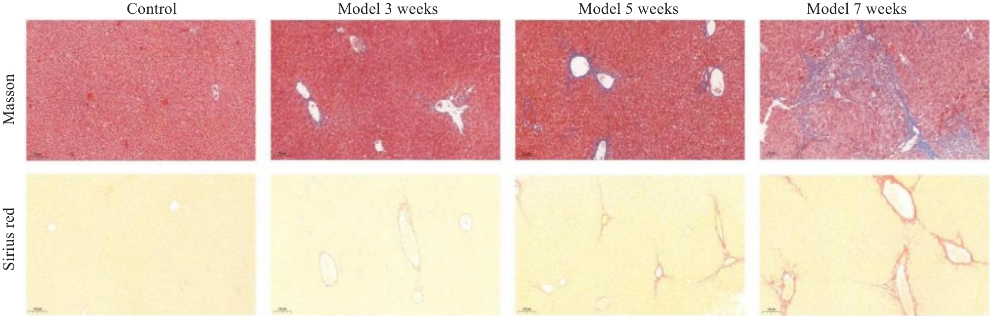
Fig.1 Histopathological changes of mouse liver with chronic iron overload induced by iron dextran (Original magnification: ×100).
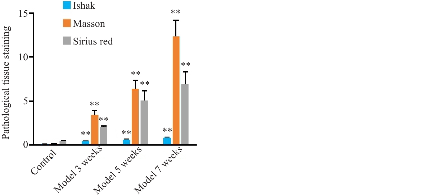
Fig.2 Histopathological changes of mouse liver with chronic iron overload induced by iron dextran. **P<0.01 vs Control.
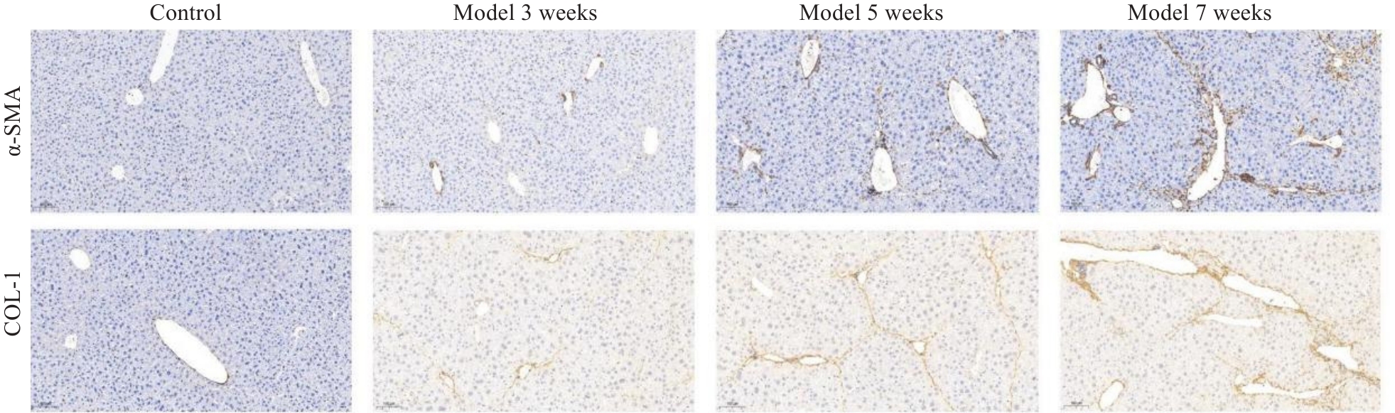
Fig.3 Changes of α-SMA and COL-1 in mouse liver with chronic iron overload induced by iron dextran (×100).
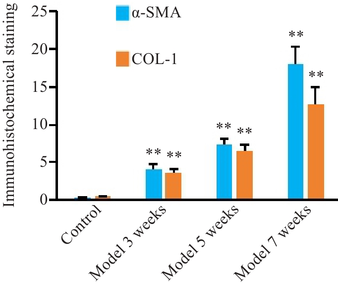
Fig.4 Changes of α-SMA and COL-1 in mouse liver with chronic iron overload induced by iron dextran. **P<0.01 vs Control.
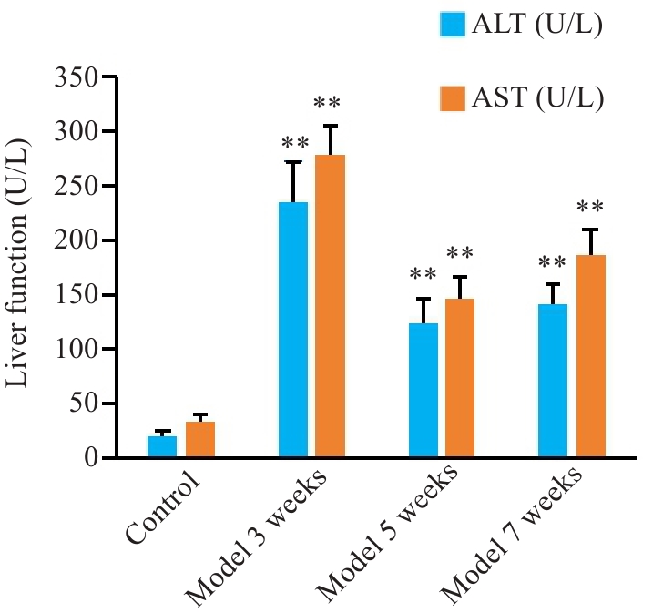
Fig.5 Changes of liver function in chronic iron overload mice induced by iron dextran. **P<0.01 vs Control.
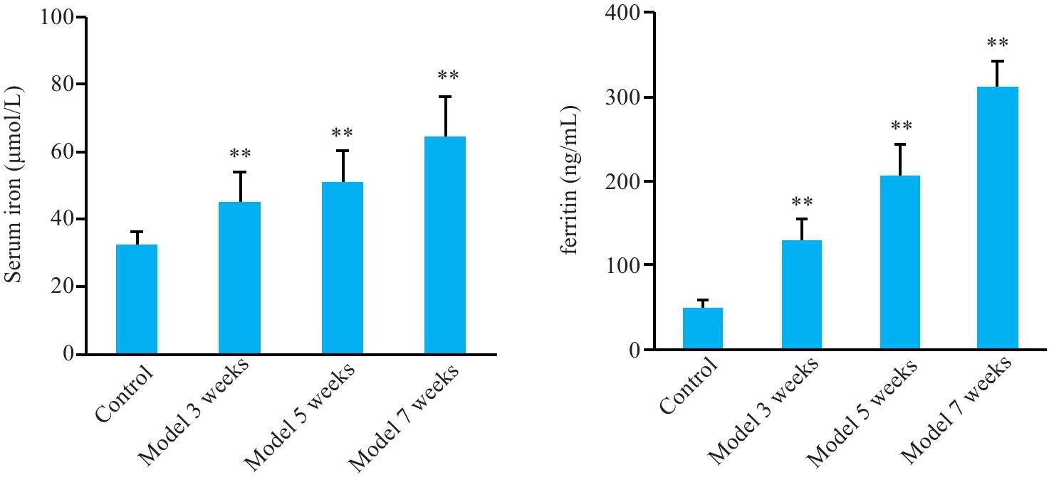
Fig.6 Changes of serum iron and ferritin contents in chronic iron overload induced by iron dextran.**P<0.01 vs Control.
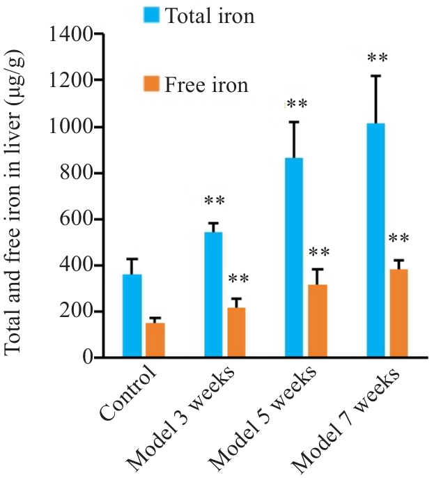
Fig.7 Changes of total iron and ferrous content in liver with chronic iron overload induced by iron dextran.**P<0.01 vs Control.
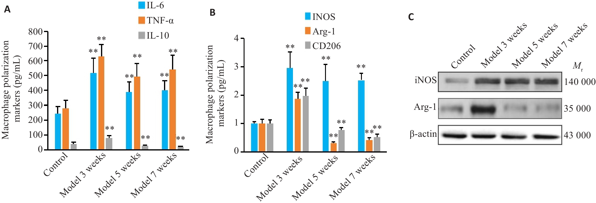
Fig.8 Changes of markers of liver macrophage polarization in chronic iron overload induced by iron dextran. A: Changes of M1 polarization markers; B, C: Changes of M2 polarization markers. **P<0.01 vs Control.
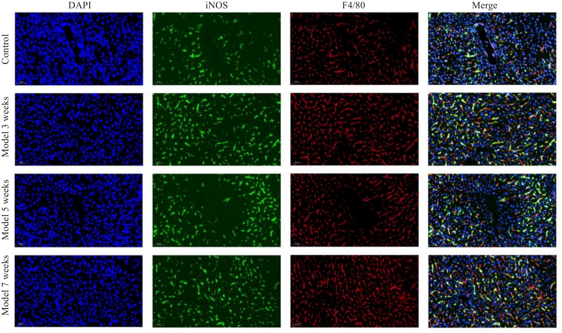
Fig.9 Changes in liver macrophage M1 polarization induced by chronic iron overload (×100).
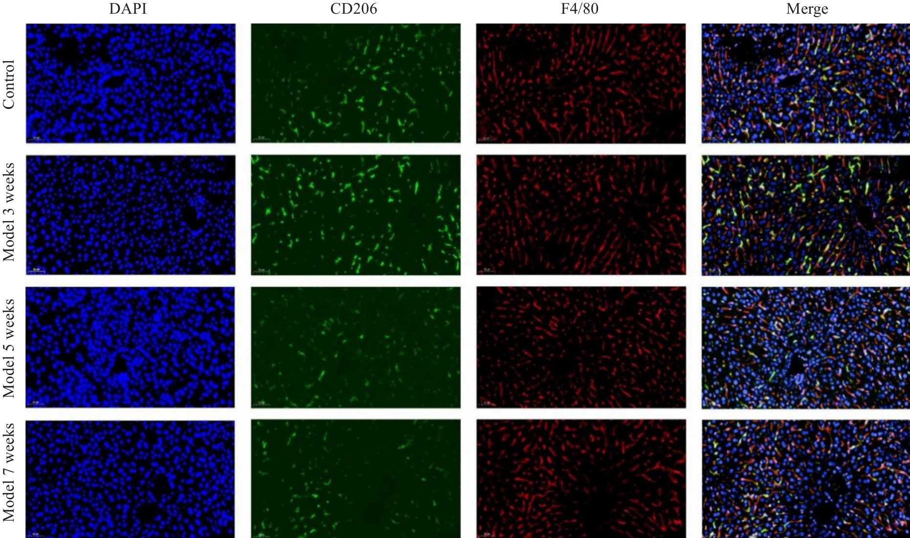
Fig.10 Changes in M2 polarization of liver macrophages in mice with chronic iron overload (×100).
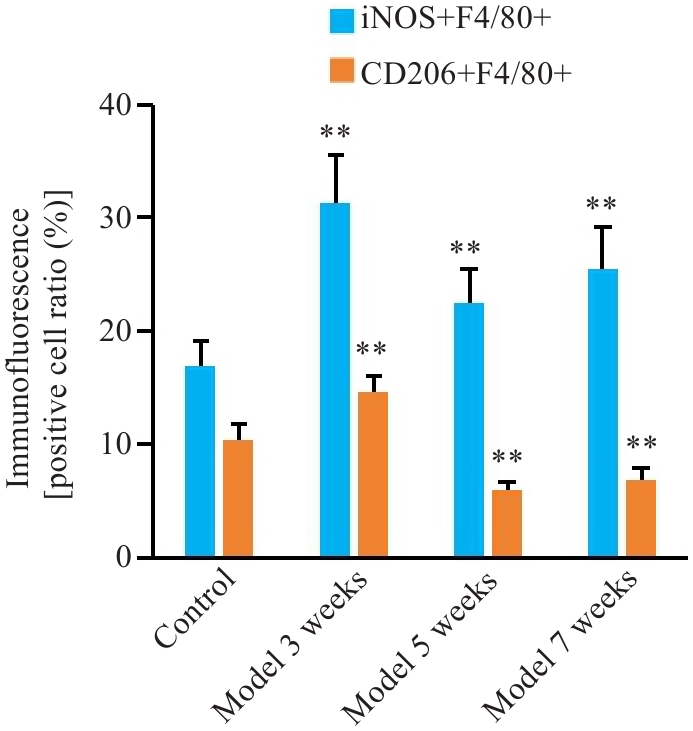
Fig.11 Changes in M1 and M2 polarization of liver macrophages in mice with chronic iron overload. **P<0.01 vs Control.
| 1 | Suresh D, Li A, Miller MJ, et al. Associations between metabolic hyperferritinaemia, fibrosis-promoting alleles and clinical outcomes in steatotic liver disease[J]. Liver Int, 2024, 44(2): 389-98. |
| 2 | Wang C, Ma C, Gong LH, et al. Macrophage polarization and its role in liver disease[J]. Front Immunol, 2021, 12: 803037. |
| 3 | Zanganeh S, Hutter G, Spitler R, et al. Iron oxide nanoparticles inhibit tumour growth by inducing pro-inflammatory macrophage polarization in tumour tissues[J]. Nat Nanotechnol, 2016, 11(11): 986-94. |
| 4 | Zhou Y, Que KT, Zhang Z, et al. Iron overloaded polarizes macrophage to proinflammation phenotype through ROS/acetyl-p53 pathway[J]. Cancer Med, 2018, 7(8): 4012-22. |
| 5 | Handa P, Thomas S, Morgan-Stevenson V, et al. Iron alters macrophage polarization status and leads to steatohepatitis and fibrogenesis[J]. J Leukoc Biol, 2019, 105(5): 1015-26. |
| 6 | Dufrusine B, Di Francesco A, Oddi S, et al. Iron-dependent trafficking of 5-lipoxygenase and impact on human macrophage activation[J]. Front Immunol, 2019, 10: 1347. |
| 7 | Liu EN, Li Z, Zhang Y, et al. Hepcidin induces M1 macrophage polarization in monocytes or THP-1 derived macrophages[J]. Iran J Immunol, 2019, 16(3): 190-9. |
| 8 | Zhang X, Fan LN, Wu JF, et al. Macrophage p38α promotes nutritional steatohepatitis through M1 polarization[J]. J Hepatol, 2019, 71(1): 163-74. |
| 9 | Shendge AK, Panja S, Basu T, et al. Ameliorating effects of white mulberry on iron-overload-induced oxidative stress and liver fibro-sis in Swiss albino mice[J]. Food Chem Toxicol, 2021, 156: 112520. |
| 10 | Philippe MA, Ruddell RG, Ramm GA. Role of iron in hepatic fibrosis: one piece in the puzzle[J]. World J Gastroenterol, 2007, 13(35): 4746-54. |
| 11 | Wood MJ, Gadd VL, Powell LW, et al. Ductular reaction in hereditary hemochromatosis: the link between hepatocyte senesc-ence and fibrosis progression[J]. Hepatology, 2014, 59(3): 848-57. |
| 12 | Pietrangelo A. Iron in NASH, chronic liver diseases and HCC: how much iron is too much[J]? J Hepatol, 2009, 50(2): 249-51. |
| 13 | Houglum K, Bedossa P, Chojkier M. TGF-beta and collagen-alpha 1 (I) gene expression are increased in hepatic acinar zone 1 of rats with iron overload[J]. Am J Physiol, 1994, 267(5 Pt 1): G908-13. |
| 14 | Carthew P, Edwards RE, Smith AG, et al. Rapid induction of hepatic fibrosis in the gerbil after the parenteral administration of iron-dextran complex[J]. Hepatology, 1991, 13(3): 534-9. |
| 15 | Ruddell RG, Hoang-Le D, Barwood JM, et al. Ferritin functions as a proinflammatory cytokine via iron-independent protein kinase C Zeta/nuclear factor kappaB-regulated signaling in rat hepatic stellate cells[J]. Hepatology, 2009, 49(3): 887-900. |
| 16 | Bridle KR, Crawford DHG, Ramm GA. Identification and characterization of the hepatic stellate cell transferrin receptor[J]. Am J Pathol, 2003, 162(5): 1661-7. |
| 17 | Shi HB, Shi HL, Ren F, et al. Naringin in Ganshuang Granule suppresses activation of hepatic stellate cells for anti-fibrosis effect by inhibition of mammalian target of rapamycin[J]. J Cell Mol Med, 2017, 21(3): 500-9. |
| 18 | Kraml P. The role of iron in the pathogenesis of atherosclerosis[J]. Physiol Res, 2017, 66(): S55-67. |
| 19 | Mesquita G, Silva T, Gomes AC, et al. H-Ferritin is essential for macrophages' capacity to store or detoxify exogenously added iron[J]. Sci Rep, 2020, 10(1): 3061. |
| 20 | Gan ZS, Wang QQ, Li JH, et al. Iron reduces M1 macrophage polarization in RAW264.7 macrophages associated with inhibition of STAT1[J]. Mediators Inflamm, 2017, 2017: 8570818. |
| 21 | Ward RJ, Wilmet S, Legssyer R, et al. Effects of marginal iron overload on iron homeostasis and immune function in alveolar macrophages isolated from pregnant and normal rats[J]. Biometals, 2009, 22(2): 211-23. |
| 22 | Lee CJ, Jeong H, Bae Y, et al. Targeting of M2-like tumor-associated macrophages with a melittin-based pro-apoptotic peptide[J]. J Immunother Cancer, 2019, 7(1): 147. |
| 23 | Kao JK, Wang SC, Ho LW, et al. M2-like polarization of THP-1 monocyte-derived macrophages under chronic iron overload[J]. Ann Hematol, 2020, 99(3): 431-41. |
| 24 | Kroner A, Greenhalgh AD, Zarruk JG, et al. TNF and increased intracellular iron alter macrophage polarization to a detrimental M1 phenotype in the injured spinal cord[J]. Neuron, 2014, 83(5): 1098-116. |
| 25 | Sumitomo R, Hirai T, Fujita M, et al. M2 tumor-associated macrophages promote tumor progression in non-small-cell lung cancer[J]. Exp Ther Med, 2019, 18(6): 4490-8. |
| 26 | Harhaji L, Vuckovic O, Miljkovic D, et al. Iron down-regulates macrophage anti-tumour activity by blocking nitric oxide production[J]. Clin Exp Immunol, 2004, 137(1): 109-16. |
| 27 | Yamaguchi T, Fushida S, Yamamoto Y, et al. Tumor-associated macrophages of the M2 phenotype contribute to progression in gastric cancer with peritoneal dissemination[J]. Gastric Cancer, 2016, 19(4): 1052-65. |
| 28 | Hoeft K, Bloch DB, Graw JA, et al. Iron loading exaggerates the inflammatory response to the toll-like receptor 4 ligand lipopo-lysaccharide by altering mitochondrial homeostasis[J]. Anesthesiology, 2017, 127(1): 121-35. |
| 29 | Sindrilaru A, Peters T, Wieschalka S, et al. An unrestrained proinflammatory M1 macrophage population induced by iron impairs wound healing in humans and mice[J]. J Clin Invest, 2011, 121(3): 985-97. |
| 30 | Agoro R, Taleb M, Quesniaux VFJ, et al. Cell iron status influences macrophage polarization[J]. PLoS One, 2018, 13(5): e0196921. |
| [1] | Yuming ZHANG, Shicheng XIA, Linlin ZHANG, Mengxi CHEN, Xiaojing LIU, Qin GAO, Hongwei YE. Protective effect of Lonicerae japonicae flos extract against doxorubicin-induced liver injury in mice [J]. Journal of Southern Medical University, 2024, 44(8): 1571-1581. |
| [2] | CAO Jiafan, SUN Yue, DING Xin, LI Shengwen, CHEN Bo, LAN Tian. Arbutin ameliorates liver fibrosis in mice by inhibiting macrophage recruitment and regulating the Akt/NF-κB and Smad signaling pathways [J]. Journal of Southern Medical University, 2024, 44(4): 652-659. |
| [3] | XU Xiaohui, FENG Jinmei, LUO Ying, HE Xinyu, ZANG Jinbao, HUANG Daochao. Adeno-associated virus-mediated hepatocyte-specific NDUFA13 overexpression protects against CCl4-induced liver fibrosis in mice by inhibiting hepatic NLRP3 activation [J]. Journal of Southern Medical University, 2024, 44(2): 201-209. |
| [4] | Wen ZHAO, Hejing RUAN, Siyuan WANG, Yuzhe CHENG, Miao LEI, Jiufa ZHAO, Chuanmiao LIU. Inhibiting Yes-associated protein alleviates CCl4 liver fibrosis in mice by reducing epithelial mesenchymal transition [J]. Journal of Southern Medical University, 2024, 44(10): 1839-1849. |
| [5] | LIN Jiayi, LOU Anni, LI Xu. Lipopolysaccharide stimulates macrophages to secrete exosomes containing miR-155-5p to promote activation and migration of hepatic stellate cells [J]. Journal of Southern Medical University, 2023, 43(6): 994-1001. |
| [6] | LI jingyi, YANG Siyuan, HAN Zhen, JIANG Tianle, ZHU Yao, ZHOU Zihang, ZHOU Jingping. Akt2 inhibitor promotes M2 macrophage polarization in rats with periapical inflammation by reducing miR-155-5p expression [J]. Journal of Southern Medical University, 2023, 43(4): 568-576. |
| [7] | ZHANG Mengying, LI Zhi, PEI Weiya, LI Xueqin, YANG Hui, ZHU Xiaolong, LÜ Kun. M2 macrophage-derived exosomal lncRNA NR_028113.1 promotes macrophage polarization possibly by activating the JAK2/STAT3 signaling pathway [J]. Journal of Southern Medical University, 2023, 43(3): 393-399. |
| [8] | YANG Xuejia, LI Yujie, WU Dengqiang, MA Yili, ZHOU Sufang. Screening and identification of key genes ATP1B3 and ENAH in the progression of hepatocellular carcinoma: based on data mining and clinical validation [J]. Journal of Southern Medical University, 2022, 42(6): 815-823. |
| [9] | DENG Ya, WANG Chunyan, FU Yiming, LI Zhongbin, JI Dong. A high relapse risk of chronic drug-induced liver injury is correlated with a greater severity of liver fibrosis [J]. Journal of Southern Medical University, 2022, 42(11): 1655-1661. |
| [10] | ZHAO Chenling, DONG Ting, SUN Lunyan, HU Huibing, WANG Qiong, TIAN Liwei, JIANG Zhangsheng. Establishment and validation of a predictive nomogram for liver fibrosis in patients with Wilson disease and abnormal lipid metabolism [J]. Journal of Southern Medical University, 2022, 42(11): 1720-1725. |
| [11] | ZHAO Zhibin, DONG Hui, LI Binghang, SHEN Bo, GUO Yuecheng, GU Tianyi, QU Ying, CAI Xiaobo, LU Lungen. Hydroxynitone suppresses hepatic stellate cell activation by inhibiting TGF-β1 phosphorylation to alleviate CCl4-induced liver fibrosis in rats [J]. Journal of Southern Medical University, 2022, 42(10): 1511-1516. |
| [12] | ZHANG Chunyan, YAN Yuxin, GAO Xiaoyang, MA Yuehong. Therapeutic mechanism of the Mongolian medicine Qiwei Qinggan Powder against liver fibrosis based on UHPLC-TOF-MS combined with network pharmacological methods [J]. Journal of Southern Medical University, 2021, 41(8): 1131-1141. |
| [13] | . Baoganning formula alleviates liver fibrosis in mice by inhibiting hepatic IDO1 expression and promoting phenotypic maturation of dendritic cells [J]. Journal of Southern Medical University, 2021, 41(7): 1002-1011. |
| [14] | . Inhibitory effect of Xinhui citrus fermentation liquor on liver fibrosis in mice [J]. Journal of Southern Medical University, 2021, 41(4): 588-592. |
| [15] | . Amentoflavone inhibits M1 polarization of THP-1-derived foam cells by activating PPAR-α/γ [J]. Journal of Southern Medical University, 2021, 41(3): 344-351. |
| Viewed | ||||||
|
Full text |
|
|||||
|
Abstract |
|
|||||