南方医科大学学报 ›› 2026, Vol. 46 ›› Issue (8): 1915-1925.doi: 10.12122/j.issn.1673-4254.2026.08.19
• • 上一篇
黄炳轶1,2,3(
), 高玉洁1,2,3, 谢靖宜1,2,3, 严晨2,3, 顾梦茹2,3, 张志郅2,3, 郭锦涛1,2,3, 刘嵩1,2,3, 王子良1,2,3, 杨玉萍4, 左芦根1,2, 陈德利1,2(
)
收稿日期:2025-12-08
出版日期:2026-08-20
发布日期:2026-08-01
通讯作者:
陈德利
E-mail:huangbingyi1005@163.com;13965295950@139.com
作者简介:黄炳轶,在读硕士研究生,E-mail: huangbingyi1005@163.com
基金资助:
Bingyi HUANG1,2,3(
), Yujie GAO1,2,3, Jingyi XIE1,2,3, Chen YAN2,3, Mengru GU2,3, Zhizhi ZHANG2,3, Jintao GUO1,2,3, Song LIU1,2,3, Ziliang WANG1,2,3, Yuping YANG4, Lugen ZUO1,2, Deli CHEN1,2(
)
Received:2025-12-08
Online:2026-08-20
Published:2026-08-01
Contact:
Deli CHEN
E-mail:huangbingyi1005@163.com;13965295950@139.com
摘要:
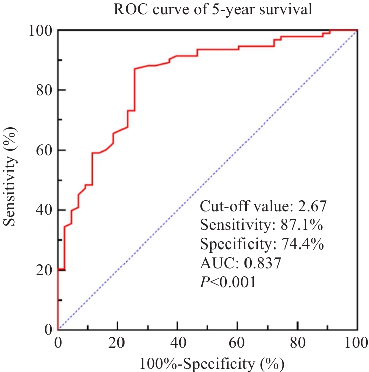
目的 探讨棕榈酰蛋白硫酯酶1(PPT1)在胃癌组织中的表达规律及其与患者临床预后的关系,并阐明其调控癌细胞脂质代谢的分子机制。 方法 联合TCGA-STAD数据库与临床样本免疫组化染色,分析PPT1在胃癌组织及癌旁组织中的表达差异。回顾性纳入136例接受根治性切除的胃癌患者,提取其病理与随访参数,运用Kaplan-Meier法、单因素及多因素Cox回归模型分析PPT1表达与术后5年生存率的关联,据此建立生存预测列线图并使用ROC曲线评估其效能。此外,结合GO及REACTOME数据库进行生物学通路富集。体外水平上,通过慢病毒转染构建胃癌细胞系模型,利用CCK-8法及Transwell小室法分别检测细胞增殖、迁移和侵袭能力。采用油红O染色、Western blotting实验以及抑制剂U0126逆转实验,探讨PPT1介导ERK信号通路对脂质代谢的调控作用。 结果 PPT1蛋白定位于细胞质,其在胃癌组织中的表达水平显著高于癌旁正常组织。PPT1高表达与患者外周血CEA、CA19-9水平的上升以及较高的T、N分期密切相关(P<0.05)。生存分析及Cox回归提示,PPT1高表达是导致患者术后5年生存率下降的独立危险因素。基于该指标构建的列线图显示出良好的预测准确度(C-index=0.812),其对应的ROC曲线AUC值为0.837。体外实验证实,下调PPT1表达可显著抑制胃癌细胞的增殖及侵袭转移能力,而过表达PPT1则起到反向的促进作用(P<0.05)。机制方面,过表达PPT1不仅诱导了细胞内脂质的异常蓄积,还显著上调了SREBP-1及FASN等脂质合成关键蛋白的表达(P<0.05)。该促脂质生成效应及其伴随的ERK通路激活,均可被U0126有效逆转。 结论 胃癌组织中PPT1呈显著的高表达状态且提示患者预后不良。PPT1可能通过激活ERK信号通路影响胃癌细胞脂质代谢,从而助推肿瘤的恶性进展。PPT1有望作为胃癌的新型预后生物标志物与潜在干预靶点。
黄炳轶, 高玉洁, 谢靖宜, 严晨, 顾梦茹, 张志郅, 郭锦涛, 刘嵩, 王子良, 杨玉萍, 左芦根, 陈德利. PPT1在胃癌中的预后价值及其对癌细胞脂质代谢的作用和机制[J]. 南方医科大学学报, 2026, 46(8): 1915-1925.
Bingyi HUANG, Yujie GAO, Jingyi XIE, Chen YAN, Mengru GU, Zhizhi ZHANG, Jintao GUO, Song LIU, Ziliang WANG, Yuping YANG, Lugen ZUO, Deli CHEN. Prognostic value of palmitoyl-protein thioesterase-1 in gastric cancer and its regulatory role in tumor cell lipid metabolism[J]. Journal of Southern Medical University, 2026, 46(8): 1915-1925.
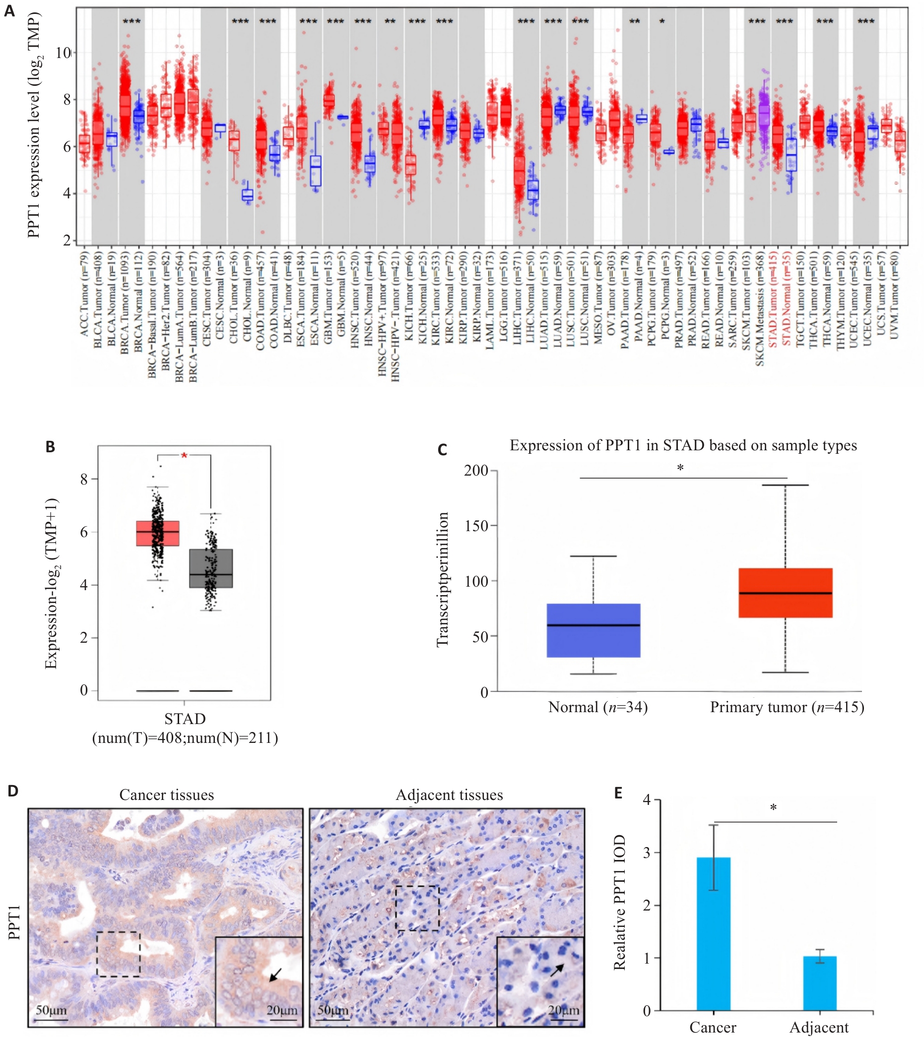
图1 PPT1在胃癌组织高表达
Fig.1 Palmitoyl-protein thioesterase-1 (PPT1) is highly expressed in gastric carcinoma (GC) tissues. A: Expression of PPT1 in human malignant tumors. B, C: Differential expression of PPT1 mRNA between gastric adenocarcinoma tissues and normal adjacent tissues (based on the TCGA-STAD cohort). D, E: Immunohistochemical (IHC) analysis of PPT1 in paired gastric carcinoma and adjacent normal tissues (n=136). *P<0.05, **P<0.01, ***P<0.001.
| Factor | n | PPT1 expression [(n (%)] | χ2 | P | |
|---|---|---|---|---|---|
| Low (n=68) | High (n=68) | ||||
| Gender | |||||
| Male | 80 | 43 (53.8%) | 37 (46.2%) | 1.093 | 0.296 |
| Female | 56 | 25 (44.6%) | 31 (55.4%) | ||
| Age (year) | |||||
| <60 | 45 | 22 (48.9%) | 23 (51.1%) | 0.033 | 0.855 |
| ≥60 | 91 | 46 (50.5%) | 45 (49.5%) | ||
| CEA (μg/L) | |||||
| <5 | 59 | 41 (69.5%) | 18 (30.5%) | 15.836 | <0.001 |
| ≥5 | 77 | 27 (35.1%) | 50 (64.9%) | ||
| CA19-9 (kU/L) | |||||
| <37 | 50 | 35 (70.0%) | 15 (30.0%) | 12.651 | <0.001 |
| ≥37 | 86 | 33 (38.4%) | 53 (61.6%) | ||
| Histological type | |||||
| Adenocarcinoma | 99 | 52 (52.5%) | 47 (47.5%) | 0.928 | 0.335 |
| Other | 37 | 16 (43.2%) | 21 (56.8%) | ||
| Pathological grading | |||||
| G1-G2 | 48 | 25 (52.1%) | 23 (47.9%) | 0.129 | 0.720 |
| G3-G4 | 88 | 43 (48.9%) | 45 (51.1%) | ||
| Tumor size (cm) | |||||
| <5 | 57 | 30 (52.6%) | 27 (47.4%) | 0.272 | 0.602 |
| ≥5 | 79 | 38 (48.1%) | 41 (51.9%) | ||
| T stage | |||||
| T1-T2 | 61 | 42 (68.9%) | 19 (31.1%) | 15.725 | <0.001 |
| T3-T4 | 75 | 26 (34.7%) | 49 (65.3%) | ||
| N stage | |||||
| N0-N1 | 63 | 41 (65.1%) | 22 (34.9%) | 10.675 | 0.001 |
| N2-N3 | 73 | 27 (37.0%) | 46 (63.0%) | ||
表1 PPT1表达水平与胃癌临床病理参数的相关性分析
Tab.1 Correlation between PPT1 expression levels and clinico-pathological parameters in gastric cancer
| Factor | n | PPT1 expression [(n (%)] | χ2 | P | |
|---|---|---|---|---|---|
| Low (n=68) | High (n=68) | ||||
| Gender | |||||
| Male | 80 | 43 (53.8%) | 37 (46.2%) | 1.093 | 0.296 |
| Female | 56 | 25 (44.6%) | 31 (55.4%) | ||
| Age (year) | |||||
| <60 | 45 | 22 (48.9%) | 23 (51.1%) | 0.033 | 0.855 |
| ≥60 | 91 | 46 (50.5%) | 45 (49.5%) | ||
| CEA (μg/L) | |||||
| <5 | 59 | 41 (69.5%) | 18 (30.5%) | 15.836 | <0.001 |
| ≥5 | 77 | 27 (35.1%) | 50 (64.9%) | ||
| CA19-9 (kU/L) | |||||
| <37 | 50 | 35 (70.0%) | 15 (30.0%) | 12.651 | <0.001 |
| ≥37 | 86 | 33 (38.4%) | 53 (61.6%) | ||
| Histological type | |||||
| Adenocarcinoma | 99 | 52 (52.5%) | 47 (47.5%) | 0.928 | 0.335 |
| Other | 37 | 16 (43.2%) | 21 (56.8%) | ||
| Pathological grading | |||||
| G1-G2 | 48 | 25 (52.1%) | 23 (47.9%) | 0.129 | 0.720 |
| G3-G4 | 88 | 43 (48.9%) | 45 (51.1%) | ||
| Tumor size (cm) | |||||
| <5 | 57 | 30 (52.6%) | 27 (47.4%) | 0.272 | 0.602 |
| ≥5 | 79 | 38 (48.1%) | 41 (51.9%) | ||
| T stage | |||||
| T1-T2 | 61 | 42 (68.9%) | 19 (31.1%) | 15.725 | <0.001 |
| T3-T4 | 75 | 26 (34.7%) | 49 (65.3%) | ||
| N stage | |||||
| N0-N1 | 63 | 41 (65.1%) | 22 (34.9%) | 10.675 | 0.001 |
| N2-N3 | 73 | 27 (37.0%) | 46 (63.0%) | ||
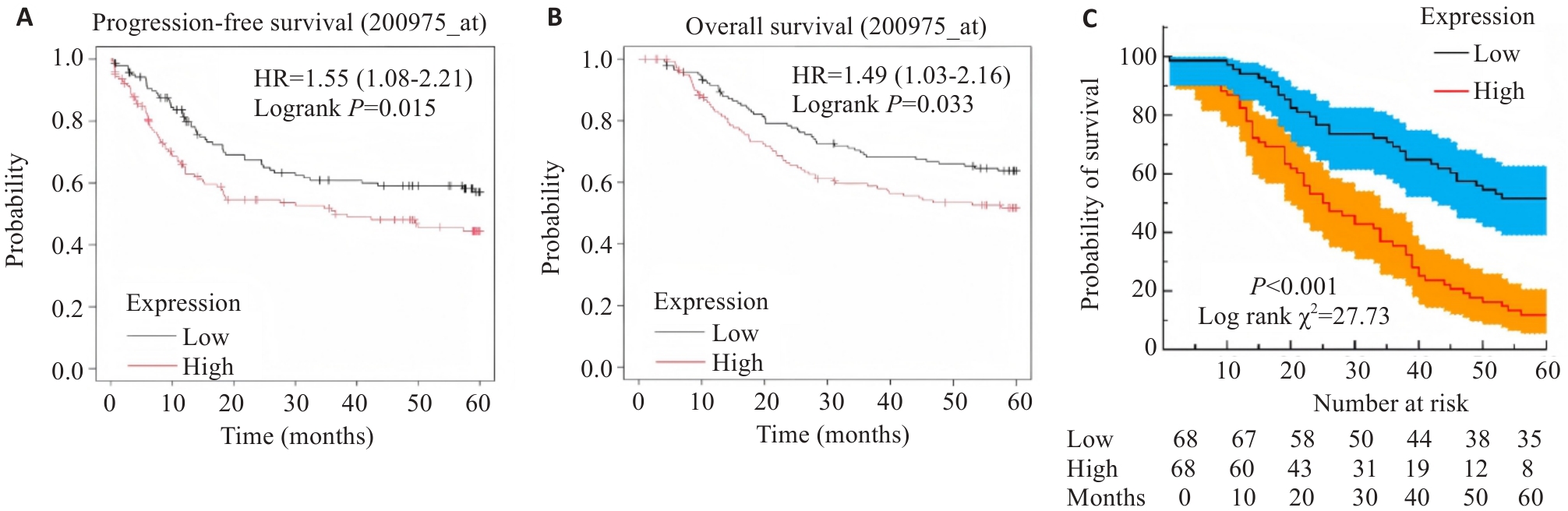
图2 PPT1分布特征与胃癌根治术后5年生存期的关系
Fig.2 Association of PPT1 expression levels with 5-year survival of GC patients after radical gastrectomy. A: Progression-free survival of the patients. B: Overall survival of the patients. C: Postoperative survival of patients with high and low PPT1 expression (n=136).
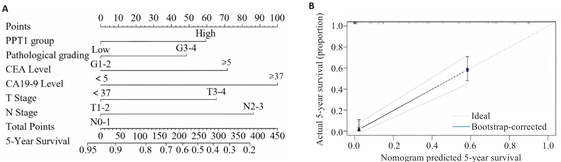
图4 胃癌根治术后5年生存率预测列线图的构建与内部验证
Fig.4 Construction and internal validation of a nomogram for predicting 5-year survival in GC patients after radical gastrectomy. A: Prognostic nomogram. B: Calibration curve (n=136).
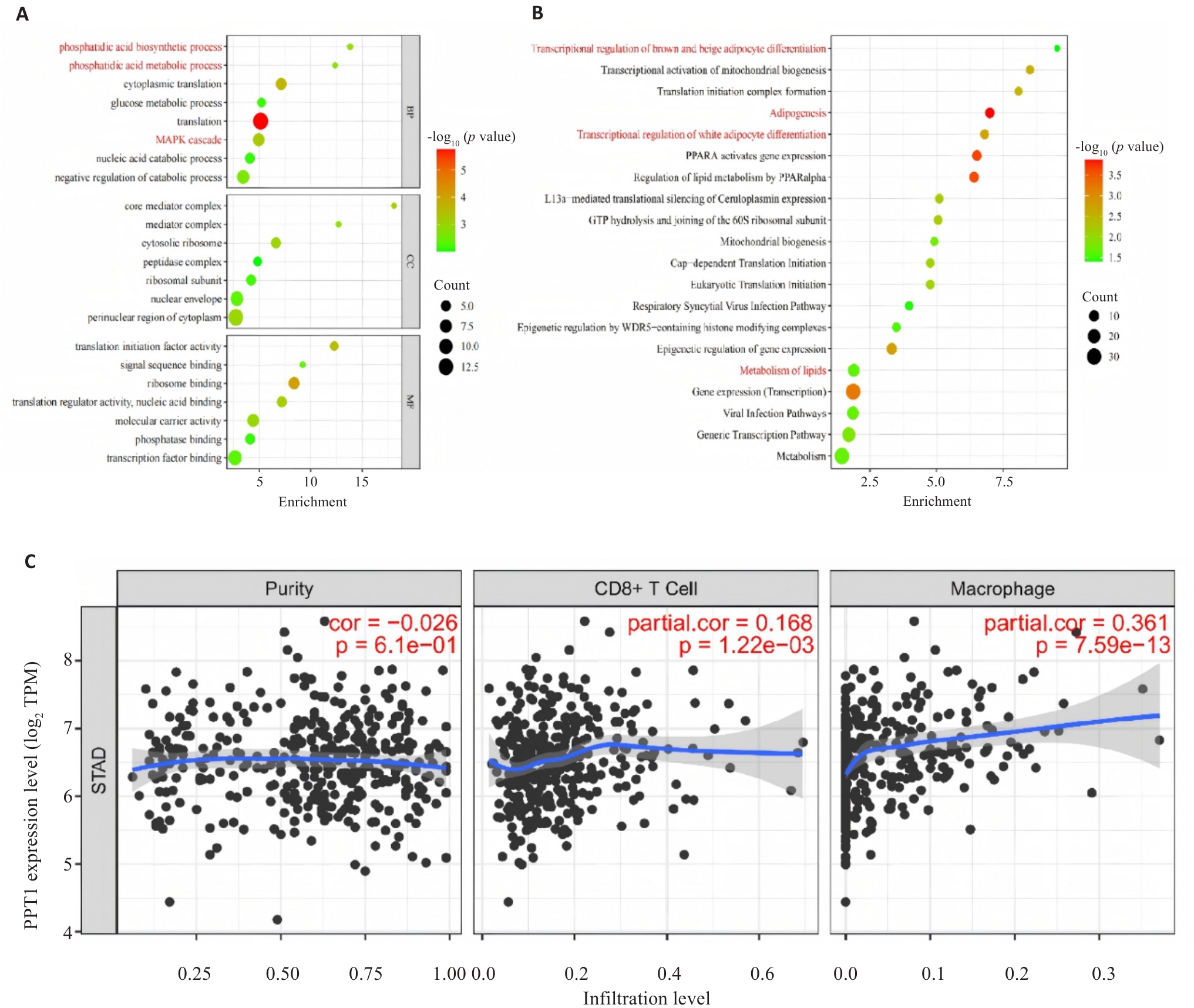
图6 PPT1的富集分析及免疫浸润相关性分析
Fig.6 Enrichment analysis of PPT1 and correlation analysis of immune infiltration. A: GO analysis of PPT1 in GC. B: REACTOME analysis of PPT1 in GC. C: Correlation analysis of PPT1 with immune cell infiltration in the TIMER database.
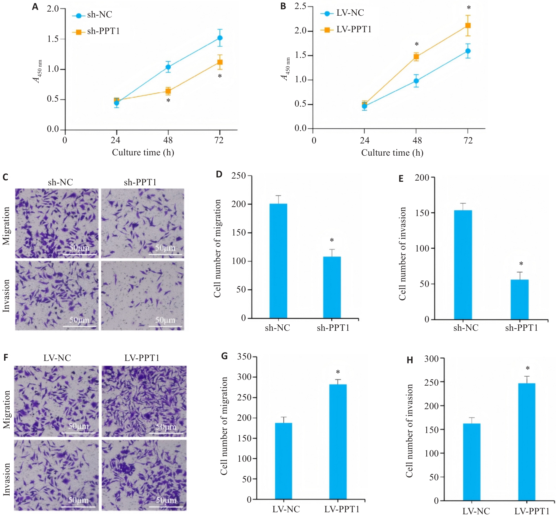
图7 PPT1表达水平对胃癌细胞的增殖、迁移和侵袭的影响
Fig.7 PPT1 overexpression enhances proliferation, migration, and invasion of GC cells. A, B: Proliferation curves showing that PPT1 knockdown inhibits (A) while PPT1 overexpression promotes (B) cell growth. C-H: Transwell assays demonstrating that PPT1 knockdown suppresses and its overexpression enhances cell migration and invasion. *P<0.05 vs sh-NC group/LV-NC group (n=3).
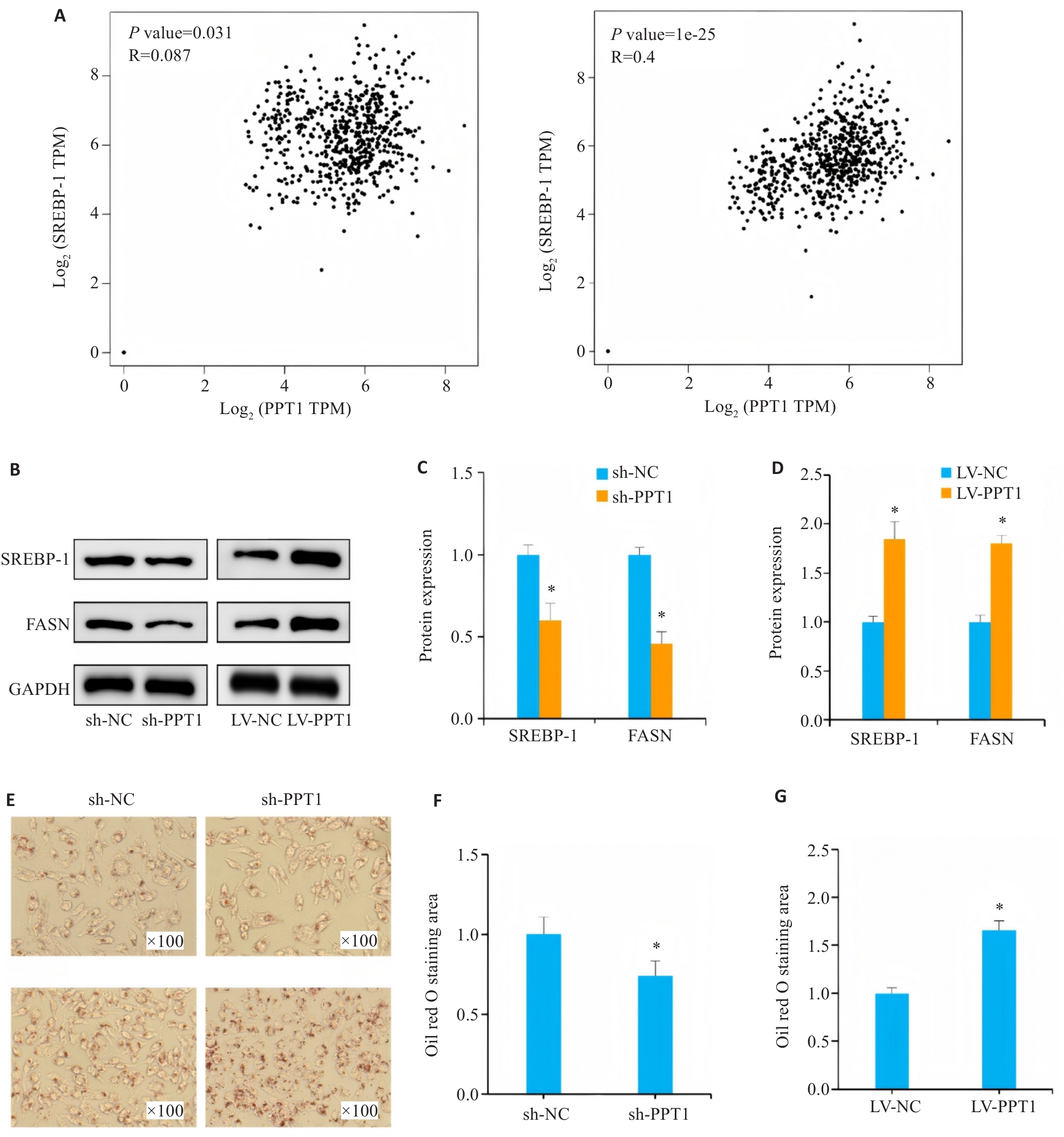
图8 PPT1表达水平对胃癌细胞脂质代谢的影响
Fig.8 Effect of PPT1 expression level on lipid metabolism in GC cells. A: Expression of PPT1 in GC tissues is positively correlated with expressions of FASN and SREBP-1 in the GEPIA database. B-D: Expressions of FASN and SREBP-1 protein in GC cells (n=3). E: Representative images of lipid droplets in GC cells from each group detected by Oil Red O staining. F, G: Statistical analysis of the area positive for Oil Red O staining (n=3). *P<0.05 vs sh-NC group/LV-NC group.
图9 PPT1表达在ERK信号通路及脂质代谢中的影响
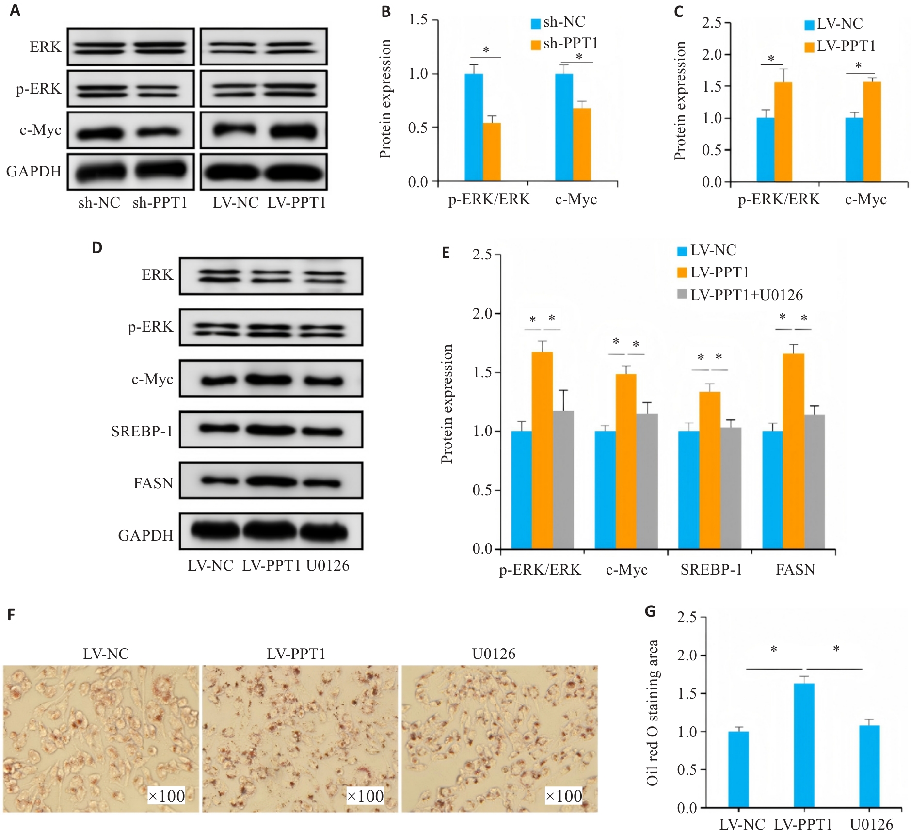
Fig.9 Effects of PPT1 on the ERK signaling pathway and lipid metabolism in GC cells (n=3). A-C: Impact of PPT1 expression on the ERK signaling pathway. D, E: Effect of U0126 on the ERK signaling pathway and lipid metabolism-related protein expressions in GC cells. F: Representative images of lipid droplets in GC cells from each group detected by Oil Red O staining. G: Statistical analysis of the area positive for Oil Red O staining. *P<0.05.
| [1] | Thrift AP, Wenker TN, El-Serag HB. Global burden of gastric cancer: epidemiological trends, risk factors, screening and prevention[J]. Nat Rev Clin Oncol, 2023, 20(5): 338-49. doi:10.1038/s41571-023-00747-0 |
| [2] | Bray F, Laversanne M, Sung H, et al. Global cancer statistics 2022: GLOBOCAN estimates of incidence and mortality worldwide for 36 cancers in 185 countries[J]. CA Cancer J Clin, 2024, 74(3): 229-63. doi:10.3322/caac.21834 |
| [3] | Leong T, Smithers BM, Michael M, et al. Preoperative chemoradiotherapy for resectable gastric cancer[J]. N Engl J Med, 2024, 391(19): 1810-21. doi:10.1056/nejmoa2405195 |
| [4] | Joshi SS, Badgwell BD. Current treatment and recent progress in gastric cancer[J]. CA Cancer J Clin, 2021, 71(3): 264-79. doi:10.3322/caac.21657 |
| [5] | Subbiah V, Gouda MA, Ryll B, et al. The evolving landscape of tissue-agnostic therapies in precision oncology[J]. CA Cancer J Clin, 2024, 74(5): 433-52. doi:10.3322/caac.21844 |
| [6] | Bhardwaj M, Lee JJ, Versace AM, et al. Lysosomal lipid peroxidation regulates tumor immunity[J]. J Clin Investig, 2023, 133(8): e164596. doi:10.1172/jci164596 |
| [7] | Brun S, Bestion E, Raymond E, et al. GNS561, a clinical-stage PPT1 inhibitor, is efficient against hepatocellular carcinoma via modulation of lysosomal functions[J]. Autophagy, 2022, 18(3): 678-94. doi:10.1080/15548627.2021.1988357 |
| [8] | Zhou BH, Hao QY, Liang YM, et al. Protein palmitoylation in cancer: molecular functions and therapeutic potential[J]. Mol Oncol, 2023, 17(1): 3-26. doi:10.1002/1878-0261.13308 |
| [9] | Rebecca VW, Nicastri MC, Fennelly C, et al. PPT1 promotes tumor growth and is the molecular target of chloroquine derivatives in cancer[J]. Cancer Discov, 2019, 9(2): 220-9. doi:10.1158/2159-8290.cd-18-0706 |
| [10] | Weng JL, Liu SQ, Zhou Q, et al. Intratumoral PPT1-positive macrophages determine immunosuppressive contexture and immunotherapy response in hepatocellular carcinoma[J]. J Immunother Cancer, 2023, 11(6): e006655. doi:10.1136/jitc-2022-006655 |
| [11] | Zhao P, Yuan F, Xu LJ, et al. HKDC1 reprograms lipid metabolism to enhance gastric cancer metastasis and cisplatin resistance via forming a ribonucleoprotein complex[J]. Cancer Lett, 2023, 569: 216305. doi:10.1016/j.canlet.2023.216305 |
| [12] | Zhou XY, Chang D, Xu HY, et al. Progress on lysosomal PPT1-mediated regulation of cellular homeostasis and pathogenesis[J]. Hereditas, 2023, 45(10): 874-86. |
| [13] | Luo Q, Hu S, Tang Y, et al. PPT1 Promotes Growth and Inhibits Ferroptosis of Oral Squamous Cell Carcinoma Cells[J]. Curr Cancer Drug Targets, 2024,24(10):1047-60. doi:10.2174/0115680096294098240123104657 |
| [14] | Nakamura Y, Kawazoe A, Lordick F, et al. Biomarker-targeted therapies for advanced-stage gastric and gastro-oesophageal junction cancers: an emerging paradigm[J]. Nat Rev Clin Oncol, 2021, 18(8): 473-87. doi:10.1038/s41571-021-00492-2 |
| [15] | Won Y, Jang B, Lee SH, et al. Oncogenic fatty acid metabolism rewires energy supply chain in gastric carcinogenesis[J]. Gastroenterology, 2024, 166(5): 772-86. e14. doi:10.1053/j.gastro.2024.01.027 |
| [16] | Bi L, Ren YD, Feng MX, et al. HDAC11 regulates glycolysis through the LKB1/AMPK signaling pathway to maintain hepatocellular carcinoma stemness[J]. Cancer Res, 2021, 81(8): 2015-28. doi:10.1158/0008-5472.can-20-3044 |
| [17] | Broadfield LA, Pane AA, Talebi A, et al. Lipid metabolism in cancer: New perspectives and emerging mechanisms[J]. Dev Cell, 2021, 56(10): 1363-93. doi:10.1016/j.devcel.2021.04.013 |
| [18] | Cao YH. Adipocyte and lipid metabolism in cancer drug resistance[J]. J Clin Investig, 2019, 129(8): 3006-17. doi:10.1172/jci127201 |
| [19] | Ebner M, Puchkov D, López-Ortega O, et al. Nutrient-regulated control of lysosome function by signaling lipid conversion[J]. Cell, 2023, 186(24): 5328-46. e26. doi:10.1016/j.cell.2023.09.027 |
| [20] | Zhang S, Peng XQ, Yang S, et al. The regulation, function, and role of lipophagy, a form of selective autophagy, in metabolic disorders[J]. Cell Death Dis, 2022, 13: 132. doi:10.1038/s41419-022-04593-3 |
| [21] | Ballabio A, Bonifacino JS. Lysosomes as dynamic regulators of cell and organismal homeostasis[J]. Nat Rev Mol Cell Biol, 2020, 21(2): 101-18. doi:10.1038/s41580-019-0185-4 |
| [22] | 陈元杏, 张 丽, 张玉荣, 等. 胃癌组织中UBE2S、FASN蛋白的表达及其临床病理意义[J]. 分子诊断与治疗杂志, 2024, 16(10): 1932-5, 1939. |
| [23] | Yu L, Wang X, Du Y, et al. FASN knockdown inhibited anoikis resistance of gastric cancer cells via P-ERK1/2/bcl-xL pathway[J]. Gastroenterol Res Pract, 2021, 2021(1): 6674204. doi:10.1155/2021/6674204 |
| [24] | Yu HY, Kou QM, Yuan H, et al. Alkannin triggered apoptosis and ferroptosis in gastric cancer by suppressing lipid metabolism mediated by the c-Fos/SREBF1 axis[J]. Phytomedicine, 2025, 140: 156604. doi:10.1016/j.phymed.2025.156604 |
| [25] | Chen ML, Li HZ, Zheng SS, et al. Nobiletin targets SREBP1/ACLY to induce autophagy-dependent cell death of gastric cancer cells through PI3K/Akt/mTOR signaling pathway[J]. Phytomedicine, 2024, 128: 155360. doi:10.1016/j.phymed.2024.155360 |
| [26] | 何卫美, 宋育林, 方 芳, 等. MEK/ERK通路在高脂饮食诱导NAFLD大鼠SREBP-1表达中的作用[J]. 胃肠病学和肝病学杂志, 2010, 19(5): 427-31. |
| [27] | Wei WX, Qin BF, Wen WJ, et al. FBXW7β loss-of-function enhances FASN-mediated lipogenesis and promotes colorectal cancer growth[J]. Signal Transduct Target Ther, 2023, 8: 187. doi:10.1038/s41392-023-01405-8 |
| [28] | Geng F, Guo DL. SREBF1/SREBP-1 concurrently regulates lipid synthesis and lipophagy to maintain lipid homeostasis and tumor growth[J]. Autophagy, 2024, 20(5): 1183-5. doi:10.1080/15548627.2023.2275501 |
| [29] | Shi J, Miao DJ, Lv QY, et al. The m6A modification-mediated OGDHL exerts a tumor suppressor role in ccRCC by downregulating FASN to inhibit lipid synthesis and ERK signaling[J]. Cell Death Dis, 2023, 14: 560. doi:10.1038/s41419-023-06090-7 |
| [30] | Liu JT, Wei YH, Jia WB, et al. Chenodeoxycholic acid suppresses AML progression through promoting lipid peroxidation via ROS/p38 MAPK/DGAT1 pathway and inhibiting M2 macrophage polarization[J]. Redox Biol, 2022, 56: 102452. doi:10.1016/j.redox.2022.102452 |
| [31] | Wen ZK, Jin K, Shen Y, et al. N-myristoyltransferase deficiency impairs activation of kinase AMPK and promotes synovial tissue inflammation[J]. Nat Immunol, 2019, 20(3): 313-25. doi:10.1038/s41590-018-0296-7 |
| [32] | Mesquita FS, Abrami L, Linder ME, et al. Mechanisms and functions of protein S-acylation[J]. Nat Rev Mol Cell Biol, 2024, 25(6): 488-509. doi:10.1038/s41580-024-00700-8 |

| [1] | 王坤, 侯顺福, 李勇, 刘清华, 尹崇高, 李洪利. miR-183-5p通过靶向调控KLHL24影响三阴性乳腺癌细胞增殖、侵袭、迁移及铁死亡[J]. 南方医科大学学报, 2026, 46(8): 1790-1798. |
| [2] | 但昱彤, 张庭辅, 刘红梅, 张鹏. 甲基结合蛋白1对人脐静脉内皮细胞迁移、增殖以及血管形成的影响[J]. 南方医科大学学报, 2026, 46(7): 1585-1592. |
| [3] | 陈镇霖, 柴鹏, 毛洋奇, 刘然新, 宋烨. ZEB2通过CYLD/FASN轴重编程脂质代谢促进胶质母细胞瘤进展[J]. 南方医科大学学报, 2026, 46(6): 1290-1300. |
| [4] | 董保龙, 彭江山, 李进茹, 孟云, 杨晓军. 过表达miR-593-5p通过靶向PLK1抑制胃癌细胞的迁移、侵袭、增殖并促进其凋亡[J]. 南方医科大学学报, 2026, 46(4): 871-879. |
| [5] | 王煜煌, 王文锐, 程淑洁, 冯云龙, 张卓, 施其英, 李雨佳, 胡茜文, 吴明彩. 槲皮素通过调控Hippo通路蛋白YAP表达抑制胃癌细胞增殖与迁移并促进其凋亡[J]. 南方医科大学学报, 2026, 46(2): 374-384. |
| [6] | 王强, 柴志欣, 邓玉露, 张志葳, 龚英, 高圣, 冯平锋. cGAS-STING激动剂cGAMP可增强自然杀伤细胞的抗胃癌效应[J]. 南方医科大学学报, 2026, 46(2): 434-442. |
| [7] | 李艳宇, 李晨, 戴传君, 郭润之, 韩浩宇, 卢林明, 周芳芳, 支慧. 皖南蝮蛇抑瘤组分-Ι通过调控RAI14抑制顺铂耐药胃癌细胞的增殖与侵袭[J]. 南方医科大学学报, 2026, 46(1): 113-121. |
| [8] | 徐嘉艺, 杨迪, 臧开来, 褚孟恩, 赵庆瑶, 李晴, 鲁森, 陈修丽, 李宁. EVA1A过表达通过调节脂质代谢和促进脂滴自噬改善非酒精性脂肪肝[J]. 南方医科大学学报, 2026, 46(1): 150-158. |
| [9] | 赵锦燕, 彭娇, 林明和, 朱晓勤, 黄彬, 林久茂. 清解扶正颗粒通过抑制线粒体依赖的凋亡、激活AMPK-PGC-1α通路缓解5-氟尿嘧啶引起的骨骼肌损伤[J]. 南方医科大学学报, 2026, 46(1): 94-103. |
| [10] | 骆碧云, 易欣, 蔡怡静, 张世卿, 王鹏, 李彤, 翁建霖, 周平正. 清心牛黄丸通过改善脂质代谢紊乱缓解小鼠非酒精性脂肪性肝病[J]. 南方医科大学学报, 2025, 45(9): 1840-1849. |
| [11] | 杨子为, 吕畅, 董柱, 计书磊, 毕生辉, 张雪花, 王晓武. 金樱子通过调控Src-AKT1轴抑制肺动脉高压平滑肌增殖[J]. 南方医科大学学报, 2025, 45(9): 1889-1902. |
| [12] | 王子良, 陈孝华, 杨晶晶, 严晨, 张志郅, 黄炳轶, 赵萌, 刘嵩, 葛思堂, 左芦根, 陈德利. 高表达SURF4通过抑制紧密连接蛋白表达促进胃癌细胞的恶性生物学行为[J]. 南方医科大学学报, 2025, 45(8): 1732-1742. |
| [13] | 陈鑫源, 吴成挺, 李瑞迪, 潘雪芹, 张耀丹, 陶俊宇, 林才志. 双术汤通过P53/SLC7A11/GPX4通路诱导胃癌细胞铁死亡[J]. 南方医科大学学报, 2025, 45(7): 1363-1371. |
| [14] | 谢婷, 王云云, 郭婷, 袁春华. 雷氏大疣蛛多肽毒素组分通过激活促凋亡通路和协同作用抑制癌细胞增殖[J]. 南方医科大学学报, 2025, 45(7): 1460-1470. |
| [15] | 吴璇, 方家敏, 韩玮玮, 陈琳, 孙菁, 金齐力. 高表达PRELID1促进胃癌细胞上皮间质转化并与不良预后相关[J]. 南方医科大学学报, 2025, 45(7): 1535-1542. |
| 阅读次数 | ||||||
|
全文 |
|
|||||
|
摘要 |
|
|||||