
南方医科大学学报 ›› 2026, Vol. 46 ›› Issue (7): 1487-1497.doi: 10.12122/j.issn.1673-4254.2026.07.03
• • 上一篇
乔通1,3( ), 张龙涛1,3, 章雨1, 黄菊2, 李晴晴1, 耿志军2, 胡建国1,2, 李静1,2()
收稿日期:2025-12-29
出版日期:2026-07-20
发布日期:2026-07-20
通讯作者:
李静
E-mail:qt1231126@163.com;lijingbyfy@bbmu.edu.cn
作者简介:乔 通,在读硕士研究生,E-mail: qt1231126@163.com
基金资助:
Tong QIAO1,3(), Longtao ZHANG1,3, Yu ZHANG1, Ju HUANG2, Qingqing LI1, Zhijun GENG2, Jianguo HU1,2, Jing Li1,2()
Received:2025-12-29
Online:2026-07-20
Published:2026-07-20
Contact:
Jing Li
E-mail:qt1231126@163.com;lijingbyfy@bbmu.edu.cn
摘要:
目的 探讨异香草酸(IVA)对葡聚糖硫酸钠(DSS)诱导的小鼠溃疡性结肠炎(UC)的保护作用及其潜在机制。 方法 将48只C57BL/6雄性小鼠随机分为6组(n=8):对照组(Con)、DSS模型组(DSS)、IVA低剂量组(IVA-L,50 mg/kg)、IVA中剂量组(IVA-M,100 mg/kg)、IVA高剂量组(IVA-H,150 mg/kg)及阳性对照组(5-ASA,100 mg/kg)。除对照组自由饮水外,其余各组自第1天起自由饮用2.5% DSS溶液至第7天,第8天更换为普通水。各干预组每日灌胃相应剂量的IVA或5-ASA(溶于含0.1% DMSO的生理盐水,100 μL/只),对照组及DSS组灌胃等体积溶剂。第10天处死动物,取结肠组织进行后续检测。实验期间监测小鼠体质量,评估疾病活动指数(DAI);处死小鼠后检测结肠长度,进行结肠组织病理学评分;采用免疫荧光、Western blotting、TUNEL染色、JC-1染色等,分别评估IVA对DSS诱导小鼠和NCM460细胞模型的屏障功能、细胞凋亡及线粒体功能的影响;并结合网络药理学分析其潜在作用通路。 结果 与Con组相比,DSS组小鼠体质量下降(P<0.05),DAI评分升高(P<0.05),结肠长度缩短(P<0.05),结肠组织中白细胞介素-6(IL-6)和白细胞介素-1β(IL-1β)水平升高(P<0.05),结肠黏膜结构破坏、炎细胞浸润增加、杯状细胞减少;而经IVA干预后上述指标呈剂量依赖性改善(P<0.05)。与DSS组相比,IVA-M组紧密连接蛋白ZO-1和Claudin-1表达升高(P<0.05),肠上皮细胞凋亡率降低(P<0.05),Bax、C-caspase3表达下调,Bcl-2表达上调(P<0.05),TOMM20阳性细胞数增加,线粒体呼吸链复合体Ⅰ、Ⅳ活性升高(P<0.05)。在NCM460细胞中,与C-Con组相比,C-DSS组线粒体膜电位下降,复合体Ⅰ、Ⅳ活性降低(P<0.05),细胞凋亡率增加(P<0.05),ZO-1、Claudin-1表达下降(P<0.05);与C-DSS组相比,C-IVA组上述指标均显著改善(P<0.05)。网络药理学分析提示PPARγ通路为潜在作用靶点。Western blotting结果显示,IVA-M组和C-IVA组PPARγ蛋白表达水平均高于相应模型组(P<0.05)。加入PPARγ拮抗剂GW9662后,与C-IVA组相比,C-IVA+GW9662组线粒体膜电位下降,复合体Ⅰ、Ⅳ活性降低(P<0.05),细胞凋亡率升高(P<0.05),Bcl-2表达降低,Bax、C-caspase3表达升高(P<0.05),ZO-1、Claudin-1表达降低(P<0.05)。 结论 IVA通过激活PPARγ通路,改善线粒体功能,抑制炎症与细胞凋亡,从而缓解DSS诱导的结肠炎,具有成为UC治疗候选药物的潜力。
乔通, 张龙涛, 章雨, 黄菊, 李晴晴, 耿志军, 胡建国, 李静. 异香草酸激活PPARγ通路改善线粒体功能缓解葡聚糖硫酸钠诱导的小鼠溃疡性结肠炎[J]. 南方医科大学学报, 2026, 46(7): 1487-1497.
Tong QIAO, Longtao ZHANG, Yu ZHANG, Ju HUANG, Qingqing LI, Zhijun GENG, Jianguo HU, Jing Li. Isovanillic acid alleviates dextran sulfate sodium-induced ulcerative colitis in mice by improving mitochondrial function via activating the PPARγ pathway[J]. Journal of Southern Medical University, 2026, 46(7): 1487-1497.
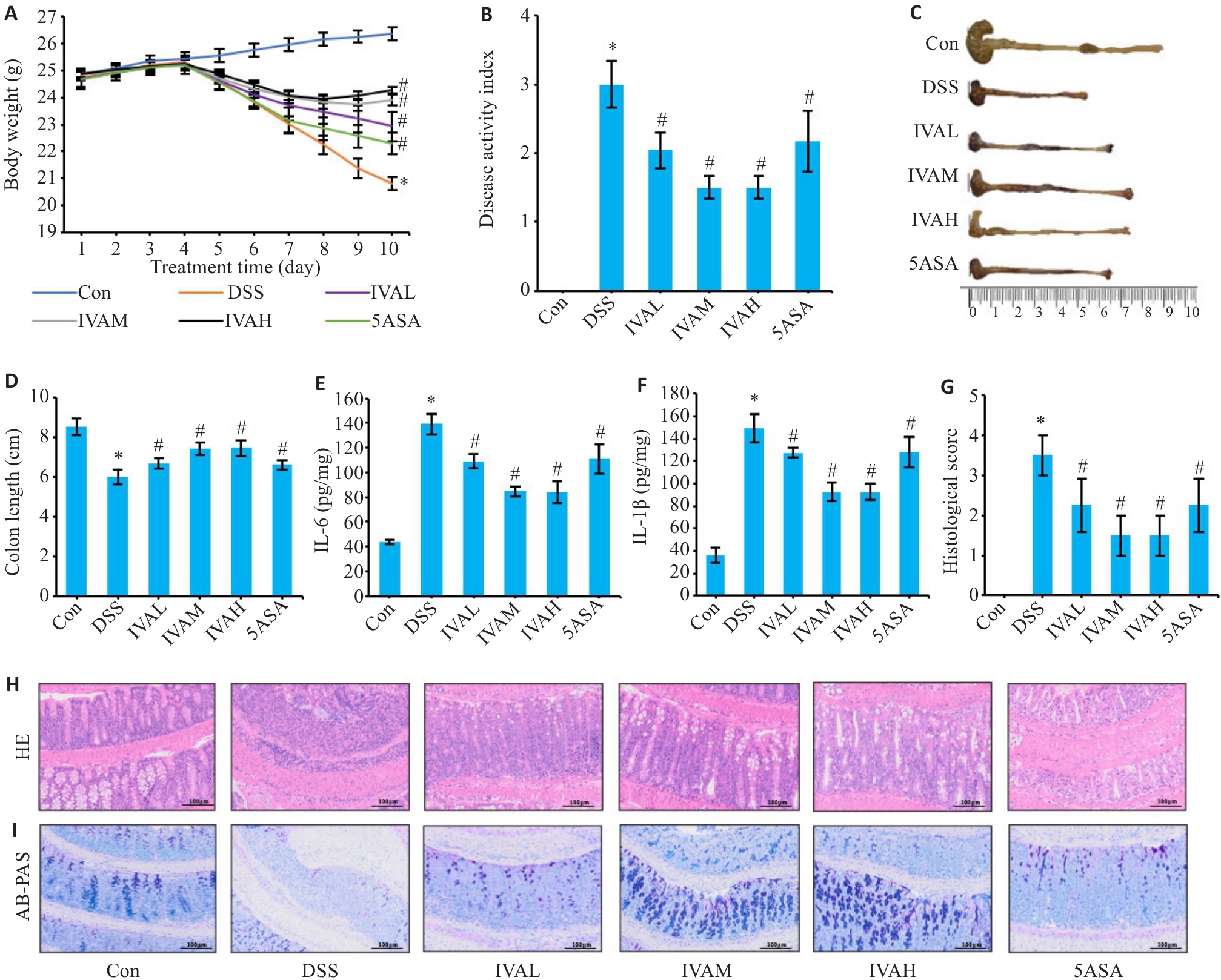
图1 IVA干预对DSS小鼠结肠炎症状的影响
Fig.1 Dose-dependent effects of IVA on DSS-induced colitis in mice. A: Body weight changes during the experiment. B: Disease activity index (DAI) scores. C: Representative images of mouse colons. D: Quantitative analysis of colon length. E, F: Concentrations of IL-6 (E) and IL-1β (F) in colonic mucosa measured by ELISA. G: Histopathological scores. H: HE staining of mouse colon sections (Original magnification: ×200). I: AB-PAS staining of mouse colon sections (×200). DSS: DSS model group; IVA-L: DSS+50 mg/kg IVA; IVA-M: DSS+100 mg/kg IVA; IVA-H: DSS+150 mg/kg IVA; 5-ASA: DSS+100 mg/kg 5-aminosalicylic acid. Data are presented as Mean±SD (n=8). *P<0.05 vs Con group; #P<0.05 vs DSS group.
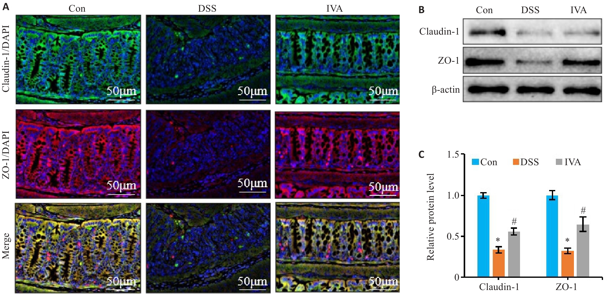
图2 IVA干预对DSS小鼠肠屏障的影响
Fig.2 IVA alleviates intestinal barrier injury in mice. A: Immunofluorescence staining of ZO-1 and claudin-1 in mouse colon tissues (×200). B, C: Quantitative analysis of ZO-1 and claudin-1 expression levels in the intestinal mucosa by Western blotting. Data are presented as Mean±SD (n=8). *P<0.05 vs Con group. #P<0.05 vs DSS group.
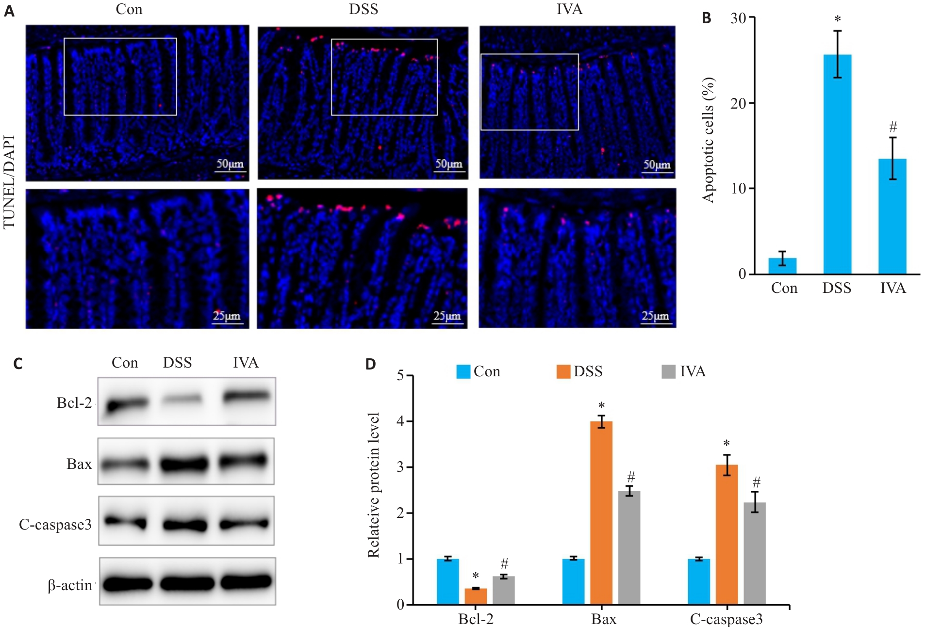
图3 IVA对DSS小鼠肠上皮细胞凋亡的影响
Fig.3 IVA ameliorates DSS-induced apoptosis of intestinal epithelial cells in mice. A: TUNEL staining of mouse colon tissues (×200). B: Quantitative analysis of intestinal epithelial cell apoptosis rate. C, D: Western blotting of Bcl-2, Bax, and cleaved caspase-3 (C-caspase3) expression levels in mouse colonic mucosa. Data are presented as Mean±SD (n=8). *P<0.05 vs Con group. #P<0.05 vs DSS group.
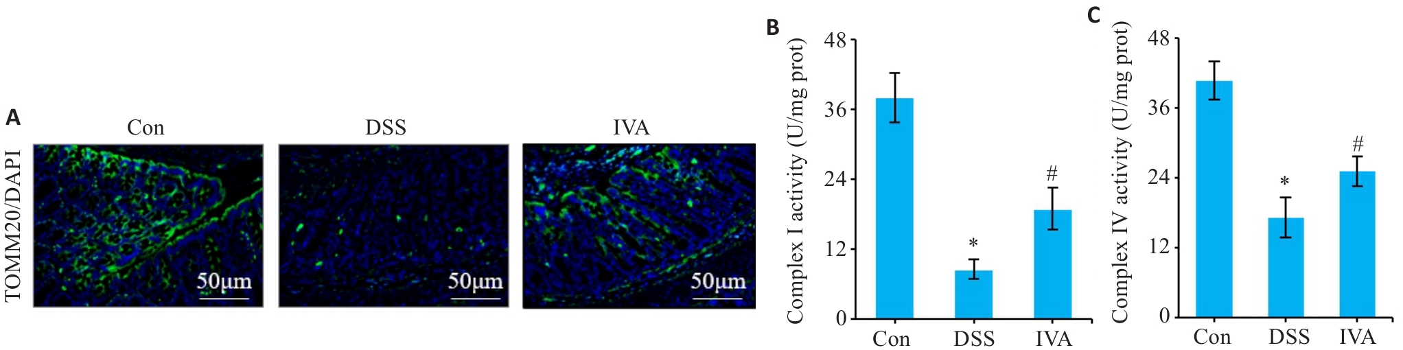
图4 IVA对DSS小鼠肠道细胞线粒体功能的影响
Fig.4 IVA alleviates DSS-induced intestinal mitochondrial dysfunction in mice. A: Immunofluorescence staining for detecting TOMM20-positive cells (×200). B: Complex I activity. C: Complex IV activity. *P<0.05 vs Con group. Data are presented as Mean±SD (n=8). #P<0.05 vs DSS group.
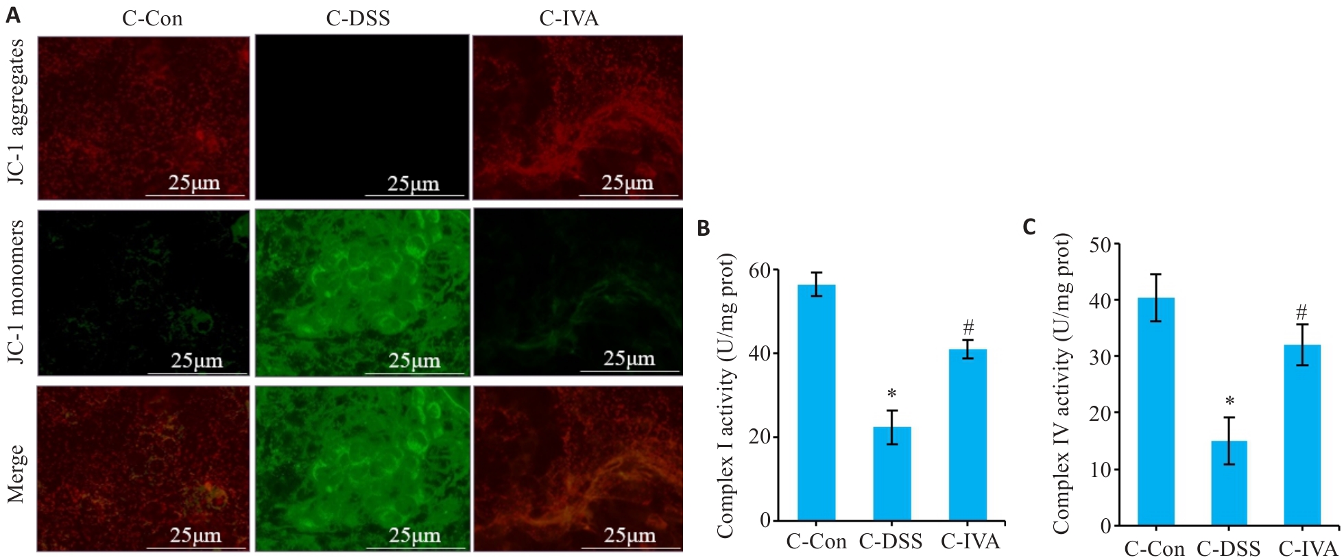
图5 IVA对NCM460线粒体功能的影响
Fig.5 IVA protects mitochondrial function in NCM460 cells with DSS-induced stress. A: Immunofluorescence staining for JC-1 (×400). B: Complex I activity. C: Complex IV activity. Data are presented as Mean±SD (n=3). *P<0.05 vs C-Con group. #P<0.05 vs C-DSS group.
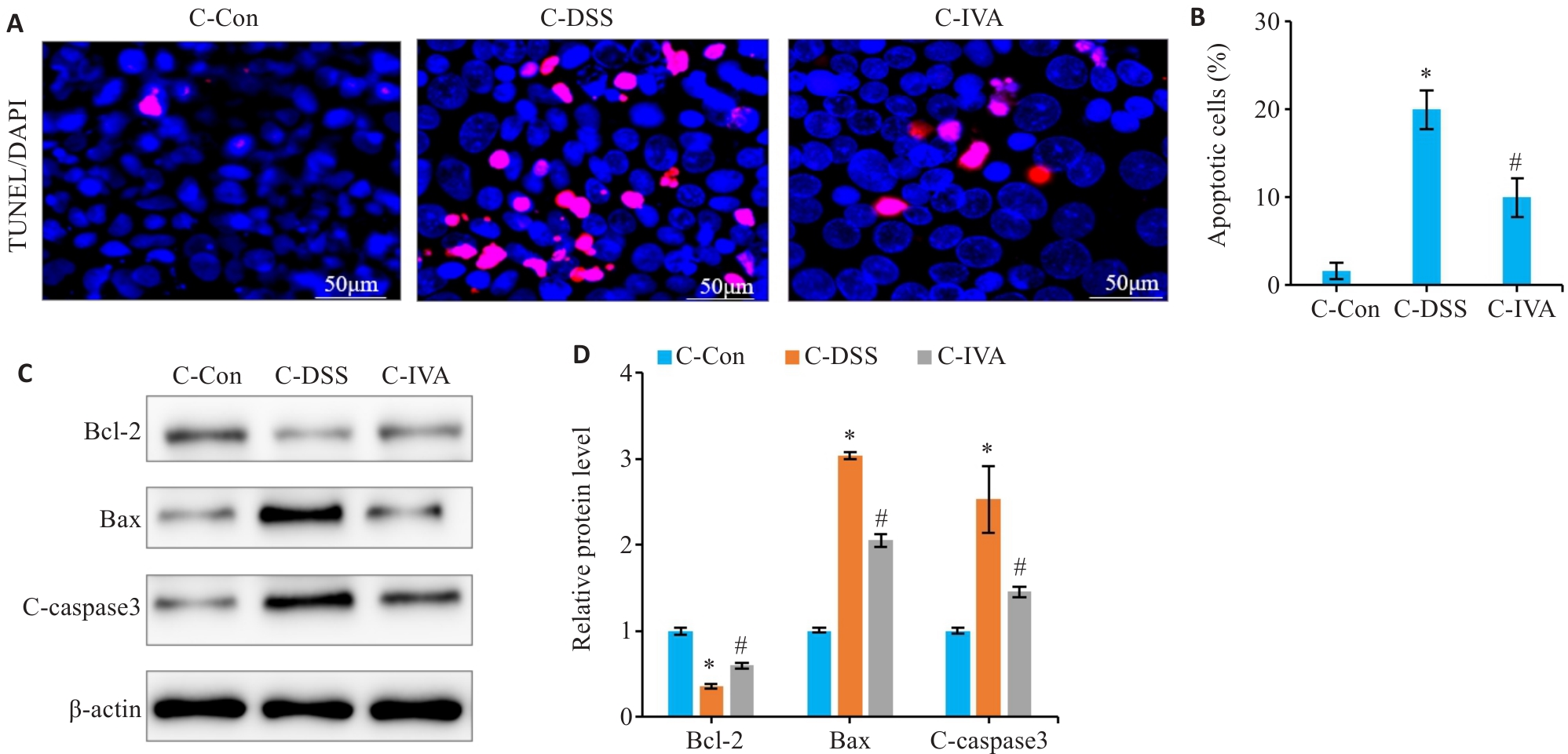
图6 IVA抑制DSS诱导的NCM460细胞凋亡
Fig. 6 Effect of IVA on DSS-induced apoptosis of NCM460 cells. A: TUNEL staining of NCM460 cells (×200). B: Quantitative analysis of apoptosis rate. C: Western blotting of Bcl-2, Bax, and C-caspase3 expression. D: Relative protein expression levels of Bcl-2, Bax, and C-caspase3. Data are presented as Mean±SD (n=3). *P<0.05 vs C-Con group. #P<0.05 vs C-DSS group.
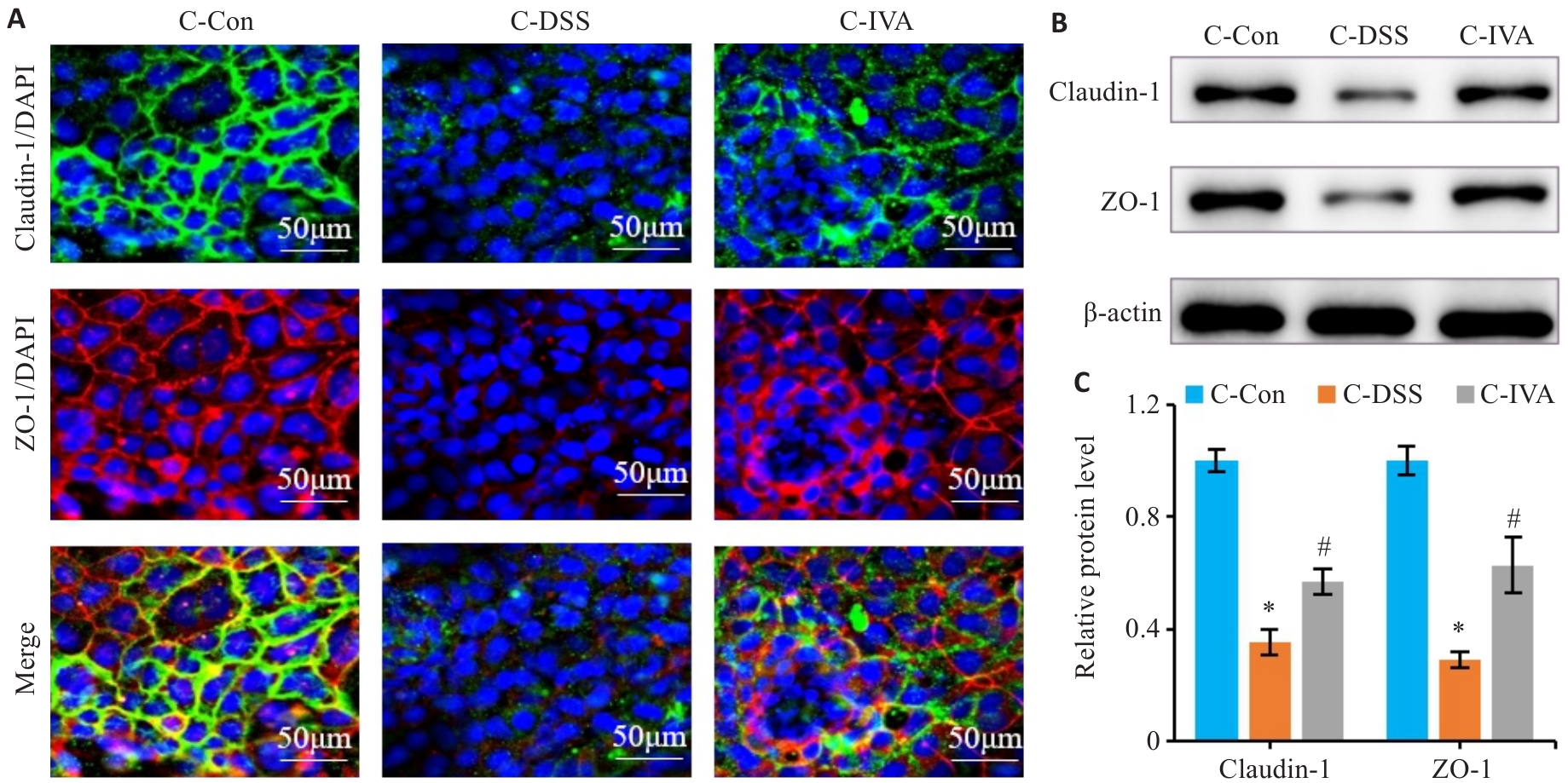
图7 IVA干预对DSS诱导的NCM460细胞屏障损伤的影响
Fig.7 IVA alleviates DSS-induced barrier damage in NCM460 cells. A: Immunofluorescence staining of ZO-1 and claudin-1 in NCM460 cells (×200). B, C: Western blotting for detecting relative protein expression levels. Data are presented as Mean±SD (n=3). *P<0.05 vs C-Con group. #P<0.05 vs C-DSS group.
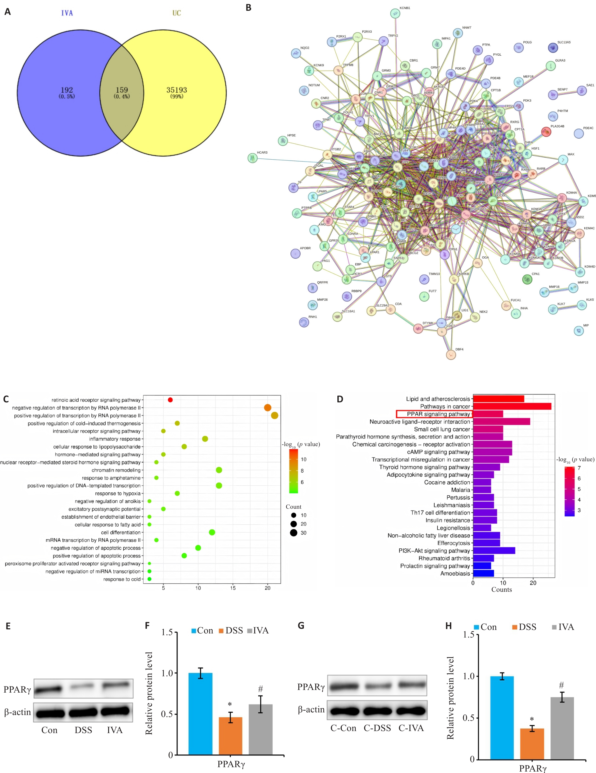
图8 IVA激活PPARγ通路来提高线粒体呼吸功能
Fig.8 IVA enhances mitochondrial respiratory function by regulating the PPARγ pathway. A: Venn diagram. B: PPI network diagram. C: GO-Biological process analysis. D: KEGG analysis. E: Western blotting of PPARγ expression in mouse intestinal epithelial cells. F: Relative protein expression levels of PPARγ in mouse colon tissues (n=8). G: Western blotting of PPARγ expression in NCM460 cells. H: Relative protein expression levels of PPARγ in NCM460 cells. Data are presented as Mean±SD (n=3). *P<0.05 vs Con/C-Con group. #P<0.05 vs DSS/C-DSS group.
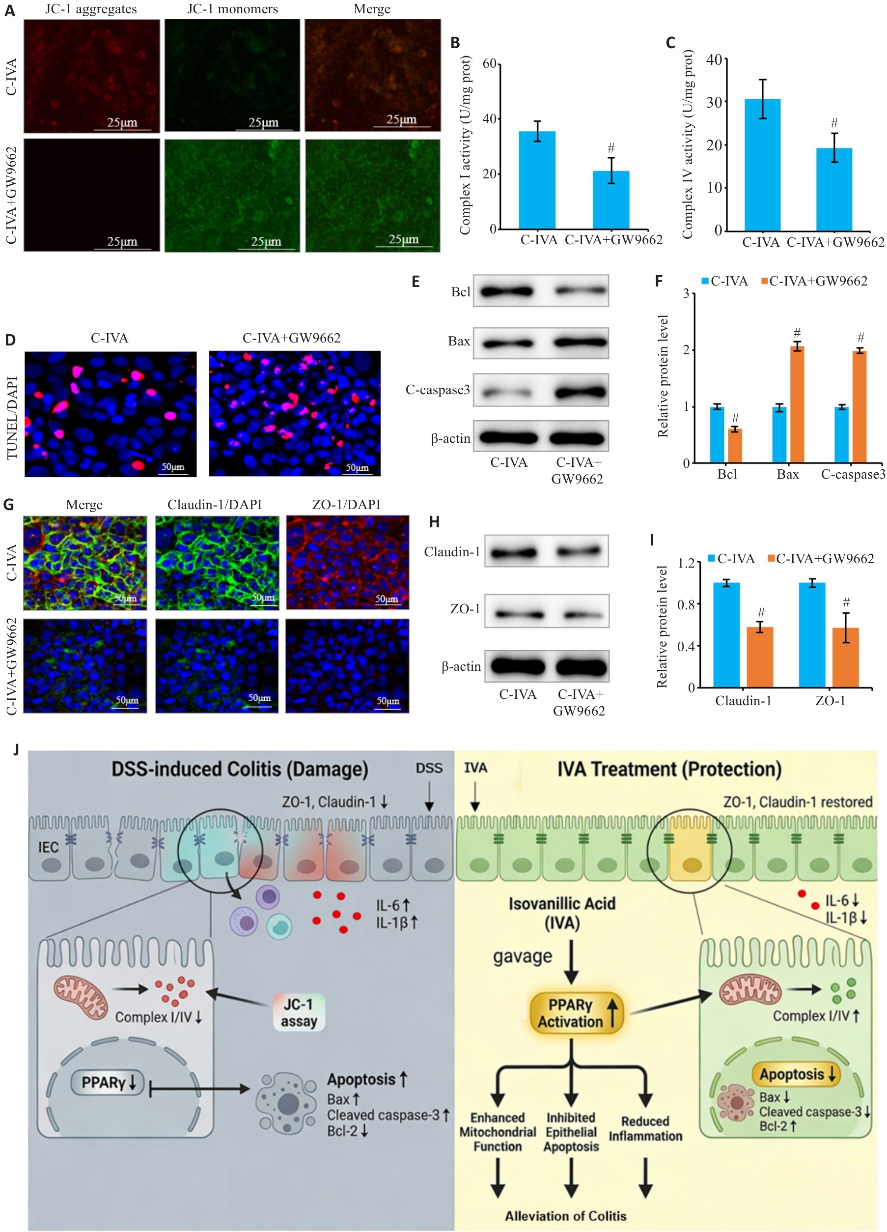
图9 IVA通过PPARγ改善DSS诱导的NCM460细胞线粒体功能障碍与凋亡
Fig.9 IVA improves mitochondrial function, apoptosis, and intestinal barrier injury via PPARγ. A: JC-1 staining showing mitochondrial membrane potential in NCM460 cells (×400). B, C: Activities of mitochondrial respiratory chain complexes I (B) and IV (C). D: TUNEL staining and quantification of apoptotic cells (×200). E, F: Western blotting of Bcl-2, Bax, and C-caspase3 expressions (E) and quantitative analysis of relative protein expression levels (F). G: Immunofluorescence staining of ZO-1 and claudin-1 in NCM460 cells (×200). H, I: Western blotting of ZO-1 and claudin-1 expressions (H) and quantitative analysis of relative protein expression levels (I). J: Schematic illustration of the proposed mechanism by which IVA ameliorates DSS-induced colitis. The cells were treated with 2% DSS in the presence or absence of IVA (20 μmol/L). C-IVA: DSS+IVA; C-IVA+GW9662: DSS+IVA+GW9662. Data are presented as Mean±SD (n=3). #P<0.05 vs C-IVA group.
| [1] | 杨翠萍, 陈 平. 全球炎症性肠病的流行趋势分析及诊治现状[J]. 诊断学理论与实践, 2025, 24(4): 373-82. doi:10.16150/j.1671-2870.2025.04.003 |
| [2] | Buie MJ, Quan J, Windsor JW, et al. Global hospitalization trends for Crohn's disease and ulcerative colitis in the 21st century: a systematic review with temporal analyses[J]. Clin Gastroenterol Hepatol, 2023, 21(9): 2211-21. |
| [3] | Hracs L, Windsor JW, Gorospe J, et al. Global evolution of inflammatory bowel disease across epidemiologic stages[J]. Nature, 2025, 642(8067): 458-66. |
| [4] | Abbas A, Di Fonzo DMP, Wetwittayakhlang P, et al. Management of ulcerative colitis: where are we at and where are we heading?[J]. Expert Rev Gastroenterol Hepatol, 2024, 18(10): 567-74. doi:10.1080/17474124.2024.2422370 |
| [5] | Vieujean S, Louis E. Precision medicine and drug optimization in adult inflammatory bowel disease patients[J]. Therap Adv Gastroenterol, 2023, 16: 17562848231173331. doi:10.1177/17562848231173331 |
| [6] | Wan Y, Yang L, Jiang S, et al. Excessive apoptosis in ulcerative colitis: crosstalk between apoptosis, ROS, ER stress, and intestinal homeostasis[J]. Inflamm Bowel Dis, 2022, 28(4): 639-48. doi:10.1093/ibd/izab277 |
| [7] | Jiang YT, Chen J, Du YY, et al. Immune modulation for the patterns of epithelial cell death in inflammatory bowel disease[J]. Int Immunopharmacol, 2025, 154: 114462. doi:10.1016/j.intimp.2025.114846 |
| [8] | Santhanam S, Venkatraman A, Ramakrishna BS. Impairment of mitochondrial acetoacetyl CoA thiolase activity in the colonic mucosa of patients with ulcerative colitis[J]. Gut, 2007, 56(11): 1543-9. doi:10.1136/gut.2006.108449 |
| [9] | Fu SC, Qu JY, Li LX, et al. Excessive mitochondrial fission suppresses mucosal repair by impairing butyrate metabolism in colonocytes[J]. Inflamm Bowel Dis, 2024, 30(1): 114-24. doi:10.1093/ibd/izad132 |
| [10] | Zhou JP, Xi YT, Wu T, et al. A potential therapeutic approach for ulcerative colitis: targeted regulation of mitochondrial dynamics and mitophagy through phytochemicals[J]. Front Immunol, 2025, 15: 1506292. doi:10.3389/fimmu.2024.1506292 |
| [11] | Choo MZY, Chua JAT, Lee SXY, et al. Privileged natural product compound classes for anti-inflammatory drug development[J]. Nat Prod Rep, 2025, 42(5): 856-75. doi:10.1039/d4np00066h |
| [12] | Cheng SY, Chen WJ, Guo ZZ, et al. Paeonol alleviates ulcerative colitis by modulating PPAR-γ and nuclear factor-κB activation[J]. Sci Rep, 2024, 14(1): 18390. doi:10.1038/s41598-024-68992-6 |
| [13] | Huang SY, Jin YM, Zhang LW, et al. PPAR gamma and PGC-1alpha activators protect against diabetic nephropathy by suppressing the inflammation and NF-kappaB activation[J]. Nephrology, 2024, 29(12): 858-72. doi:10.1111/nep.14381 |
| [14] | Kim JT, Li C, Weiss HL, et al. Regulation of ketogenic enzyme HMGCS2 by Wnt/β-catenin/PPARγ pathway in intestinal cells[J]. Cells, 2019, 8(9): 1106. doi:10.3390/cells8091106 |
| [15] | Dou XT, Xiao JH, Jin ZL, et al. Peroxisome proliferator-activated receptor-γ is downregulated in ulcerative colitis and is involved in experimental colitis-associated neoplasia[J]. Oncol Lett, 2015, 10(3): 1259-66. doi:10.3892/ol.2015.3397 |
| [16] | Bashiri K, Mattar MC, Meighani A, et al. Peroxisome proliferator-activated receptors in inflammatory bowel disease: linking immunometabolism, lipid signaling, and therapeutic potential[J]. Intest Res, 2026, 24(1): 11-26. doi:10.5217/ir.2025.00090 |
| [17] | Chen XQ, Hu CJ, Shu ZH, et al. Isovanillic acid protects mice against Staphylococcus aureus by targeting vWbp and Coa[J]. Future Microbiol, 2023, 18: 735-49. doi:10.2217/fmb-2022-0219 |
| [18] | Swislocka R, Kowalczyk N, Dabrowska A, et al. Spectroscopic, antioxidant and cytotoxicity studies of vanillic acids[J]. Sci Rep, 2025, 15(1): 31396. doi:10.1038/s41598-025-13965-6 |
| [19] | Bharat D, Cavalcanti RRM, Petersen C, et al. Blueberry metabolites attenuate lipotoxicity-induced endothelial dysfunction[J]. Mol Nutr Food Res, 2018, 62(2). DOI:10.1002/mnfr.201700601 . |
| [20] | Lei L, Wang J, Wang JJ, et al. Quercetin boosts gut microbiota-driven production of isovanillic acid to alleviate colitis via enhancing intestinal barrier function[J]. Curr Res Food Sci, 2025, 11: 101183. doi:10.1016/j.crfs.2025.101183 |
| [21] | Roselli M, Maruszak A, Grimaldi R, et al. Galactooligosaccharide treatment alleviates DSS-induced colonic inflammation in caco-2 cell model[J]. Front Nutr, 2022, 9: 862974. doi:10.3389/fnut.2022.862974 |
| [22] | Wirtz S, Popp V, Kindermann M, et al. Chemically induced mouse models of acute and chronic intestinal inflammation[J]. Nat Protoc, 2017, 12(7): 1295-309. doi:10.1038/nprot.2017.044 |
| [23] | 张 敏, 刘生宝, 张 诺, 等. 改进型“瑞士卷”法在小鼠肠道组织切片中的应用[J]. 中华病理学杂志, 2024, 53(4): 393-7. |
| [24] | Chougule PR, Sangaraju R, Patil PB, et al. Effect of ethyl gallate and propyl gallate on dextran sulfate sodium (DSS)-induced ulcerative colitis in C57BL/6 J mice: preventive and protective[J]. Inflammopharmacology, 2023, 31(4): 2103-20. doi:10.1007/s10787-023-01254-5 |
| [25] | Niu YX, Li AH, Xu WH, et al. Platelet activation stimulates macrophages to enhance ulcerative colitis through PF4/CXCR3 signaling[J]. Int J Mol Med, 2025, 55(5). DOI:10.3892/ijmm.2025.5519 . |
| [26] | Li RF, Sun Y, Kelsang D, et al. Butyrate ameliorates DSS-induced ulcerative colitis in mice by facilitating autophagy in intestinal epithelial cells and modulating the gut microbiota through blocking the PI3K-AKT-mTOR pathway[J]. PLoS One, 2025, 20(12): e0337214. doi:10.1371/journal.pone.0337214 |
| [27] | Yu KQ, Peng H, Zhang ZC, et al. Long non-coding RNA ANRIL/p65 negative feedback loop protects intestinal barrier function in inflammatory bowel disease[J]. Noncoding RNA Res, 2025, 12: 167-79. doi:10.1016/j.ncrna.2025.03.002 |
| [28] | Jiang ZH, Wang Y, Gong JF, et al. An Aeromonas variant that produces aerolysin promotes susceptibility to ulcerative colitis[J]. Science, 2025, 390(6775): eadz4712. doi:10.1126/science.adz4712 |
| [29] | Huang PX, Yang YQ, Lian JW, et al. Neutrophils disrupt the intestinal barrier via IL-22/TGF-β/Mmp9 axis in the zebrafish model of inflammatory bowel disease[J]. J Genet Genomics, 2025, 52(7): 962-5. doi:10.1016/j.jgg.2025.04.012 |
| [30] | Zeng ZW, Shi YM, Cai YH, et al. PHLDA1 protects intestinal barrier function via restricting intestinal epithelial cells apoptosis in inflammatory bowel disease[J]. Exp Cell Res, 2024, 443(1): 114322. doi:10.1016/j.yexcr.2024.114322 |
| [31] | Wu JC, Xu XQ, Duan JQ, et al. EFHD2 suppresses intestinal inflammation by blocking intestinal epithelial cell TNFR1 internalization and cell death[J]. Nat Commun, 2024, 15(1): 1282. doi:10.1038/s41467-024-45539-x |
| [32] | Huang J, Li J, Geng ZJ, et al. Cynaroside ameliorates TNBS-induced colitis by inhibiting intestinal epithelial cell apoptosis via the PI3K/AKT signalling pathway[J]. Front Pharmacol, 2025, 15: 1496068. doi:10.3389/fphar.2024.1496068 |
| [33] | 牛民主, 殷丽霞, 段 婷, 等. 川续断皂苷Ⅵ通过抑制PI3K/AKT/NF-κB通路拮抗肠上皮细胞凋亡缓解TNBS诱导的小鼠克罗恩病样结肠炎[J]. 南方医科大学学报, 2024, 44(12): 2335-46. |
| [34] | Zhang YF, Fan MY, Bai QR, et al. Precision therapy for ulcerative colitis: insights from mitochondrial dysfunction interacting with the immune microenvironment[J]. Front Immunol, 2024, 15: 1396221. doi:10.3389/fimmu.2024.1396221 |
| [35] | Liu L, Song FH, Gao SJ, et al. Peroxisome proliferator-activated receptor gamma: a promising therapeutic target for the treatment of chronic pain[J]. Brain Res, 2025, 1850: 149366. doi:10.1016/j.brainres.2024.149366 |
| [36] | Zang RX, Zhou R, Li YD, et al. The probiotic Lactobacillus plantarum alleviates colitis by modulating gut microflora to activate PPARγ and inhibit MAPKs/NF-κB[J]. Eur J Nutr, 2024, 64(1): 32. doi:10.1007/s00394-024-03520-w |
| [37] | Xu F, Hu JY, Li YN, et al. Qin-Yu-Qing-Chang decoction reshapes colonic metabolism by activating PPAR‑γ signaling to inhibit facultative anaerobes against DSS-induced colitis[J]. Chin Med, 2024, 19(1): 130. doi:10.1186/s13020-024-01006-9 |
| [1] | 尹林, 章雨, 张可妮, 乔通, 张龙涛, 黄菊, 李静, 胡建国, 耿志军. 1,3-二咖啡酰奎宁酸通过抑制PI3K/Akt通路减轻氧化应激缓解葡聚糖硫酸钠诱导的小鼠结肠炎[J]. 南方医科大学学报, 2026, 46(6): 1244-1255. |
| [2] | 温贺新, 林洁, 左芦根, 刘牧林. 珠子草素通过拮抗肠上皮细胞凋亡与调控肠道Th1/Th2免疫平衡改善克罗恩病样小鼠结肠炎[J]. 南方医科大学学报, 2026, 46(3): 655-665. |
| [3] | 李斌杰, 周晓芳, 朗晓猛, 康欣, 刘建平. 泄浊解毒方通过调节Th17/Treg免疫平衡改善大鼠溃疡性结肠炎[J]. 南方医科大学学报, 2026, 46(2): 335-344. |
| [4] | 尹林, 张可妮, 乔通, 牛民主, 殷丽霞, 刘馨悦, 耿志军, 李静, 胡建国. 藜芦酸通过激活Nrf2/HO-1信号通路减轻氧化应激缓解葡聚糖硫酸钠诱导的小鼠实验性结肠炎[J]. 南方医科大学学报, 2026, 46(2): 403-411. |
| [5] | 黄林林, 郑旺, 胡建国, 宋雪, 陶露, 耿志军, 李静, 左芦根, 葛思堂. 马鞭草苷通过抑制PI3K-AKT通路减轻肠上皮炎症改善小鼠克罗恩病样结肠炎[J]. 南方医科大学学报, 2026, 46(2): 423-433. |
| [6] | 乔通, 尹林, 张可妮, 牛民主, 黄菊, 耿志军, 李静, 胡建国. 茯苓新酸A通过调节AMPK/mTOR介导的自噬来减轻葡聚糖硫酸钠诱导的小鼠结肠炎[J]. 南方医科大学学报, 2026, 46(1): 131-140. |
| [7] | 何榕茂, 方泽扬, 张芸芸, 吴友谅, 梁世秀, 计涛, 陈科全, 王斯琪. 铁死亡相关基因对溃疡性结肠炎具有诊断预测价值[J]. 南方医科大学学报, 2025, 45(9): 1927-1937. |
| [8] | 刘辰菲, 张玮, 曾尧, 梁艳, 王梦婷, 张明芳, 李新元, 王凤超, 杨燕青. 2,6-二甲氧基-1,4-苯醌通过抑制NLRP3炎症小体活化改善葡聚糖硫酸钠诱导的小鼠溃疡性结肠炎[J]. 南方医科大学学报, 2025, 45(8): 1654-1662. |
| [9] | 常笑语, 张瀚文, 曹红亭, 侯玲, 孟鑫, 陶虹, 罗彦, 李光华. 热应激对大鼠胸主动脉内皮细胞生物钟基因 Bmal1和细胞周期蛋白表达水平的影响[J]. 南方医科大学学报, 2025, 45(7): 1353-1362. |
| [10] | 牛民主, 殷丽霞, 乔通, 尹林, 张可妮, 胡建国, 宋传旺, 耿志军, 李静. 旱莲苷A通过调控JAK2/STAT3通路抑制M1型巨噬细胞极化改善葡聚糖硫酸钠诱导的小鼠结肠炎[J]. 南方医科大学学报, 2025, 45(6): 1297-1306. |
| [11] | 张璐, 丁焕章, 许浩燃, 陈珂, 许博文, 杨勤军, 吴迪, 童佳兵, 李泽庚. 参芪补中方通过激活AMPK/SIRT1/PGC-1α改善COPD肺脾气虚证大鼠线粒体功能障碍[J]. 南方医科大学学报, 2025, 45(5): 969-976. |
| [12] | 储菲, 陈孝华, 宋博文, 杨晶晶, 左芦根. 苏荠宁黄酮通过抑制PI3K/AKT信号通路拮抗肠上皮细胞凋亡改善小鼠实验性结肠炎[J]. 南方医科大学学报, 2025, 45(4): 819-828. |
| [13] | 申琳, 宋翠豪, 王聪敏, 高西, 安俊红, 李承新, 梁斌, 李霞. 溃疡性结肠炎并发坏疽性脓皮病患者发生营养风险的因素及预测模型[J]. 南方医科大学学报, 2025, 45(3): 514-521. |
| [14] | 殷丽霞, 牛民主, 张可妮, 耿志军, 胡建国, 李江艳, 李静. 升麻素抑制MAPK通路调节辅助性T细胞免疫平衡改善小鼠克罗恩病样结肠炎[J]. 南方医科大学学报, 2025, 45(3): 595-602. |
| [15] | 马思雨, 陈美庆, 吴天宇, 赵文红. 膳食亚麻木酚素通过调控Bcl2/Bax/caspase3信号轴通道改善大鼠母体反式脂肪酸暴露所导致的子代肾损伤[J]. 南方医科大学学报, 2025, 45(12): 2658-2666. |
| 阅读次数 | ||||||
|
全文 |
|
|||||
|
摘要 |
|
|||||
