
南方医科大学学报 ›› 2026, Vol. 46 ›› Issue (8): 1861-1869.doi: 10.12122/j.issn.1673-4254.2026.08.13
• • 上一篇
史士恒1( ), 花代平1, 项尚1, 孙兰婷1, 宣巧玉1, 杨文明1,2, 汪瀚1,2()
收稿日期:2025-12-22
出版日期:2026-08-20
发布日期:2026-08-01
通讯作者:
汪瀚
E-mail:156767269@qq.com;neuwhah@126.com
作者简介:史士恒,在读博士研究生,E-mail: 156767269@qq.com
基金资助:
Shiheng SHI1(), Daiping HUA1, Shang XIANG1, Lanting SUN1, Qiaoyu XUAN1, Wenming YANG1,2, Han WANG1,2()
Received:2025-12-22
Online:2026-08-20
Published:2026-08-01
Contact:
Han WANG
E-mail:156767269@qq.com;neuwhah@126.com
Supported by:摘要:
目的 构建基于机器学习的Wilson病(WD)患者合并脂肪肝的预测模型,并评估其效能。 方法 回顾性收集安徽中医药大学第一附属医院1862例WD患者的临床资料,按7∶3分为训练集(n=1296)和验证集(n=566),采用最小绝对收缩和选择算子(LASSO)筛选关键预测变量,运用逻辑回归(LR)、决策树(DT)、随机森林(RF)、极端梯度提升(XGBoost)、轻量梯度提升机(LightGBM)、支持向量机(SVM)和人工神经网络(ANN)7种算法构建预测模型,结合受试者工作特征曲线下面积(AUC)、精准-召回曲线(PR)、校准曲线、决策曲线分析(DCA)评估模型性能,通过SHAP分析每个特征对模型预测的贡献。 结果 本研究纳入的1862例WD患者中1296 例(69.60%)合并脂肪肝。LASSO回归筛选出血小板计数(PLT)、红细胞分布宽度(RDW)、丙氨酸氨基转移酶(ALT)、总胆汁酸(TBA)、IV型胶原(CIV)、间接胆红素(IBIL)6项关键预测变量。在7种模型中,LightGBM模型综合效能最优:训练集AUC为0.826(95% CI:0.801~0.849),校准度良好(Brier评分0.143);验证集AUC为 0.815(95% CI:0.776~0.852),PR平均精确率高(AP=0.903),校准度良好(Brier评分0.138),DCA证实其具有临床净获益。SHAP分析表明ALT、IBIL、TBA、CIV对模型输出有显著的正向影响,RDW、PLT对预测结果的累积影响极小。 结论 本研究构建并验证了7种WD脂肪肝的预测模型,其中LightGBM模型更具优势,可为WD患者脂肪肝的早期筛查与风险分层提供一种可靠的评估工具。
史士恒, 花代平, 项尚, 孙兰婷, 宣巧玉, 杨文明, 汪瀚. 基于机器学习的Wilson病脂肪肝预测模型的开发与验证[J]. 南方医科大学学报, 2026, 46(8): 1861-1869.
Shiheng SHI, Daiping HUA, Shang XIANG, Lanting SUN, Qiaoyu XUAN, Wenming YANG, Han WANG. Development and validation of a machine learning-based prediction model for fatty liver in Wilson disease[J]. Journal of Southern Medical University, 2026, 46(8): 1861-1869.
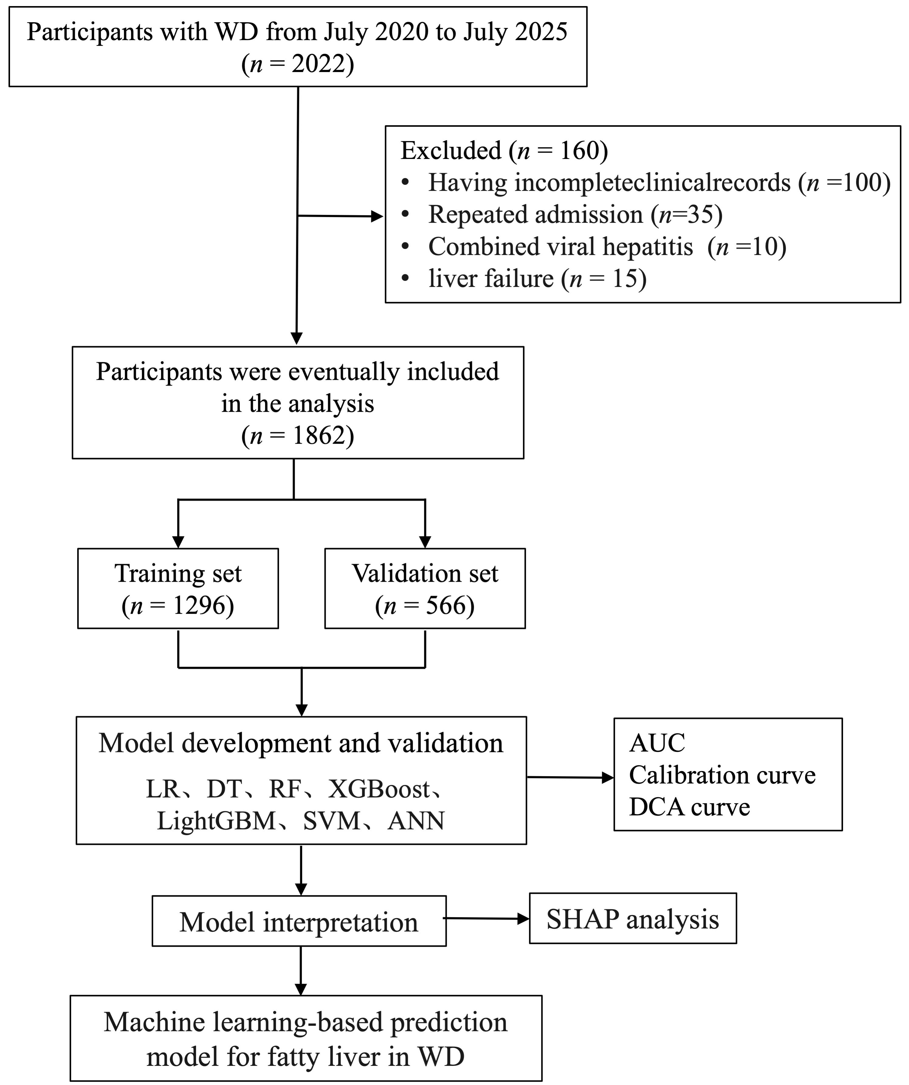
图1 WD患者脂肪肝预测模型的开发与验证流程图
Fig.1 Flowchart of the development and validation of the fatty liver prediction model in WD patients.
| Variable | Overall (n=1862) | Non-fatty liver group (n=566) | Fatty liver group (n=1296) | P |
|---|---|---|---|---|
| Male[n (%)] | 1057 (56.77) | 312 (55.12) | 745 (57.48) | 0.371 |
| Age (years) | 30.00 (23.00, 37.00) | 29.00 (23.00, 36.00) | 30.00 (23.00, 37.00) | 0.170 |
| BMI (kg/m2) | 21.26 (19.05, 23.79) | 21.22 (19.16, 23.61) | 21.30 (19.05, 23.92) | 0.600 |
| WBC (×10⁹/L) | 4.60 (3.61, 5.83) | 4.72 (3.84, 5.96) | 4.52 (3.51, 5.73) | 0.003 |
| RBC (×10¹²/L) | 4.37 (3.99, 4.79) | 4.39 (4.07, 4.78) | 4.36 (3.95, 4.79) | 0.038 |
| Hb (g/L) | 130.00 (118.00, 142.00) | 130.00 (119.00, 142.00) | 130.00 (117.00, 142.00) | 0.377 |
| PLT (×10⁹/L) | 144.00 (94.00, 203.00) | 159.50(111.00, 217.00) | 138.00 (87.75, 196.25) | <0.001 |
| RDW (%) | 13.30 (12.70, 14.30) | 13.10 (12.50, 13.90) | 13.40 (12.70, 14.50) | <0.001 |
| ALT (U/L) | 30.40 (20.02, 53.10) | 24.10 (17.00, 40.30) | 34.00 (21.98, 59.12) | <0.001 |
| AST (U/L) | 29.00 (22.00, 43.60) | 26.00 (20.40, 36.00) | 30.65(23.00, 46.00) | <0.001 |
| TP (g/L) | 63.60 (60.00, 67.60) | 64.10 (60.60, 67.77) | 63.40 (59.80, 67.43) | 0.048 |
| GLB (g/L) | 24.40 (21.90, 27.70) | 24.20 (21.90, 27.08) | 24.50 (21.90, 27.90) | 0.141 |
| A/G | 1.61 (1.36, 1.86) | 1.64 (1.41, 1.87) | 1.59 (1.34, 1.86) | 0.014 |
| GGT(U/L) | 31.00 (19.00, 58.00) | 28.00 (18.00, 52.00) | 32.00 (20.00, 61.25) | <0.001 |
| ALP (U/L) | 96.00 (76.00, 124.00) | 93.00 (75.00, 118.00) | 98.00 (78.00, 127.00) | 0.002 |
| LDH (U/L) | 168.00 (146.00, 198.00) | 166.00 (145.25, 192.00) | 170.00 (146.75, 201.25) | 0.010 |
| GLU (mmol/L) | 4.61 (4.32, 4.93) | 4.62 (4.34, 4.95) | 4.60 (4.31, 4.92) | 0.454 |
| TG (mmol/L) | 0.92 (0.71, 1.22) | 0.97 (0.75, 1.29) | 0.88 (0.69, 1.19) | <0.001 |
| TC (mmol/L) | 4.05 (3.51, 4.66) | 4.08 (3.61, 4.66) | 4.04 (3.47, 4.65) | 0.131 |
| HDL-C (mmol/L) | 1.26 (1.07, 1.48) | 1.27 (1.08, 1.52) | 1.26 (1.06, 1.47) | 0.008 |
| LDL-C (mmol/L) | 2.38 (2.01, 2.82) | 2.38 (2.01, 2.79) | 2.38 (2.00, 2.83) | 0.748 |
| TG/HDL-C | 0.72 (0.52, 1.02) | 0.74 (0.53, 1.02) | 0.70 (0.51, 1.02) | 0.349 |
| TBA (μmol/L) | 7.80 (4.50, 16.20) | 5.80 (3.70, 9.70) | 9.15 (5.00, 21.92) | <0.001 |
| Lp(a) (mg/L) | 53.75 (26.70, 115.50) | 58.45 (27.50, 129.10) | 51.65 (26.17, 110.93) | 0.079 |
| Hcy (μmol/L) | 11.30 (8.30, 15.80) | 11.10 (8.20, 15.88) | 11.30 (8.30, 15.72) | 0.718 |
| UA (μmol/L) | 222.50 (173.25, 285.75) | 221.50 (174.00, 282.00) | 223.00 (172.75, 286.00) | 0.860 |
| BUN (mmol/) | 4.88 (3.99, 5.90) | 4.93 (4.11, 5.98) | 4.87 (3.95, 5.85) | 0.076 |
| Cr (μmol/L) | 65.20 (54.00, 79.18) | 65.55 (53.12, 82.07) | 64.95 (54.68, 78.23) | 0.541 |
| PⅢNP (ng/mL) | 14.24 (9.92, 23.26) | 12.37 (9.18, 19.11) | 15.21 (10.20, 24.79) | <0.001 |
| LN (ng/mL) | 104.36 (80.90, 140.61) | 98.98 (76.04, 126.21) | 108.03 (82.45, 151.14) | <0.001 |
| HA (ng/mL) | 123.04 (71.61, 226.08) | 102.69 (61.83, 176.16) | 134.66 (77.42, 248.96) | <0.001 |
| CIV (ng/mL) | 66.87 (49.04, 101.60) | 57.42 (40.35, 79.25) | 72.22 (51.76, 109.73) | <0.001 |
| INR | 1.01 (0.95, 1.09) | 1.00 (0.94, 1.07) | 1.02 (0.96, 1.11) | <0.001 |
| FIB (g/L) | 2.08 (1.78, 2.42) | 2.17 (1.86, 2.54) | 2.04 (1.74, 2.38) | <0.001 |
| PT (s) | 31.00 (29.10, 33.10) | 30.70 (28.92, 32.68) | 31.00 (29.10, 33.40) | 0.002 |
| APTT (s) | 11.50 (10.80, 12.40) | 11.30 (10.60, 12.00) | 11.50 (10.90, 12.60) | <0.001 |
| TBIL (μmol/L) | 15.20 (11.30, 20.58) | 13.60 (9.10, 17.98) | 15.91 (12.00, 21.70) | <0.001 |
| DBIL (μmol/L) | 3.20 (2.40, 4.50) | 2.80 (1.90, 3.90) | 3.40 (2.50, 4.73) | <0.001 |
| IBIL (μmol/L) | 11.80 (8.80, 16.00) | 10.30 (7.00, 13.90) | 12.40 (9.47, 17.00) | <0.001 |
| 24-h UCu (μg/24 h) | 748.37 (423.13, 1151.57) | 703.16 (420.82, 1108.14) | 762.30 (427.12, 1162.81) | 0.294 |
表1 WD患者脂肪肝组与非脂肪肝组的基线特征
Tab.1 Baseline characteristics of WD patients with and without fatty liver [Median (IQR)]
| Variable | Overall (n=1862) | Non-fatty liver group (n=566) | Fatty liver group (n=1296) | P |
|---|---|---|---|---|
| Male[n (%)] | 1057 (56.77) | 312 (55.12) | 745 (57.48) | 0.371 |
| Age (years) | 30.00 (23.00, 37.00) | 29.00 (23.00, 36.00) | 30.00 (23.00, 37.00) | 0.170 |
| BMI (kg/m2) | 21.26 (19.05, 23.79) | 21.22 (19.16, 23.61) | 21.30 (19.05, 23.92) | 0.600 |
| WBC (×10⁹/L) | 4.60 (3.61, 5.83) | 4.72 (3.84, 5.96) | 4.52 (3.51, 5.73) | 0.003 |
| RBC (×10¹²/L) | 4.37 (3.99, 4.79) | 4.39 (4.07, 4.78) | 4.36 (3.95, 4.79) | 0.038 |
| Hb (g/L) | 130.00 (118.00, 142.00) | 130.00 (119.00, 142.00) | 130.00 (117.00, 142.00) | 0.377 |
| PLT (×10⁹/L) | 144.00 (94.00, 203.00) | 159.50(111.00, 217.00) | 138.00 (87.75, 196.25) | <0.001 |
| RDW (%) | 13.30 (12.70, 14.30) | 13.10 (12.50, 13.90) | 13.40 (12.70, 14.50) | <0.001 |
| ALT (U/L) | 30.40 (20.02, 53.10) | 24.10 (17.00, 40.30) | 34.00 (21.98, 59.12) | <0.001 |
| AST (U/L) | 29.00 (22.00, 43.60) | 26.00 (20.40, 36.00) | 30.65(23.00, 46.00) | <0.001 |
| TP (g/L) | 63.60 (60.00, 67.60) | 64.10 (60.60, 67.77) | 63.40 (59.80, 67.43) | 0.048 |
| GLB (g/L) | 24.40 (21.90, 27.70) | 24.20 (21.90, 27.08) | 24.50 (21.90, 27.90) | 0.141 |
| A/G | 1.61 (1.36, 1.86) | 1.64 (1.41, 1.87) | 1.59 (1.34, 1.86) | 0.014 |
| GGT(U/L) | 31.00 (19.00, 58.00) | 28.00 (18.00, 52.00) | 32.00 (20.00, 61.25) | <0.001 |
| ALP (U/L) | 96.00 (76.00, 124.00) | 93.00 (75.00, 118.00) | 98.00 (78.00, 127.00) | 0.002 |
| LDH (U/L) | 168.00 (146.00, 198.00) | 166.00 (145.25, 192.00) | 170.00 (146.75, 201.25) | 0.010 |
| GLU (mmol/L) | 4.61 (4.32, 4.93) | 4.62 (4.34, 4.95) | 4.60 (4.31, 4.92) | 0.454 |
| TG (mmol/L) | 0.92 (0.71, 1.22) | 0.97 (0.75, 1.29) | 0.88 (0.69, 1.19) | <0.001 |
| TC (mmol/L) | 4.05 (3.51, 4.66) | 4.08 (3.61, 4.66) | 4.04 (3.47, 4.65) | 0.131 |
| HDL-C (mmol/L) | 1.26 (1.07, 1.48) | 1.27 (1.08, 1.52) | 1.26 (1.06, 1.47) | 0.008 |
| LDL-C (mmol/L) | 2.38 (2.01, 2.82) | 2.38 (2.01, 2.79) | 2.38 (2.00, 2.83) | 0.748 |
| TG/HDL-C | 0.72 (0.52, 1.02) | 0.74 (0.53, 1.02) | 0.70 (0.51, 1.02) | 0.349 |
| TBA (μmol/L) | 7.80 (4.50, 16.20) | 5.80 (3.70, 9.70) | 9.15 (5.00, 21.92) | <0.001 |
| Lp(a) (mg/L) | 53.75 (26.70, 115.50) | 58.45 (27.50, 129.10) | 51.65 (26.17, 110.93) | 0.079 |
| Hcy (μmol/L) | 11.30 (8.30, 15.80) | 11.10 (8.20, 15.88) | 11.30 (8.30, 15.72) | 0.718 |
| UA (μmol/L) | 222.50 (173.25, 285.75) | 221.50 (174.00, 282.00) | 223.00 (172.75, 286.00) | 0.860 |
| BUN (mmol/) | 4.88 (3.99, 5.90) | 4.93 (4.11, 5.98) | 4.87 (3.95, 5.85) | 0.076 |
| Cr (μmol/L) | 65.20 (54.00, 79.18) | 65.55 (53.12, 82.07) | 64.95 (54.68, 78.23) | 0.541 |
| PⅢNP (ng/mL) | 14.24 (9.92, 23.26) | 12.37 (9.18, 19.11) | 15.21 (10.20, 24.79) | <0.001 |
| LN (ng/mL) | 104.36 (80.90, 140.61) | 98.98 (76.04, 126.21) | 108.03 (82.45, 151.14) | <0.001 |
| HA (ng/mL) | 123.04 (71.61, 226.08) | 102.69 (61.83, 176.16) | 134.66 (77.42, 248.96) | <0.001 |
| CIV (ng/mL) | 66.87 (49.04, 101.60) | 57.42 (40.35, 79.25) | 72.22 (51.76, 109.73) | <0.001 |
| INR | 1.01 (0.95, 1.09) | 1.00 (0.94, 1.07) | 1.02 (0.96, 1.11) | <0.001 |
| FIB (g/L) | 2.08 (1.78, 2.42) | 2.17 (1.86, 2.54) | 2.04 (1.74, 2.38) | <0.001 |
| PT (s) | 31.00 (29.10, 33.10) | 30.70 (28.92, 32.68) | 31.00 (29.10, 33.40) | 0.002 |
| APTT (s) | 11.50 (10.80, 12.40) | 11.30 (10.60, 12.00) | 11.50 (10.90, 12.60) | <0.001 |
| TBIL (μmol/L) | 15.20 (11.30, 20.58) | 13.60 (9.10, 17.98) | 15.91 (12.00, 21.70) | <0.001 |
| DBIL (μmol/L) | 3.20 (2.40, 4.50) | 2.80 (1.90, 3.90) | 3.40 (2.50, 4.73) | <0.001 |
| IBIL (μmol/L) | 11.80 (8.80, 16.00) | 10.30 (7.00, 13.90) | 12.40 (9.47, 17.00) | <0.001 |
| 24-h UCu (μg/24 h) | 748.37 (423.13, 1151.57) | 703.16 (420.82, 1108.14) | 762.30 (427.12, 1162.81) | 0.294 |
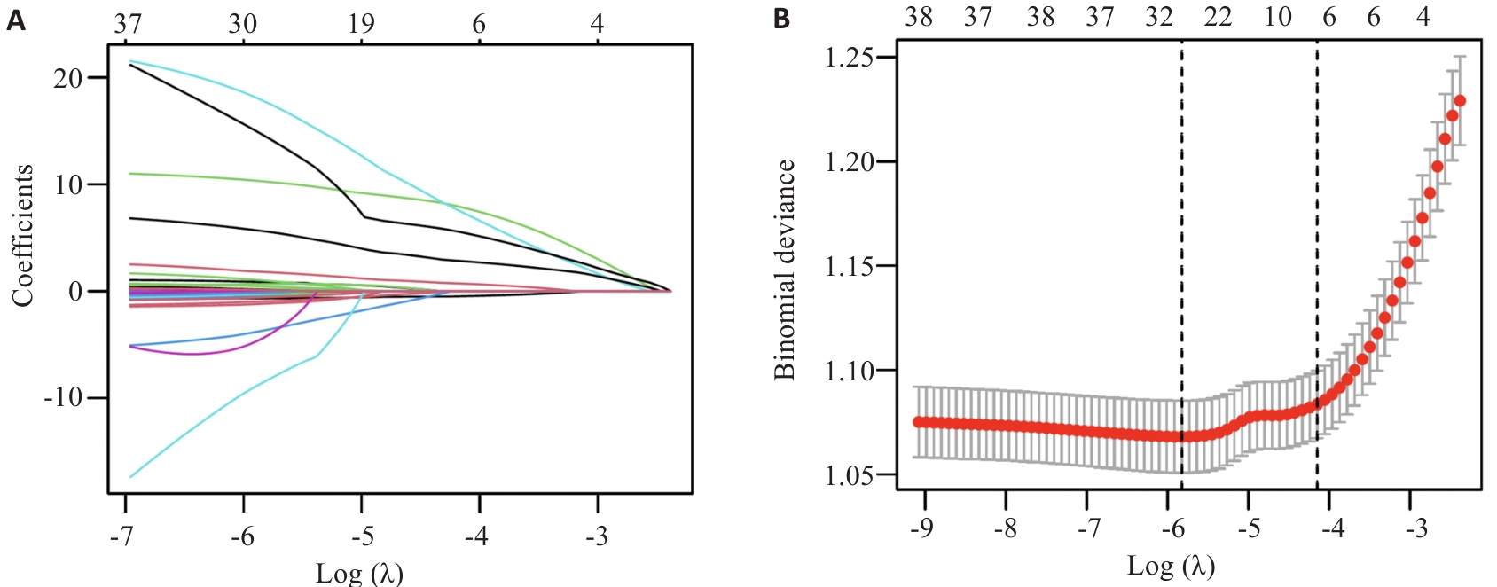
图2 LASSO回归筛选WD患者脂肪肝预测变量的特征选择图
Fig.2 Feature selection plot of LASSO regression for screening predictors of fatty liver in WD patients. A: Distribution of LASSO regression coefficients. B: Determination of the regularization parameter (λ) for the LASSO model by 10-fold cross-validation. The left dashed line represents the λmin, and the right dashed line represents λ1se.
| Model | AUC (95% CI) | Accuracy | Precision | Sensitivity | Specificity | F1 Score | Youden's J | PPV | NPV |
|---|---|---|---|---|---|---|---|---|---|
| Training set | |||||||||
| LR | 0.738 (0.710-0.765) | 0.728 | 0.752 | 0.910 | 0.312 | 0.823 | 0.222 | 0.752 | 0.602 |
| DT | 0.785 (0.759-0.811) | 0.800 | 0.777 | 0.999 | 0.345 | 0.874 | 0.344 | 0.777 | 0.993 |
| RF | 0.840 (0.816-0.863) | 0.802 | 0.780 | 0.994 | 0.360 | 0.875 | 0.355 | 0.780 | 0.966 |
| XGBoost | 0.811 (0.785-0.837) | 0.785 | 0.767 | 0.992 | 0.310 | 0.865 | 0.302 | 0.767 | 0.946 |
| LightGBM | 0.826 (0.801-0.849) | 0.797 | 0.778 | 0.991 | 0.353 | 0.872 | 0.344 | 0.778 | 0.946 |
| SVM | 0.735 (0.706-0.762) | 0.741 | 0.739 | 0.970 | 0.217 | 0.839 | 0.187 | 0.739 | 0.761 |
| ANN | 0.771 (0.744-0.795) | 0.759 | 0.778 | 0.915 | 0.403 | 0.841 | 0.318 | 0.778 | 0.675 |
| Validation set | |||||||||
| LR | 0.729 (0.684-0.772) | 0.713 | 0.751 | 0.879 | 0.331 | 0.810 | 0.210 | 0.751 | 0.544 |
| DT | 0.785 (0.744-0.824) | 0.808 | 0.786 | 0.995 | 0.379 | 0.878 | 0.374 | 0.786 | 0.970 |
| RF | 0.809 (0.770-0.848) | 0.813 | 0.793 | 0.990 | 0.408 | 0.881 | 0.398 | 0.793 | 0.945 |
| XGBoost | 0.804 (0.764-0.843) | 0.799 | 0.780 | 0.990 | 0.361 | 0.873 | 0.351 | 0.780 | 0.938 |
| LightGBM | 0.815 (0.776-0.852) | 0.820 | 0.798 | 0.995 | 0.420 | 0.885 | 0.415 | 0.798 | 0.973 |
| SVM | 0.722 (0.677-0.767) | 0.731 | 0.743 | 0.938 | 0.254 | 0.829 | 0.193 | 0.743 | 0.642 |
| ANN | 0.734 (0.691-0.780) | 0.754 | 0.777 | 0.907 | 0.402 | 0.837 | 0.310 | 0.777 | 0.654 |
表2 不同机器学习模型预测WD患者脂肪肝的性能表现对比分析
Tab.2 Performance comparison of different machine learning models for predicting fatty liver in WD patients
| Model | AUC (95% CI) | Accuracy | Precision | Sensitivity | Specificity | F1 Score | Youden's J | PPV | NPV |
|---|---|---|---|---|---|---|---|---|---|
| Training set | |||||||||
| LR | 0.738 (0.710-0.765) | 0.728 | 0.752 | 0.910 | 0.312 | 0.823 | 0.222 | 0.752 | 0.602 |
| DT | 0.785 (0.759-0.811) | 0.800 | 0.777 | 0.999 | 0.345 | 0.874 | 0.344 | 0.777 | 0.993 |
| RF | 0.840 (0.816-0.863) | 0.802 | 0.780 | 0.994 | 0.360 | 0.875 | 0.355 | 0.780 | 0.966 |
| XGBoost | 0.811 (0.785-0.837) | 0.785 | 0.767 | 0.992 | 0.310 | 0.865 | 0.302 | 0.767 | 0.946 |
| LightGBM | 0.826 (0.801-0.849) | 0.797 | 0.778 | 0.991 | 0.353 | 0.872 | 0.344 | 0.778 | 0.946 |
| SVM | 0.735 (0.706-0.762) | 0.741 | 0.739 | 0.970 | 0.217 | 0.839 | 0.187 | 0.739 | 0.761 |
| ANN | 0.771 (0.744-0.795) | 0.759 | 0.778 | 0.915 | 0.403 | 0.841 | 0.318 | 0.778 | 0.675 |
| Validation set | |||||||||
| LR | 0.729 (0.684-0.772) | 0.713 | 0.751 | 0.879 | 0.331 | 0.810 | 0.210 | 0.751 | 0.544 |
| DT | 0.785 (0.744-0.824) | 0.808 | 0.786 | 0.995 | 0.379 | 0.878 | 0.374 | 0.786 | 0.970 |
| RF | 0.809 (0.770-0.848) | 0.813 | 0.793 | 0.990 | 0.408 | 0.881 | 0.398 | 0.793 | 0.945 |
| XGBoost | 0.804 (0.764-0.843) | 0.799 | 0.780 | 0.990 | 0.361 | 0.873 | 0.351 | 0.780 | 0.938 |
| LightGBM | 0.815 (0.776-0.852) | 0.820 | 0.798 | 0.995 | 0.420 | 0.885 | 0.415 | 0.798 | 0.973 |
| SVM | 0.722 (0.677-0.767) | 0.731 | 0.743 | 0.938 | 0.254 | 0.829 | 0.193 | 0.743 | 0.642 |
| ANN | 0.734 (0.691-0.780) | 0.754 | 0.777 | 0.907 | 0.402 | 0.837 | 0.310 | 0.777 | 0.654 |
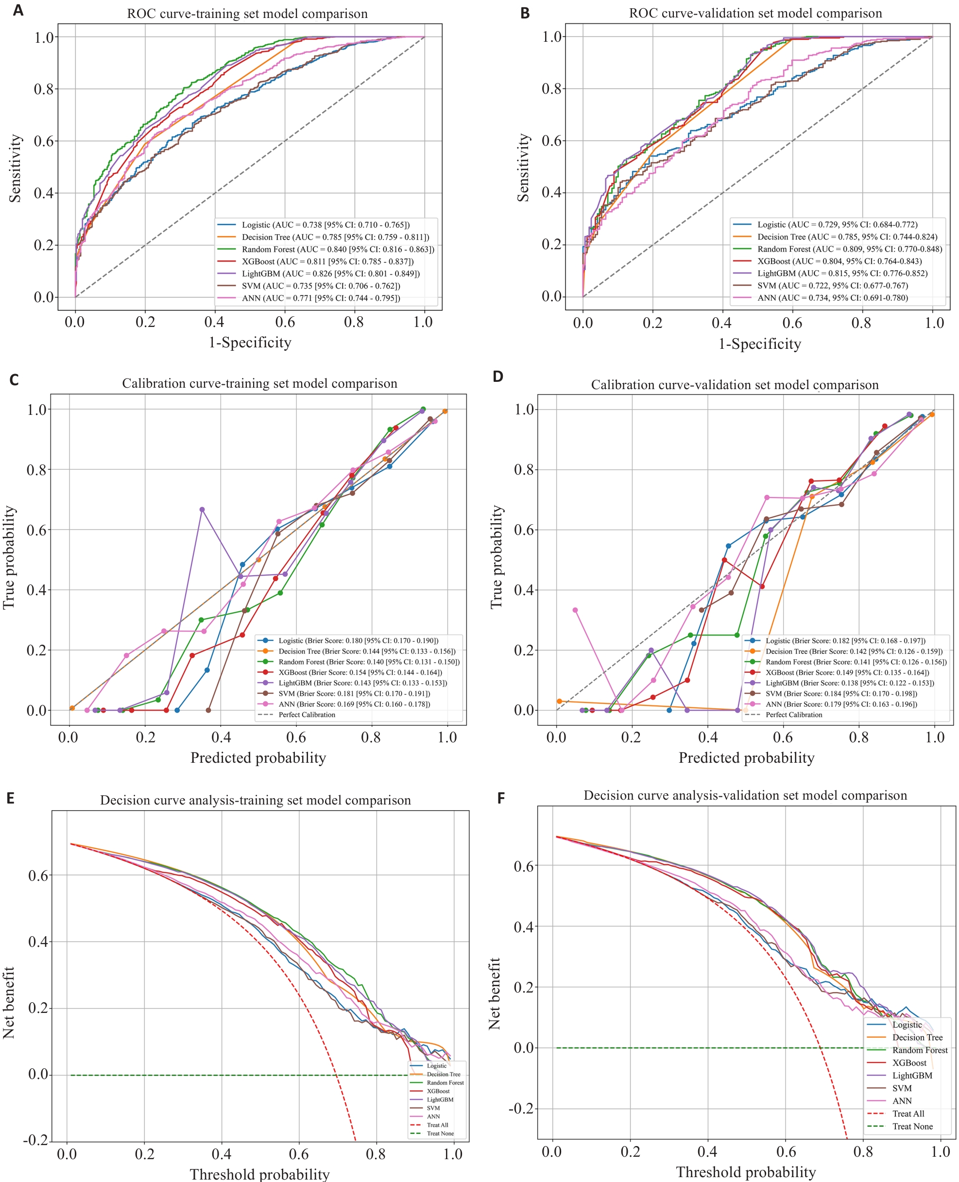
图3 WD患者脂肪肝预测模型在训练集与验证集的AUC、校准曲线及决策曲线
Fig.3 AUC, calibration curves and decision curves of fatty liver prediction models for WD patients in the training and validation sets. A: Training-set AUC. B: Validation-set AUC. C: Training-set calibration curves. D: Validation-set calibration curves. E: Training-set decision curves. F: Validation-set decision curves.
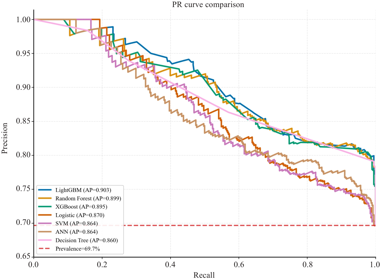
图4 WD患者脂肪肝预测模型在验证集的PR曲线图
Fig.4 PR curve of the prediction model for fatty liver in WD patients in the validation set. X-axis: Recall; Y-axis: Precision. Colored curves: machine learning models (annotated with AP values, higher = better performance). Red dashed line: sample prevalence (69.7%, random guessing baseline).
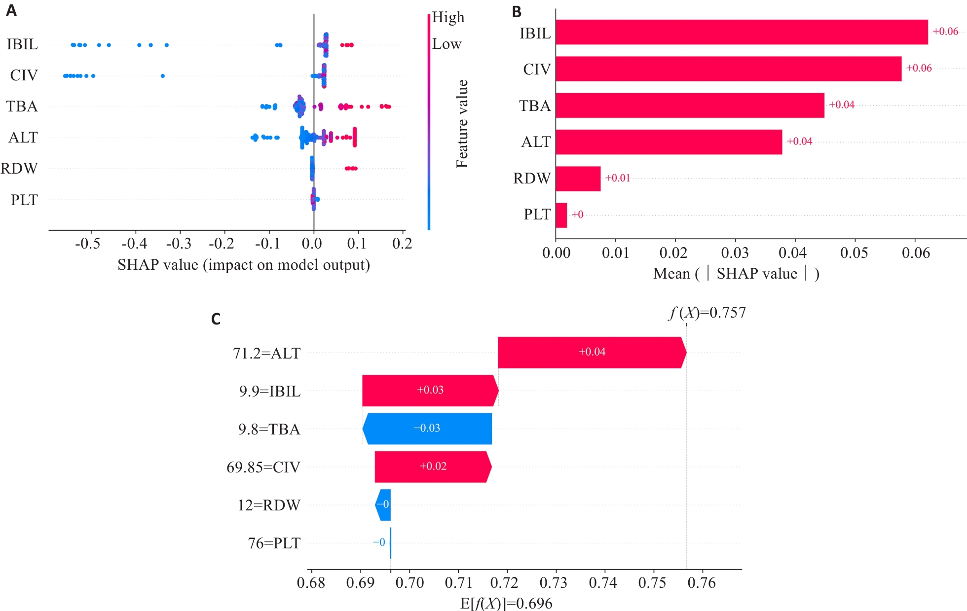
图5 LightGBM模型预测WD患者脂肪肝的SHAP可解释性分析
Fig.5 SHAP interpretability analysis of the LightGBM model for predicting fatty liver in WD patients. A: Beeswarm plot of SHAP values for each feature. Each point represents a sample. Greater dispersion of points indicates a more significant impact on the model output. Colors represent feature values (red=high, blue=low). B: Bar plot of mean absolute SHAP values. Each row represents a feature, with higher values indicating greater overall contribution to the model prediction. C: Waterfall plot of SHAP values for a single sample. Baseline E[f(X)] denotes the expected model output, and final f(x)denotes the sample-specific prediction. Red bars represent positive SHAP contributions (pushing the prediction upward), while blue bars represent negative contributions (pushing the prediction downward). Labels show each feature and its corresponding SHAP value.
| [1] | Członkowska A, Litwin T, Dusek P, et al. Wilson disease[J]. Nat Rev Dis Primers, 2018, 4: 21. doi:10.1038/s41572-018-0018-3 |
| [2] | Mazi TA, Shibata NM, Medici V. Lipid and energy metabolism in Wilson disease[J]. Liver Res, 2020, 4(1): 5-14. doi:10.1016/j.livres.2020.02.002 |
| [3] | Medici V, Shibata NM, Kharbanda KK, et al. Wilson's disease: changes in methionine metabolism and inflammation affect global DNA methylation in early liver disease[J]. Hepatology, 2013, 57(2): 555-65. doi:10.1002/hep.26047 |
| [4] | Shamout F, Zhu TT, Clifton DA. Machine learning for clinical outcome prediction[J]. IEEE Rev Biomed Eng, 2021, 14: 116-26. doi:10.1109/rbme.2020.3007816 |
| [5] | Greener JG, Kandathil SM, Moffat L, et al. A guide to machine learning for biologists[J]. Nat Rev Mol Cell Biol, 2022, 23(1): 40-55. doi:10.1038/s41580-021-00407-0 |
| [6] | Khene ZE, Bigot P, Doumerc N, et al. Application of machine learning models to predict recurrence after surgical resection of nonmetastatic renal cell carcinoma[J]. Eur Urol Oncol, 2023, 6(3): 323-30. doi:10.1016/j.euo.2022.07.007 |
| [7] | Yu YD, Lee KS, Man Kim J, et al. Artificial intelligence for predicting survival following deceased donor liver transplantation: Retrospective multi-center study[J]. Int J Surg, 2022, 105: 106838. doi:10.1016/j.ijsu.2022.106838 |
| [8] | Rao ZH, Yang WM, Yang YL, et al. Revolutionizing Wilson disease prognosis: a machine learning approach to predict acute-on-chronic liver failure[J]. J Transl Med, 2025, 23(1): 999. doi:10.1186/s12967-025-06987-1 |
| [9] | Chen K, Wan Y, Mao J, et al. Liver cirrhosis prediction for patients with Wilson disease based on machine learning: a case-control study from southwest China[J]. Eur J Gastroenterol Hepatol, 2022, 34(10): 1067-73. doi:10.1097/meg.0000000000002424 |
| [10] | 程琳傑, 袁 晴, 李宛凇, 等. 基于机器学习的非酒精性脂肪性肝病预测模型的构建及验证[J].中华疾病控制杂志, 2025, 29(6):682-7, 696. |
| [11] | European Association for the Study of the Liver. EASL-ERN Clinical Practice Guidelines on Wilson' s disease[J]. J Hepatol, 2025, 8278(24):2706-15. |
| [12] | Ferenci P, Caca K, Loudianos G, et al. Diagnosis and phenotypic classification of Wilson disease 1[J]. Liver Int, 2003, 23(3): 139-42. doi:10.1034/j.1600-0676.2003.00824.x |
| [13] | 中华医学会肝病学分会. 代谢相关(非酒精性)脂肪性肝病防治指南(2024年版)[J]. 中华肝脏病杂志, 2024, 32(5): 418-34. |
| [14] | Sanchez-Pinto LN, Venable LR, Fahrenbach J, et al. Comparison of variable selection methods for clinical predictive modeling[J]. Int J Med Inform, 2018, 116: 10-7. doi:10.1016/j.ijmedinf.2018.05.006 |
| [15] | Kim JH. Multicollinearity and misleading statistical results[J]. Korean J Anesthesiol, 2019, 72(6): 558-69. doi:10.4097/kja.19087 |
| [16] | Huang CX, Li SX, Caraballo C, et al. Performance metrics for the comparative analysis of clinical risk prediction models employing machine learning[J]. Circ Cardiovasc Qual Outcomes, 2021, 14(10): 7526-38. doi:10.1161/circoutcomes.120.007526 |
| [17] | Ralston A, Liu P, Warrener K, et al. Small field diode correction factors derived using an air core fibre optic scintillation dosimeter and EBT2 film[J]. Phys Med Biol, 2012, 57(9): 2587. doi:10.1088/0031-9155/57/9/2587 |
| [18] | Van Calster B, Wynants L, Verbeek JFM, et al. Reporting and interpreting decision curve analysis: a guide for investigators[J]. Eur Urol, 2018, 74(6): 796-804. doi:10.1016/j.eururo.2018.08.038 |
| [19] | Collins GS, Dhiman P, Andaur Navarro CL, et al. Protocol for development of a reporting guideline (TRIPOD-AI) and risk of bias tool (PROBAST-AI) for diagnostic and prognostic prediction model studies based on artificial intelligence[J]. BMJ Open, 2021, 11(7): e048008. doi:10.1136/bmjopen-2020-048008 |
| [20] | Islam MM, Rahman MJ, Rabby MS, et al. Predicting the risk of diabetic retinopathy using explainable machine learning algorithms[J]. Diabetes Metab Syndr, 2023, 17(12): 102919. doi:10.1016/j.dsx.2023.102919 |
| [21] | Petch J, Di S, Nelson W. Opening the black box: the promise and limitations of explainable machine learning in cardiology[J]. Can J Cardiol, 2022, 38(2): 204-13. doi:10.1016/j.cjca.2021.09.004 |
| [22] | Poujois A, Woimant F. Wilson’s disease: a 2017 update[J]. Clin Res Hepatol Gastroenterol, 2018, 42(6): 512-20. doi:10.1016/j.clinre.2018.03.007 |
| [23] | 张梦影,赵晨玲,田丽伟,等. 肝豆扶木汤通过GPX4/ACSL4/ALOX15通路抑制铁死亡改善Wilson病小鼠的肝脏脂肪变性[J]. 南方医科大学学报, 2025, 45(7): 1471-8. |
| [24] | Zou LX, Wang X, Hou ZL, et al. Machine learning algorithms for diabetic kidney disease risk predictive model of Chinese patients with type 2 diabetes mellitus. Ren Fail. 2025, 47(1): 2486558. doi:10.1080/0886022x.2025.2486558 |
| [25] | Tang S, Ren F, Hou W, et al. Uncovering the critical role of cuproptosis in Wilson disease: insights into potential therapeutic targets[J]. J Cell Mol Med, 2025, 29(21): e70946. doi:10.1111/jcmm.70946 |
| [26] | Zischka H, Einer C. Mitochondrial copper homeostasis and its derailment in Wilson disease[J]. Int J Biochem Cell Biol, 2018, 102: 71-5. doi:10.1016/j.biocel.2018.07.001 |
| [27] | Stefano JT, Guedes LV, de Souza AAA, et al. Usefulness of collagen type IV in the detection of significant liver fibrosis in nonalcoholic fatty liver disease[J]. Ann Hepatol, 2021, 20: 100253. doi:10.1016/j.aohep.2020.08.070 |
| [28] | Ge HM, Ge H, Tian M, et al. Extracellular matrix stiffness: new areas affecting cell metabolism[J]. Front Oncol, 2021, 11: 631991. doi:10.3389/fonc.2021.631991 |
| [29] | 李 楠, 王雪莹, 郭佳桐, 等. 中青年人群非酒精性脂肪肝发生风险预测模型的建立[J]. 中国慢性病预防与控制, 2021, 29(3): 167-71. doi:10.16386/j.cjpccd.issn.1004-6194.2021.03.002 |
| [30] | Tutunchi H, Saghafi-Asl M, Asghari-Jafarabadi M, et al. The relationship between severity of liver steatosis and metabolic parameters in a sample of Iranian adults[J]. BMC Res Notes, 2020, 13(1): 218. doi:10.1186/s13104-020-05059-5 |
| [31] | Gao DJ, Zeng T, Chong YT, et al. Copper and hepatic lipid dysregulation: Mechanisms and implications[J]. World J Hepatol, 2025, 17(8): 107803-12. doi:10.4254/wjh.v17.i8.107803 |
| [32] | 田 盟, 严 俊, 李 汛. 胆汁酸受体在非酒精性脂肪性肝病中的作用[J]. 中国生物化学与分子生物学报, 2022, 38(5): 587-94. |
| [33] | Gillard J, Clerbaux LA, Nachit M, et al. Bile acids contribute to the development of non-alcoholic steatohepatitis in mice[J]. JHEP Rep, 2022, 4(1): 100387. doi:10.1016/j.jhepr.2021.100387 |
| [34] | Michalak A, Guz M, Kozicka J, et al. Red blood cell distribution width derivatives in alcohol-related liver cirrhosis and metabolic-associated fatty liver disease[J]. World J Gastroenterol, 2022, 28(38): 5636-47. doi:10.3748/wjg.v28.i38.5636 |
| [35] | Aslam H, Oza F, Ahmed K, et al. The role of red cell distribution width as a prognostic marker in chronic liver disease: a literature review[J]. Int J Mol Sci, 2023, 24(4): 3487. doi:10.3390/ijms24043487 |
| [36] | Li L, Yu JX, Zhou ZW. Association between platelet indices and non-alcoholic fatty liver disease: a systematic review and meta-analysis[J]. Rev Esp Enferm Dig, 2024,116(5): : 264-73. |
| [37] | Deng L, Lu KJ, Hu HH. An interpretable LightGBM model for predicting coronary heart disease: Enhancing clinical decision-making with machine learning[J]. PLoS One, 2025, 20(9): e0330377. doi:10.1371/journal.pone.0330377 |
| [1] | 李壮, 石欢, 陈洁婷, 梁欲琪, 吴迎朝, 左谦, 陈前军. 芍药苷靶向EZH2/CCNE1调控细胞周期与炎症微环境改善乳腺癌化疗期癌因性疲乏[J]. 南方医科大学学报, 2026, 46(8): 1835-1849. |
| [2] | 朱梦欣, 刘晓, 赵珊, 张新, 刘超, 刘献孔, 綦浩辰, 韩铁生, 马冬. 可解释性机器学习模型对宫颈癌周围神经浸润的术前精准预测及临床决策支持:一项多中心回顾性研究[J]. 南方医科大学学报, 2026, 46(7): 1509-1519. |
| [3] | 陈萌萌, 郭润芳, 赵文洁, 王咏琪, 左芦根, 刘牧林, 李彬. 多维特征下的消化道恶性肿瘤患者焦虑情绪识别模型构建:基于多种可解释性机器学习算法[J]. 南方医科大学学报, 2026, 46(7): 1660-1670. |
| [4] | 陈浩, 李振汉, 纪梦佳, 汪鑫诚, 陈博峰, 管谦, 武嫚, 卢林明. 全球非酒精性脂肪性肝病负担的关键决定因素:基于GBD数据的机器学习联合孟德尔随机化验证[J]. 南方医科大学学报, 2026, 46(4): 770-784. |
| [5] | 杨新梅, 王咏琪, 莫晓华, 朱智航, 洪丹萍, 胡樱子, 彭凯琪, 徐艺轩, 刘锦岚, 郭文娟, 林绮文, 麦俊杰, 麦思茗, 卢京洁, 杨婵娟, 周燕玲, 程道猛, 余萌, 殷炜珍. 患儿个体与家庭特征对注意缺陷/多动障碍团体干预效果的预测:基于多维模型构建与分析[J]. 南方医科大学学报, 2026, 46(4): 794-802. |
| [6] | 徐陈陈, 韩永升, 程楠, 董健健. 通腑养髓治法通过抑制PKR介导的突触损伤改善Wilson病TX小鼠认知功能[J]. 南方医科大学学报, 2026, 46(4): 880-889. |
| [7] | 陶红成, 梁富凯, 黄文波, 范思奇, 曾平. 酒精暴露与股骨头坏死的潜在关联:基于机器学习构建诊断模型[J]. 南方医科大学学报, 2026, 46(3): 559-569. |
| [8] | 向科霖, 章晓云, 黎征鹏, 徐志为, 刘素杰, 柴源. 胞葬相关基因UCP2、EGLN3、IL1B对骨关节炎的诊断价值及中药治疗预测:基于生物信息学及机器学习[J]. 南方医科大学学报, 2026, 46(3): 693-706. |
| [9] | 陈君尧, 陈泽宇, 林钊杰, 方梦浩, 沈超英, 许琦, 张晓怡, 卢鲁. 饮茶对胃肠道疾病风险的双重作用:基于可解释机器学习与大语言模型的联合预测辅助模型[J]. 南方医科大学学报, 2026, 46(2): 353-361. |
| [10] | 耿召碟, 胡丽, 代云莉, 罗荣刚, 徐韬, 廖旭阳, 袁质平, 王建塔, 肖瑛. 树豆酮酸A衍生物XJ-60通过抑制SP1/TGF-β/Smad3信号轴改善非酒精性脂肪肝病小鼠的肝纤维化[J]. 南方医科大学学报, 2026, 46(2): 362-373. |
| [11] | 崔运能, 冯敏清, 姚亮凤, 严杰文, 李闻瀚, 黄燕平. 基于欠采样的影像组学机器学习模型术前预测子宫肌瘤高强度聚焦超声消融效果[J]. 南方医科大学学报, 2026, 46(1): 141-149. |
| [12] | 徐嘉艺, 杨迪, 臧开来, 褚孟恩, 赵庆瑶, 李晴, 鲁森, 陈修丽, 李宁. EVA1A过表达通过调节脂质代谢和促进脂滴自噬改善非酒精性脂肪肝[J]. 南方医科大学学报, 2026, 46(1): 150-158. |
| [13] | 黄启智, 谢戴鹏, 姚霖彤, 李洽轩, 吴少伟, 周海榆. 肿瘤微环境特异性CT影像组学标签预测非小细胞肺癌免疫治疗疗效[J]. 南方医科大学学报, 2025, 45(9): 1903-1918. |
| [14] | 姜君, 封硕, 孙银贵, 安燕. 经尿道前列腺钬激光剜除术后低体温风险预测模型:基于逻辑回归、决策树和支持向量机[J]. 南方医科大学学报, 2025, 45(9): 2019-2025. |
| [15] | 张梦影, 赵晨玲, 田丽伟, 余郭芳, 杨文明, 董婷. 肝豆扶木汤通过GPX4/ACSL4/ALOX15通路抑制铁死亡改善Wilson病小鼠的肝脏脂肪变性[J]. 南方医科大学学报, 2025, 45(7): 1471-1478. |
| 阅读次数 | ||||||
|
全文 |
|
|||||
|
摘要 |
|
|||||
