
南方医科大学学报 ›› 2026, Vol. 46 ›› Issue (8): 1823-1834.doi: 10.12122/j.issn.1673-4254.2026.08.10
• • 上一篇
钟瑶1,2( ), 蔡晓诗1,2, 李欣媛1,2, 王素英1,2, 吴忧1,2, 陈朝玺3, 王悦1,2, 沈冰榕1,2, 龚琳1,2, 沈建英1,2, 闵莉1,2, 梁文娜1,2()
收稿日期:2026-01-07
出版日期:2026-08-20
发布日期:2026-08-01
通讯作者:
梁文娜
E-mail:1603553346@qq.com;18960868801@163.com
作者简介:钟 瑶,在读硕士研究生,E-mail: 1603553346@qq.com
基金资助:
Yao ZHONG1,2(), Xiaoshi CAI1,2, Xinyuan LI1,2, Suying WANG1,2, You WU1,2, Zhaoxi CHEN3, Yue WANG1,2, Bingrong SHEN1,2, Lin GONG1,2, Jianying SHEN1,2, Li MIN1,2, Wenna LIANG1,2()
Received:2026-01-07
Online:2026-08-20
Published:2026-08-01
Contact:
Wenna LIANG
E-mail:1603553346@qq.com;18960868801@163.com
Supported by:摘要:
目的 探讨柴胡疏肝散调节C3/C3aR介导M1型小胶质细胞极化改善绝经综合征(MPS)肝郁证的作用机制。 方法 将56只SPF级雌性C57BL/6小鼠按照体质量分层随机分为假手术组(n=13)和去势手术组(n=43)。手术过程中假手术组小鼠死亡1只,去势手术组小鼠死亡4只、3只造模未成功,故予以剔除。去势手术组小鼠摘除双侧卵巢建立绝经期模型后,再分为绝经组、绝经肝郁组和柴胡疏肝散组,12只/组。其中,绝经肝郁组和柴胡疏肝散组均采用双侧卵巢去势联合慢性温和不可预知应激(CUMS)法建立MPS肝郁证模型。柴胡疏肝散组给予6.47 g/kg柴胡疏肝散混悬液灌胃28 d,其余各组小鼠灌胃等剂量生理盐水。通过阴道脱落细胞涂片和行为学评价模型;ELISA法检测血清性激素、神经递质,血清和海马炎症因子水平;HE染色法观察海马组织形态;高尔基染色法观察海马神经元树突棘形态;免疫荧光法检测海马CA1区小胶质细胞相关标志物、突触可塑性相关蛋白;Western blotting检测海马补体C3(C3)/补体C3a受体(C3aR)通路相关蛋白。 结果 与假手术组比较,去势小鼠动情周期紊乱,绝经肝郁组小鼠表现出抑郁样行为(P<0.01);血清性激素和神经递质水平异常(P<0.01),血清和海马炎症因子含量增加(P<0.01);海马CA1区神经元细胞数量减少、排列紊乱且核固缩,树突棘数量减少;海马CA1区小胶质细胞相关标志物表达增加(P<0.01)、突触可塑性相关蛋白荧光表达降低(P<0.01);C3/C3aR通路相关蛋白表达增加(P<0.05,P<0.01)。与绝经肝郁组比较,柴胡疏肝散干预后可改善MPS肝郁小鼠的抑郁样行为(P<0.05,P<0.01);血清性激素和神经递质水平逆转(P<0.05,P<0.01),血清和海马炎症因子含量降低(P<0.01);海马CA1区神经元细胞数量增加、排列有序,树突棘数量增加、形态改善;海马CA1区小胶质细胞相关标志物表达逆转,突触可塑性相关蛋白荧光强度增强(P<0.01);海马C3/C3aR通路相关蛋白表达下调(P<0.05,P<0.01)。 结论 柴胡疏肝散可以改善MPS肝郁模型小鼠海马突触可塑性,其作用机制与抑制C3/C3aR信号通路介导小胶质细胞M1极化有关。
钟瑶, 蔡晓诗, 李欣媛, 王素英, 吴忧, 陈朝玺, 王悦, 沈冰榕, 龚琳, 沈建英, 闵莉, 梁文娜. 柴胡疏肝散抑制C3/C3aR介导小胶质细胞M1极化改善绝经综合征肝郁证模型小鼠的海马突触可塑性[J]. 南方医科大学学报, 2026, 46(8): 1823-1834.
Yao ZHONG, Xiaoshi CAI, Xinyuan LI, Suying WANG, You WU, Zhaoxi CHEN, Yue WANG, Bingrong SHEN, Lin GONG, Jianying SHEN, Li MIN, Wenna LIANG. Chaihu Shugan San inhibits C3/C3aR-mediated microglial M1 polarization to improve hippocampal synaptic plasticity in mice with menopausal syndrome with liver qi stagnation[J]. Journal of Southern Medical University, 2026, 46(8): 1823-1834.
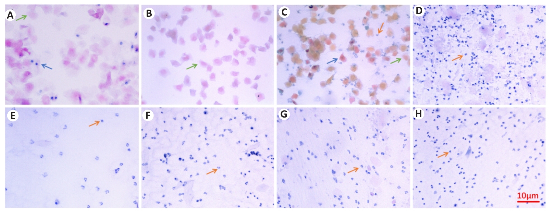
图1 小鼠阴道脱落细胞巴氏染色结果
Fig.1 Papanicolaou staining results of mouse vaginal exfoliated cells (Original magnification: ×100). A: Proestrus. B: Estrus. C: Late estrus. D-H: Interestrous. Blue arrows indicate nucleated cells, green arrows indicate keratinized cells, and red arrows indicate leukocytes.
| Group | E2 (pg/mL) | FSH (ng/mL) | LH (mU/mL) |
|---|---|---|---|
| Sham | 10.57±1.31 | 7.62±0.97 | 18.97±0.76 |
| MPS | 5.77±0.74** | 9.38±0.26** | 24.58±0.72** |
| MPS+CUMS | 4.89±0.71** | 11.20±0.92** | 33.13±1.48** |
| CSS | 6.98±1.31## | 9.68±0.89# | 22.88±1.42## |
表1 各组小鼠血清E2、FSH、LH含量比较
Tab.1 Comparison of serum E2, FSH and LH levels in the mice among the 4 groups (Mean±SD)
| Group | E2 (pg/mL) | FSH (ng/mL) | LH (mU/mL) |
|---|---|---|---|
| Sham | 10.57±1.31 | 7.62±0.97 | 18.97±0.76 |
| MPS | 5.77±0.74** | 9.38±0.26** | 24.58±0.72** |
| MPS+CUMS | 4.89±0.71** | 11.20±0.92** | 33.13±1.48** |
| CSS | 6.98±1.31## | 9.68±0.89# | 22.88±1.42## |
| Group | Weight (g) | OFT | SPT Sucrose solution preference rate (%) | FST Immobile time (s) | TST Immobile time (s) | |
|---|---|---|---|---|---|---|
| Total distance (m) | Time in the center (s) | |||||
| Sham | 18.88±0.87 | 14.26±2.61 | 32.21±11.78 | 62.21±8.58 | 33.00±7.07 | 139.09±5.05 |
| MPS | 18.52±0.57 | 14.05±2.76 | 32.67±13.02 | 62.37±6.70 | 32.43±8.06 | 138.83±8.97 |
| MPS+CUMS | 19.52±0.98 | 12.80±2.29 | 32.67±10.91 | 61.70±2.58 | 33.69±4.16 | 139.56±11.17 |
| CSS | 18.86±0.95 | 12.90±1.54 | 32.41±14.05 | 65.82±4.58 | 31.44±7.46 | 136.19±5.83 |
表2 各组小鼠基线及行为学测试比较
Tab.2 Comparison of baseline general condition and behavioral test results of the mice among the 4 groups (Mean±SD)
| Group | Weight (g) | OFT | SPT Sucrose solution preference rate (%) | FST Immobile time (s) | TST Immobile time (s) | |
|---|---|---|---|---|---|---|
| Total distance (m) | Time in the center (s) | |||||
| Sham | 18.88±0.87 | 14.26±2.61 | 32.21±11.78 | 62.21±8.58 | 33.00±7.07 | 139.09±5.05 |
| MPS | 18.52±0.57 | 14.05±2.76 | 32.67±13.02 | 62.37±6.70 | 32.43±8.06 | 138.83±8.97 |
| MPS+CUMS | 19.52±0.98 | 12.80±2.29 | 32.67±10.91 | 61.70±2.58 | 33.69±4.16 | 139.56±11.17 |
| CSS | 18.86±0.95 | 12.90±1.54 | 32.41±14.05 | 65.82±4.58 | 31.44±7.46 | 136.19±5.83 |
| Group | Weight (g) | OFT | SPT Sucrose solution preference rate (%) | FST Immobilization time(s) | TST Immobilization time(s) | |
|---|---|---|---|---|---|---|
| Total distance (m) | Time in the center(s) | |||||
| Sham | 24.27±1.03 | 12.22±0.55 | 30.05±7.77 | 74.34±14.33 | 27.38±4.31 | 117.29±10.06 |
| MPS | 25.20±1.21 | 10.44±1.17* | 26.25±9.67 | 52.80±12.72** | 45.72±8.14** | 149.60±10.98** |
| MPS+CUMS | 22.48±0.84** | 7.71±1.16** | 12.44±6.55** | 49.91±5.78** | 88.91±10.65** | 188.66±12.86** |
| CSS | 23.13±0.70 | 14.20±2.18## | 25.76±9.19# | 72.18±7.36## | 39.95±7.74## | 150.36±14.17## |
表3 各组小鼠造模、干预后行为学测试比较
Tab.3 Comparison of behavioral tests after modeling and intervention among the 4 groups (Mean±SD)
| Group | Weight (g) | OFT | SPT Sucrose solution preference rate (%) | FST Immobilization time(s) | TST Immobilization time(s) | |
|---|---|---|---|---|---|---|
| Total distance (m) | Time in the center(s) | |||||
| Sham | 24.27±1.03 | 12.22±0.55 | 30.05±7.77 | 74.34±14.33 | 27.38±4.31 | 117.29±10.06 |
| MPS | 25.20±1.21 | 10.44±1.17* | 26.25±9.67 | 52.80±12.72** | 45.72±8.14** | 149.60±10.98** |
| MPS+CUMS | 22.48±0.84** | 7.71±1.16** | 12.44±6.55** | 49.91±5.78** | 88.91±10.65** | 188.66±12.86** |
| CSS | 23.13±0.70 | 14.20±2.18## | 25.76±9.19# | 72.18±7.36## | 39.95±7.74## | 150.36±14.17## |
| Group | Glu (μmol/L) | GABA (ng/mL) |
|---|---|---|
| Sham | 22.07±1.20 | 9.92±0.48 |
| MPS | 25.21±1.48** | 9.26±0.37* |
| MPS+CUMS | 29.53±1.83** | 7.08±0.35** |
| CSS | 25.47±1.45## | 9.89±0.55## |
表4 各组小鼠血清Glu、GABA含量
Tab.4 Serum levels of Glu and GABA in each group (Mean±SD)
| Group | Glu (μmol/L) | GABA (ng/mL) |
|---|---|---|
| Sham | 22.07±1.20 | 9.92±0.48 |
| MPS | 25.21±1.48** | 9.26±0.37* |
| MPS+CUMS | 29.53±1.83** | 7.08±0.35** |
| CSS | 25.47±1.45## | 9.89±0.55## |
| Group | Serum | Hippocampal | ||
|---|---|---|---|---|
| IL-1β (pg/mL) | TNF-α (pg/mL) | IL-1β (pg/mg.pro) | TNF-α (pg/mg.pro) | |
| Sham | 1.02±0.53 | 0.33±0.20 | 12.45±0.78 | 87.04±3.23 |
| MPS | 2.96±0.45** | 1.87±0.65** | 22.76±1.21** | 105.10±1.25** |
| MPS+CUMS | 8.29±1.58** | 5.00±0.74** | 26.85±1.47** | 194.16±9.43** |
| CSS | 3.59±1.09## | 3.16±0.67## | 18.41±1.92## | 116.13±4.58## |
表5 各组小鼠血清与海马IL-1β、TNF-α含量
Tab.5 Serum and hippocampal levels of IL-1β and TNF-α in each group (Mean±SD)
| Group | Serum | Hippocampal | ||
|---|---|---|---|---|
| IL-1β (pg/mL) | TNF-α (pg/mL) | IL-1β (pg/mg.pro) | TNF-α (pg/mg.pro) | |
| Sham | 1.02±0.53 | 0.33±0.20 | 12.45±0.78 | 87.04±3.23 |
| MPS | 2.96±0.45** | 1.87±0.65** | 22.76±1.21** | 105.10±1.25** |
| MPS+CUMS | 8.29±1.58** | 5.00±0.74** | 26.85±1.47** | 194.16±9.43** |
| CSS | 3.59±1.09## | 3.16±0.67## | 18.41±1.92## | 116.13±4.58## |
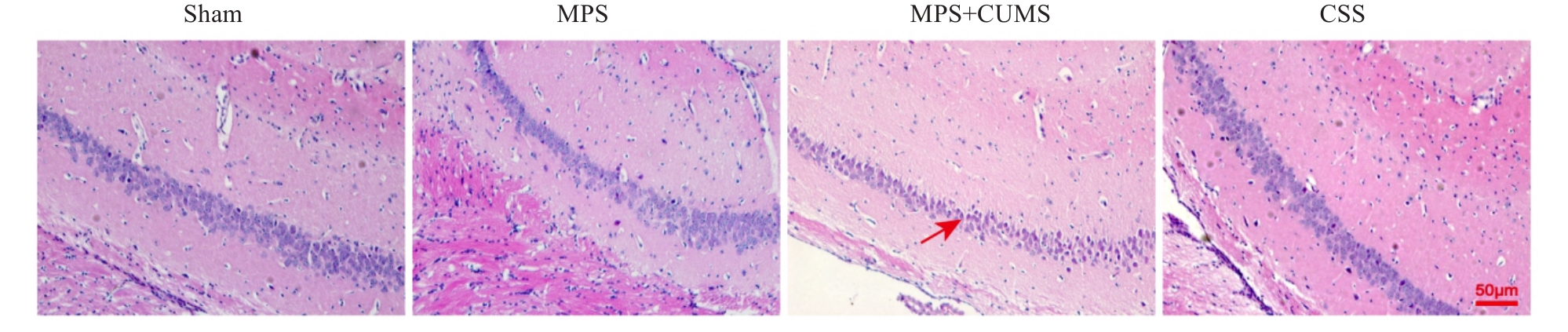
图2 各组小鼠海马CA1区HE染色结果
Fig.2 HE staining of the hippocampal CA1 region in each group (Original magnification: ×200). The red arrows in the figure indicate necrotic cells with karyopyknosis. MPS: Menopausal syndrome; CUMS: Chronic unpredictable mild stress; CSS: Chaihu Shugan San.
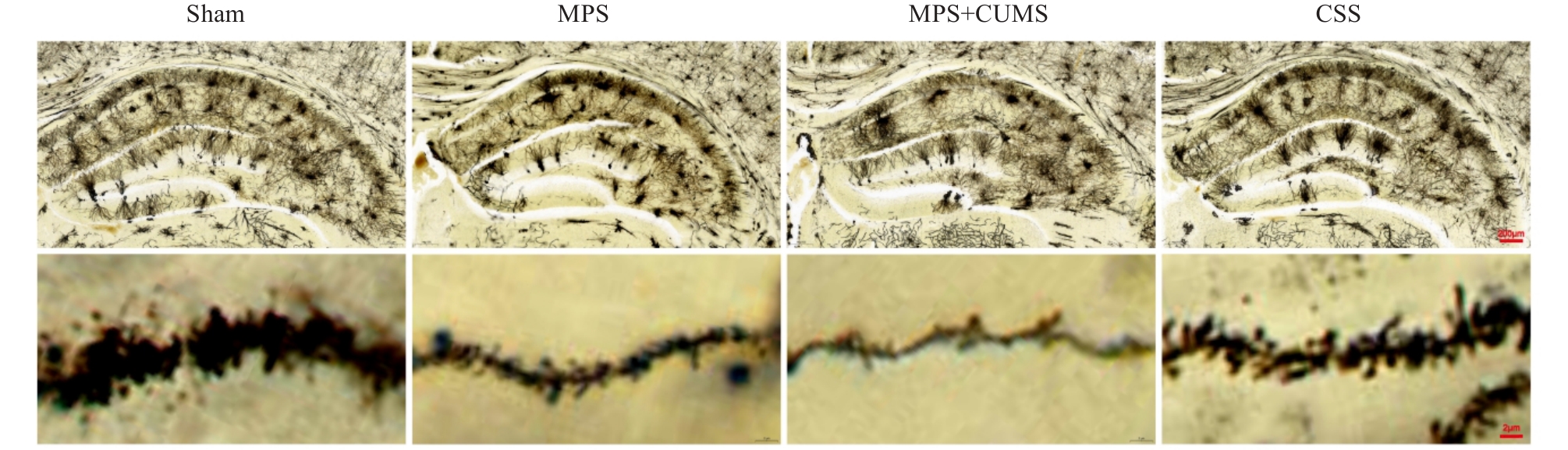
图3 各组小鼠海马CA1区高尔基染色结果
Fig.3 Results of Golgi staining in the hippocampal CA1 region of the mice in each group (Upper panel: ×10; Lower panel: ×1000).
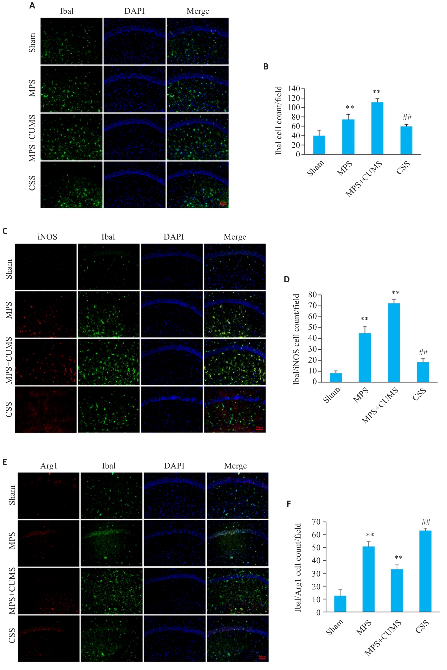
图4 柴胡疏肝散对MPS肝郁证小鼠海马CA1区小胶质细胞活化相关蛋白Iba1、Iba1/iNOS、Iba1/Arg1表达的影响
Fig.4 Effect of Chaihu Shugan San on expressions of microglial activation related proteins Iba1, Iba1/iNOS, and Iba1/Arg1 in the hippocampal CA1 region of the mice. A, B: Immunofluorescence staining of Iba1 expression (×200) and Iba1+ cell counts in the hippocampal CA1 region. C, D: Immunofluorescence staining of Iba1/iNOS expression (×200) and Iba1+/iNOS+ cell counts in the hippocampal CA1 region. E, F: Immunofluorescence staining of Iba1/Arg1 expression (×200) and Iba1+/Arg1+ cell counts in the hippocampal CA1 region. Data are presented as Mean±SD (n=3). Statistical analysis was performed using one-way ANOVA followed by Tukey's post hoc test. Iba1: Ionized calcium-binding adapter molecule 1; iNOS: Inducible nitric oxide synthase; Arg1: Arginase 1. **P<0.01 vs Sham group, ##P<0.01 vs MPS+CUMS group.
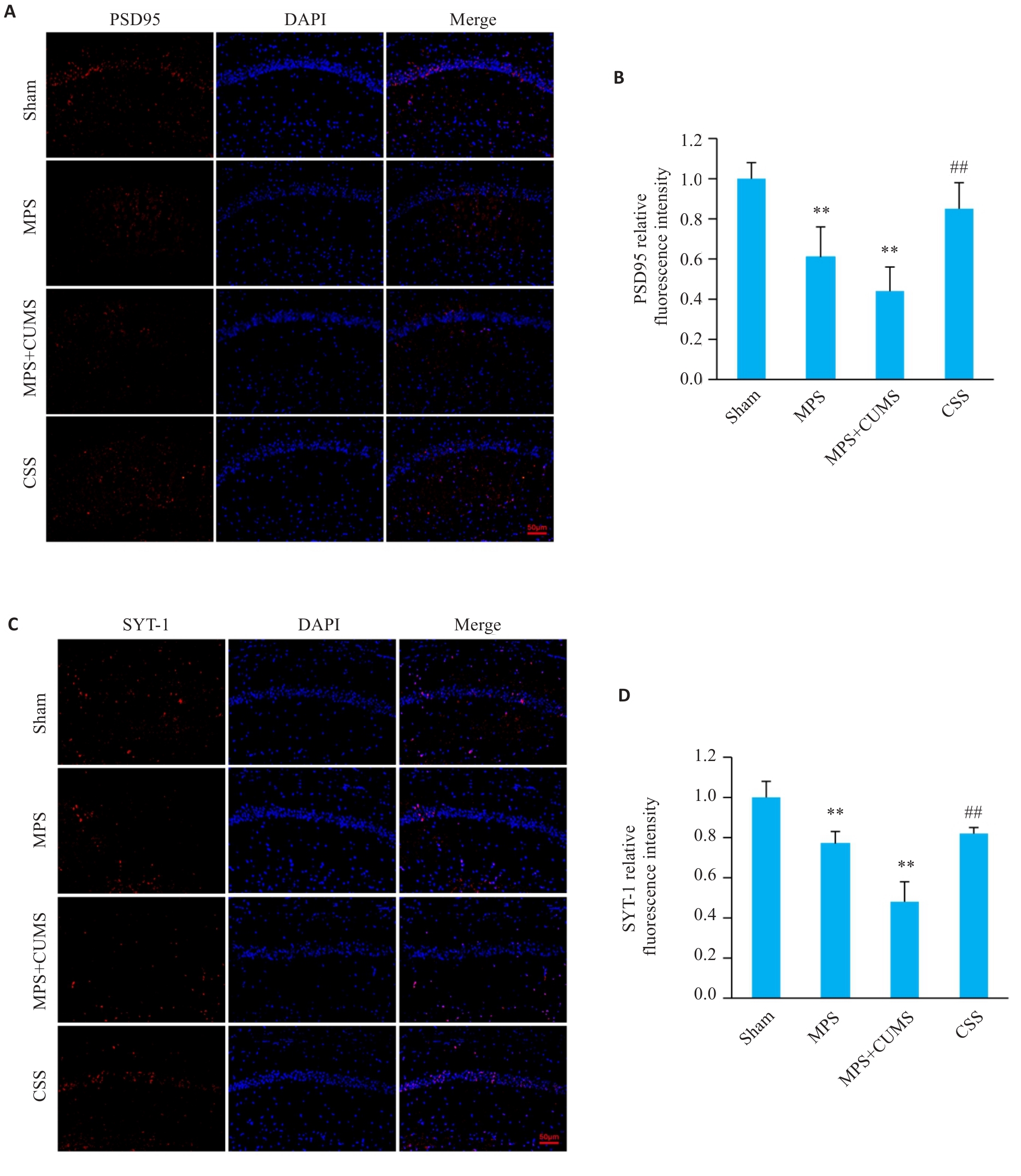
图5 柴胡疏肝散对MPS肝郁证小鼠海马区突触相关蛋白PSD95、SYT-1表达的影响
Fig.5 Effect of Chaihu Shugan San on expressions of synapse related proteins PSD95 and SYT-1 in the hippocampal CA1 region of the mice. A, B: Immunofluorescence staining of PSD95 expression (×200) and relative fluorescence intensity in the CA1 region. C, D: Immunofluorescence staining of SYT-1 expression (×200) and relative fluorescence intensity in the CA1 region. Data are presented as Mean±SD (n=3). Statistical analysis was performed using one-way ANOVA followed by Tukey's post hoc test. PSD95: Postsynaptic density protein 95; SYT-1: Synaptotagmin 1. **P<0.01 vs Sham group, ##P<0.01 vs MPS+CUMS group.
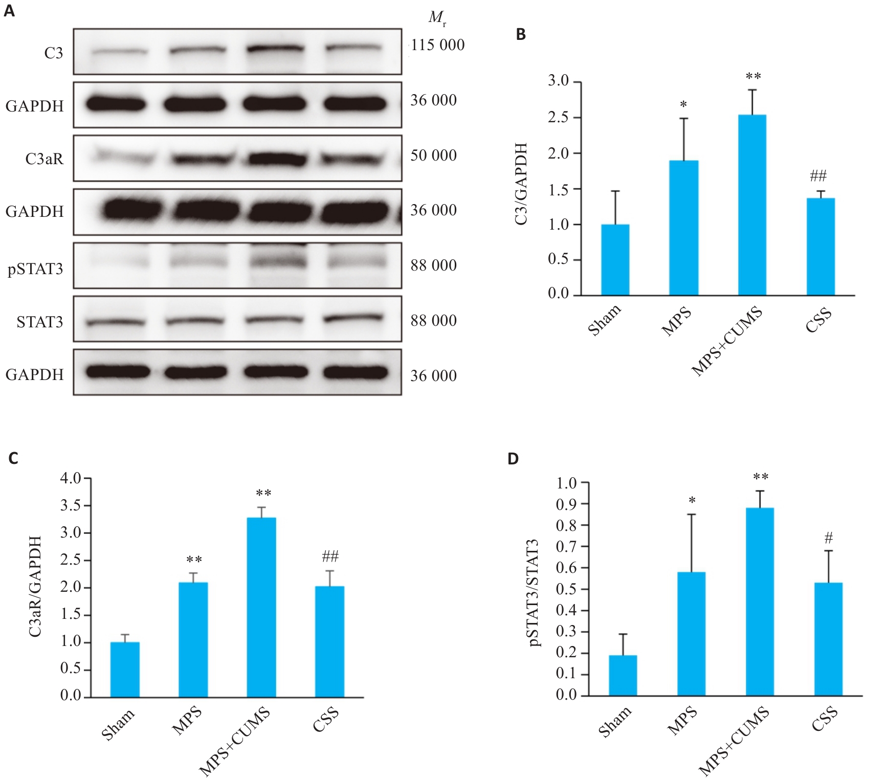
图6 柴胡疏肝散对MPS肝郁证小鼠海马经典补体通路相关蛋白C3、C3aR、pSTAT3/STAT3表达的影响
Fig.6 Effect of Chaihu Shugan San on expression of classical complement pathway-related proteins C3, C3aR and pSTAT3/STAT3 in the hippocampus of the mice. A: Protein bands of C3, C3aR, p-STAT3 and STAT3 detected by Western blotting. B-D: Quantitative analysis of C3, C3aR, p-STAT3, and STAT3 protein expression levels. Data are presented as Mean±SD (n=3). Statistical analysis was performed using one-way ANOVA followed by Tukey's post hoc test. C3: Component 3; C3aR: Complement C3a receptor; pSTAT3: Phosphorylated signal transducer and activator of transcription 3; STAT3: Signal transducer and activator of transcription 3; GAPDH: Glyceraldehyde-3-phosphate dehydrogenase. *P<0.05, **P<0.01 vs Sham group; #P<0.05, ##P<0.01 vs MPS+CUMS group.
| [1] | 倪 青, 卜祥伟, 庞 晴. 更年期综合征(围绝经期综合征)病证结合诊疗指南[J]. 世界中医药, 2025, 20(10): 1633-43. |
| [2] | Li Z, Guo JP, Huang L. Perimenopausal syndrome and hypertension during perimenopause in South China: prevalence, relationships and risk factors[J]. BMC Women's Health, 2024, 24(1): 215. doi:10.1186/s12905-024-03056-5 |
| [3] | Sander B, Gordon JL. Premenstrual mood symptoms in the perimenopause[J]. Curr Psychiatry Rep, 2021, 23(11): 73. doi:10.1007/s11920-021-01285-1 |
| [4] | Cybulska AM, Rachubińska K, Grochans E, et al. Systemic inflammation indices, chemokines, and metabolic markers in perimenopausal women[J]. Nutrients, 2025, 17(17): 2885. doi:10.3390/nu17172885 |
| [5] | McCarthy M, Raval AP. The peri-menopause in a woman's life: a systemic inflammatory phase that enables later neurodegenerative disease[J]. J Neuroinflammation, 2020, 17(1): 317. doi:10.1186/s12974-020-01998-9 |
| [6] | Pronier É, Morici JF, Girardeau G. The role of the hippocampus in the consolidation of emotional memories during sleep[J]. Trends Neurosci, 2023, 46(11): 912-25. doi:10.1016/j.tins.2023.08.003 |
| [7] | Picard K, Bisht K, Poggini S, et al. Microglial-glucocorticoid receptor depletion alters the response of hippocampal microglia and neurons in a chronic unpredictable mild stress paradigm in female mice[J]. Brain Behav Immun, 2021, 97: 423-39. doi:10.1016/j.bbi.2021.07.022 |
| [8] | You Y, Chen Z, Hu WW. The role of microglia heterogeneity in synaptic plasticity and brain disorders: Will sequencing shed light on the discovery of new therapeutic targets[J]? Pharmacol Ther, 2024, 255: 108606. doi:10.1016/j.pharmthera.2024.108606 |
| [9] | Park DS, Kozaki T, Tiwari SK, et al. iPS-cell-derived microglia promote brain organoid maturation via cholesterol transfer[J]. Nature, 2023, 623(7986): 397-405. doi:10.1038/s41586-023-06713-1 |
| [10] | Hong S, Beja-Glasser VF, Nfonoyim BM, et al. Complement and microglia mediate early synapse loss in Alzheimer mouse models[J]. Science, 2016, 352(6286): 712-6. doi:10.1126/science.aad8373 |
| [11] | 严 灿, 徐志伟. 肝主疏泄调畅情志功能的中枢神经生物学机制探讨[J]. 中国中西医结合杂志, 2005, 25(5): 459-62. |
| [12] | Zhang H, Gao Z, Sun Y, et al. Profiling GABA(A) receptor subunit expression in the hippocampus of PMDD rat models based on TCM theories[J]. Mol Neurobiol, 2023, 60(8): 4418-28. doi:10.1007/s12035-023-03354-3 |
| [13] | 陈惠芳, 龚 琳, 李巧琳, 等. 基于“肝脑相维”探讨肝失疏泄对不同月龄雌性大鼠学习记忆能力及海马突触可塑性的影响[J]. 中华中医药杂志, 2024, 39(5): 2548-53. |
| [14] | 高洁茹. 基于情志致病理论的围绝经期综合征相关因素及致病机制研究[D]. 天津: 天津中医药大学, 2021. |
| [15] | 胡永伟, 应 翔, 张俊英, 等. 柴胡疏肝散联合氟哌噻醇美利曲辛治疗围绝经期综合征肝郁型66例[J]. 浙江中医杂志, 2021, 56(11): 824-5. |
| [16] | 梁文娜, 阮璐薇, 张鹏横, 等. 柴胡疏肝散调节围绝经期肝郁证大鼠ERK1/2-CREB-BDNF信号通路的机制[J]. 中华中医药杂志, 2022, 37(6): 3147-52. |
| [17] | 王素英, 蔡晓诗, 陈朝玺, 等. 柴胡疏肝散调节SIK2-CRTC1通路改善围绝经期肝郁证的机制[J]. 中华中医药杂志, 2024, 39(11): 5869-74. |
| [18] | Huang RT, Gong M, Tan X, et al. Effects of Chaihu Shugan San on brain functional network connectivity in the hippocampus of a perimenopausal depression rat model[J]. Mol Neurobiol, 2024, 61(3): 1655-72. doi:10.1007/s12035-023-03615-1 |
| [19] | 徐莲薇, 张淑君, 孙卓君. 围绝经期综合征病因病机特点的思考[J]. 中医杂志, 2008, 49(11): 1031-3. |
| [20] | 吴杨乔, 刘雁峰, 李冰冰, 等. 围绝经期综合征情绪症状相关因素研究概况[J]. 中国妇幼保健, 2019, 34(8): 1919-22. |
| [21] | 张晓丹, 任亚萍, 乔丽平. 浅谈肝与围绝经期综合征的关系[J]. 中医杂志, 2010, 51(S1): 75-7. |
| [22] | 梁文娜, 李灿东, 李西海. 围绝经期综合征中医证素规律的临床研究[J]. 福建中医学院学报, 2009, 19(6): 3-5. |
| [23] | Lyu HY, Li T, Chen YJ, et al. Exploring the effective substances and mechanisms of action of ChaiHu ShuGan San in treating perimenopausal syndrome based on in vivo exposure profiles and steroid hormone metabolic networks[J]. J Ethnopharmacol, 2026, 355: 120648. doi:10.1016/j.jep.2025.120648 |
| [24] | 韩 悦, 于丽媛, 卢桂兰, 等. 柴胡疏肝散对围绝经期抑郁模型大鼠焦虑抑郁的改善作用及机制研究[J]. 时珍国医国药, 2024, 35(6): 1316-9. |
| [25] | Guo ZY, Long TJ, Yao JP, et al. Potential antidepressant effects of Traditional Chinese botanical drug formula Chaihu-Shugan-San and its active ingredients[J]. Front Pharmacol, 2024, 15: 1337876. doi:10.3389/fphar.2024.1337876 |
| [26] | He MC, Xia SH, Pan H, et al. Chaihu-Shugan-San ameliorated osteoporosis of mice with depressive behavior caused by chronic unpredictable mild stress via repressing neuroinflammation and HPA activity[J]. Drug Des Dev Ther, 2024, 18: 5997-6015. doi:10.2147/dddt.s480077 |
| [27] | 于 眉, 王颖超, 李 玲, 等. 腹针配合柴胡疏肝散治疗气郁型围绝经期失眠的临床效果[J]. 中国医药导报, 2020, 17(20): 160-3. |
| [28] | Itoh N, Itoh Y, Meyer CE, et al. Estrogen receptor beta in astrocytes modulates cognitive function in mid-age female mice[J]. Nat Commun, 2023, 14: 6044. doi:10.1038/s41467-023-41723-7 |
| [29] | 刘 洋, 程来洋, 郭中坤. 中医病症结合的围绝经期综合征动物模型研究进展[J]. 实验动物与比较医学, 2025, 45(5): 586-95. |
| [30] | Lei HJ, Liu J, Deng J, et al. Behavior, hormone, and gut microbiota change by YYNS intervention in an OVX mouse model[J]. Front Cell Infect Microbiol, 2024, 14: 1445741. doi:10.3389/fcimb.2024.1445741 |
| [31] | 匡洛逸, 罗紫龙, 贾 庆, 等. 肝郁证动物模型中枢生物学改变异同研究进展[J]. 中国比较医学杂志, 2026, 36(1): 92-102. |
| [32] | Zheng ZH, Tu JL, Li XH, et al. Neuroinflammation induces anxiety- and depressive-like behavior by modulating neuronal plasticity in the basolateral amygdala[J]. Brain Behav Immun, 2021, 91: 505-18. doi:10.1016/j.bbi.2020.11.007 |
| [33] | Kong Y, Zhang X, Li L, et al. Microglia-derived vitamin D binding protein mediates synaptic damage and induces depression by binding to the neuronal receptor megalin[J]. Adv Sci, 2025, 12(6): 2410273. doi:10.1002/advs.202410273 |
| [34] | 苏 虹, 粟胜勇, 李 竞, 等. 麦粒灸对围绝经期抑郁大鼠海马ERK/CREB-BDNF/TrkB信号通路及神经递质水平的影响[J]. 中国中医药信息杂志, 2025, 32(8): 98-104. |
| [35] | Wu AB, Zhang JY. Neuroinflammation, memory, and depression: new approaches to hippocampal neurogenesis[J]. J Neuroinflam-mation, 2023, 20(1): 283. doi:10.1186/s12974-023-02964-x |
| [36] | Jia XN, Gao ZH, Hu HL. Microglia in depression: current perspectives[J]. Sci China Life Sci, 2021, 64(6): 911-25. doi:10.1007/s11427-020-1815-6 |
| [37] | 莫申申, 程 爽, 张 芹, 等. 滋肾清心安神汤对围绝经期抑郁模型小鼠行为学的改善作用及对神经炎症的影响[J]. 浙江中西医结合杂志, 2025, 35(12): 1113-8. |
| [38] | Liang GF, Kow ASF, Yusof R, et al. Menopause-associated depression: impact of oxidative stress and neuroinflammation on the central nervous system: a review[J]. Biomedicines, 2024, 12(1): 184. doi:10.3390/biomedicines12010184 |
| [39] | Gonzalez KC, Negrean A, Liao ZR, et al. Synaptic basis of feature selectivity in hippocampal neurons[J]. Nature, 2025, 637(8048): 1152-60. doi:10.1038/s41586-024-08325-9 |
| [40] | Tartt AN, Mariani MB, Hen RE, et al. Dysregulation of adult hippocampal neuroplasticity in major depression: pathogenesis and therapeutic implications[J]. Mol Psychiatry, 2022, 27(6): 2689-99. doi:10.1038/s41380-022-01520-y |
| [41] | Deng D, Cui YF, Gan S, et al. Sinisan alleviates depression-like behaviors by regulating mitochondrial function and synaptic plasticity in maternal separation rats[J]. Phytomedicine, 2022, 106: 154395. doi:10.1016/j.phymed.2022.154395 |
| [42] | Mehta N, Mondal S, Watson ET, et al. The juxtamembrane linker of synaptotagmin 1 regulates Ca2+ binding via liquid-liquid phase separation[J]. Nat Commun, 2024, 15: 262. doi:10.1038/s41467-023-44414-5 |
| [43] | Song QL, Huang WM, Ye WB, et al. Neuroprotective effects of estrogen through BDNF-transient receptor potential channels 6 signaling pathway in the hippocampus in a rat model of perimenopausal depression[J]. Front Aging Neurosci, 2022, 14: 869274. doi:10.3389/fnagi.2022.869274 |
| [44] | Lin YX, Wang LY, Meng SN, et al. 3-hydroxykynurenine increase in kynurenine pathway driven bisphenol F induced anxiety- and depression-like behaviors[J]. Sci Total Environ, 2025, 995: 180073. doi:10.1016/j.scitotenv.2025.180073 |
| [45] | 黄楚瑶, 张祉思, 陈淑敏, 等. 电针改善围绝经期抑郁模型小鼠内侧前额叶皮层Glu/GABA平衡介导的突触可塑性[J]. 针刺研究,2026,[Epub ahead of print]. |
| [46] | Salvador AF, de Lima KA, Kipnis J. Neuromodulation by the immune system: a focus on cytokines[J]. Nat Rev Immunol, 2021, 21(8): 526-41. doi:10.1038/s41577-021-00508-z |
| [47] | Jiang X, Yi SN, Liu Q, et al. Asperosaponin VI ameliorates the CMS-induced depressive-like behaviors by inducing a neuroprotective microglial phenotype in hippocampus via PPAR‑γ pathway[J]. J Neuroinflammation, 2022, 19(1): 115. doi:10.1186/s12974-022-02478-y |
| [48] | Zhang JQ, Rong PJ, Zhang LJ, et al. IL4-driven microglia modulate stress resilience through BDNF-dependent neurogenesis[J]. Sci Adv, 2021, 7(12): eabb9888. doi:10.1126/sciadv.abb9888 |
| [49] | Wang XM, Li SS, Yu JY, et al. Saikosaponin B2 ameliorates depression-induced microglia activation by inhibiting ferroptosis-mediated neuroinflammation and ER stress[J]. J Ethnopharmacol, 2023, 316: 116729. doi:10.1016/j.jep.2023.116729 |
| [50] | Cao MM, Guo Z, Wang J, et al. Astragalin alleviates lipopolysaccharide-induced depressive-like behavior in mice by preserving blood-brain barrier integrity and suppressing neuroinflammation[J]. Free Radic Biol Med, 2025, 232: 340-52. doi:10.1016/j.freeradbiomed.2025.03.014 |
| [51] | Wang J, Chen HS, Li HH, et al. Microglia-dependent excessive synaptic pruning leads to cortical underconnectivity and behavioral abnormality following chronic social defeat stress in mice[J]. Brain Behav Immun, 2023, 109: 23-36. doi:10.1016/j.bbi.2022.12.019 |
| [52] | Zhang AM, Zhang XX, Tang XH, et al. Activation of the C3 complement pathway in the hippocampus produces anxiodepressive effects in a mouse model of inflammation pain[J]. Mol Neurobiol, 2025, 62(11): 14032-49. doi:10.1007/s12035-025-05187-8 |
| [53] | Zhang MM, Guo MX, Zhang QP, et al. IL-1R/C3aR signaling regulates synaptic pruning in the prefrontal cortex of depression[J]. Cell Biosci, 2022, 12(1): 90. doi:10.1186/s13578-022-00832-4 |
| [54] | 唐璎娟, 郭海鹏, 邹蔓姝, 等. 柴芍开郁汤通过补体C3/C3aR通路改善抑郁模型大鼠海马神经炎症的研究[J]. 中国中药杂志, 2025, 50(1): 1-9. doi:10.19540/j.cnki.cjcmm.20240712.705 |
| [55] | Hao WZ, Ma QY, Wang L, et al. Gut dysbiosis induces the development of depression-like behavior through abnormal synapse pruning in microglia-mediated by complement C3[J]. Microbiome, 2024, 12(1): 34. doi:10.1186/s40168-024-01756-6 |
| [1] | 梁仁杰, 王荷颖, 李兆艳, 陈惠萍, 王奕淳, 闵莉. 柴胡疏肝散加减可改善胆囊胆固醇结石肝郁证小鼠肝郁和结石程度[J]. 南方医科大学学报, 2026, 46(3): 615-628. |
| [2] | 冯鹏, 王洁, 褚江伟, 王鹤霖, 田永衍, 张杰昌, 王明露, 王宇彤. 石菖蒲挥发油通过抑制TLR4/MyD88/NF-κB通路介导的小胶质细胞极化改善大鼠抽动障碍[J]. 南方医科大学学报, 2026, 46(3): 646-654. |
| [3] | 夏金枝, 陈悦, 任侣, 李静, 宋雪, 陶露, 胡建国. 咖啡豆醇通过调控IκBα/NF-κB通路抑制小胶质细胞活化改善脊髓损伤后小鼠的运动功能[J]. 南方医科大学学报, 2025, 45(12): 2561-2572. |
| [4] | 孙一鸣, 徐昕冉, 卓雪瑞, 蔡慧, 王艳. C1q中和抗体可通过C1q/C3通路改善小鼠的产后抑郁样行为[J]. 南方医科大学学报, 2025, 45(10): 2111-2117. |
| [5] | 左涵珺, 段兆达, 王朝, 郭涛, 石金沙, 石浩龙, 李娟娟. 天麻素经PI3K/AKT通路改善新生大鼠缺氧缺血性脑损伤后小胶质细胞介导的炎症反应[J]. 南方医科大学学报, 2024, 44(9): 1712-1719. |
| [6] | 石金沙, 张皓南, 张幸霖, 石浩龙, 左涵珺, 郭涛, 王朝, 余航, 李娟娟. 天麻素通过调节CCR5/AKT信号传导缓解新生小鼠缺血缺氧后小胶质细胞介导的炎症反应[J]. 南方医科大学学报, 2024, 44(10): 1850-1857. |
| [7] | 孙晓鹏, 史 航, 张 磊, 刘 中, 李克威, 钱玲玲, 朱星宇, 杨康佳, 付 强, 丁 华. 外胚层间充质干细胞来源的外泌体通过控制炎症和氧化损伤减少M1型小胶质细胞并促进H2O2处理后PC12细胞的存活[J]. 南方医科大学学报, 2024, 44(1): 119-128. |
| [8] | 孙 洋, 许轶博, 肖林雨, 朱国庆, 李 静, 宋 雪, 徐 磊, 胡建国. 乙酰紫堇灵促进大鼠脊髓损伤后的功能恢复:基于调控EGFR/MAPK信号通路抑制小胶质细胞活化[J]. 南方医科大学学报, 2023, 43(6): 915-923. |
| [9] | 张嘉发, 杨灿洪, 张淑芬, 曹婷婷, 彭 瑞, 郭蔚泓, 严予苹, 谢淑婷, 彭晓佳, 吕田明, 黄添容. 莱菔硫烷通过下调MAPK/NF-κB信号通路逆转Aβ纤维介导的M1型小胶质细胞极化和神经炎症介导的神经干细胞程序性坏死[J]. 南方医科大学学报, 2023, 43(12): 2132-2138. |
| [10] | 陈丽旋, 黄定帮, 郑 刚, 孟晓静. 铅暴露加重Aβ1-42处理的小胶质细胞活化和小胶质细胞中的铜离子蓄积[J]. 南方医科大学学报, 2023, 43(10): 1752-1760. |
| [11] | 赵佳佳, 杨荷雨, 王招娣, 朱海丽, 谢 敏. ANA-12通过靶向阻断BDNF/TrkB信号通路降低大鼠的脊髓炎症和缓解病理性疼痛[J]. 南方医科大学学报, 2022, 42(2): 232-237. |
| [12] | 黄河灵, 高玉元, 聂 坤, 王丽娟. 巨噬细胞移动抑制因子介导MPP+/MPTP诱导的小胶质细胞NLRP3炎症小体的激活[J]. 南方医科大学学报, 2021, 41(7): 972-979. |
| [13] | 刘杨,蒋朴,徐颖. 脂多糖可致敏高浓度氧对新生小鼠未成熟脑的损伤[J]. 南方医科大学学报, 2014, 34(02): 214-. |
| [14] | 缪星宇,刘晓斌,岳青,秋楠,黄卫东,王继军,许彦钢,张越林,杨军,陈新林. 去铁胺对大鼠脑出血后小胶质细胞活化的抑制及其继发性神经损伤的保护作用[J]. 南方医科大学学报, 2012, 32(07): 970-. |
| [15] | 蒋朴,徐颖,胡良安,刘杨,邓世雄. 高浓度氧暴露对N9小胶质细胞功能的影响[J]. 南方医科大学学报, 2012, 32(01): 71-. |
| 阅读次数 | ||||||
|
全文 |
|
|||||
|
摘要 |
|
|||||
