
南方医科大学学报 ›› 2026, Vol. 46 ›› Issue (7): 1622-1633.doi: 10.12122/j.issn.1673-4254.2026.07.16
• • 上一篇
孙宇航1,2( ), 卢兴珍2, 张馨月1,3, 郭建鹏1,2,3(), 金鑫1,3()
收稿日期:2025-12-23
出版日期:2026-07-20
发布日期:2026-07-20
通讯作者:
郭建鹏,金鑫
E-mail:m15543555820@163.com;gjp807@ybu.edu.cn;jinchenlcn@163.com
作者简介:孙宇航,在读硕士研究生,E-mail: m15543555820@163.com
基金资助:
Yuhang SUN1,2(), Xingzhen LU2, Xinyue ZHANG1,3, Jianpeng GUO1,2,3(), Xin JIN1,3()
Received:2025-12-23
Online:2026-07-20
Published:2026-07-20
Contact:
Jianpeng GUO, Xin JIN
E-mail:m15543555820@163.com;gjp807@ybu.edu.cn;jinchenlcn@163.com
摘要:
目的 探究熊胆粉(BBP)通过调控NF-κB和Nrf-2/HO-1信号通路对脂多糖诱导的急性肺损伤(ALI)小鼠的保护作用。 方法 体外实验:采用脂多糖(LPS)诱导RAW264.7细胞建立体外模型,设置对照组、模型组、地塞米松(Dex,25 μg/mL)、BBP低剂量组(6.25 μg/mL)和BBP高剂量组(12.5 μg/mL)。体内实验:将60只C57BL/6小鼠随机分为对照组、模型组、Dex组(5 mg/kg)、BBP低剂量组(30 mg/kg)和BBP高剂量组(120 mg/kg),12只/组。通过ELISA和相关试剂盒检测细胞和肺组织中炎症与氧化应激指标;通过肺组织病理分析、免疫细胞计数和肺湿/干质量比等指标评价BBP对ALI的干预作用。通过qRT-PCR、Western blotting和免疫组化法分析NF-κB和Nrf-2/HO-1信号通路相关蛋白与基因的表达变化。 结果 细胞实验结果显示,与模型组相比,BBP组(12.5μg/mL)显著降低TNF-α、COX-2、IL-1β、IL-6、丙二醛(MDA)、一氧化氮(NO)和活性氧水平,升高SOD水平,且IκB-α、Nrf-2和HO-1的mRNA表达水平升高,而TNF-α、IL-1β、Keap-1和NF-κB(p65)的mRNA表达水平降低(P<0.05,P<0.01,P<0.001)。动物实验结果显示,与对照组相比,模型组小鼠肺组织结构紊乱,肺泡壁增厚,肺泡内可见大量炎症细胞浸润,肺组织湿/干质量比及支气管肺泡灌洗液(BALF)总细胞数升高(P<0.01);肺组织中的TNF-α、COX-2、IL-1β、IL-6、MDA和NO含量升高,而SOD浓度降低(P<0.01,P<0.001);且肺组织中IκB-α、Nrf-2和HO-1的蛋白表达水平降低,Keap-1和NF-κB(p65)的蛋白表达水平升高(P<0.01)。与模型组相比,BBP(120 mg/kg)组能改善小鼠肺组织病理损伤状态,改善肺肿胀程度,降低BALF总细胞数、TNF-α、COX-2、IL-1β、IL-6、MDA和NO水平,升高SOD水平(P<0.05,P<0.01);同时,BBP上调了肺组织中IκB-α、Nrf-2和HO-1的蛋白表达水平,并抑制了Keap-1和NF-κB(p65)的蛋白表达水平(P<0.05,P<0.01)。 结论 BBP可能通过作用于Nrf-2/HO-1和NF-κB信号通路对LPS诱导的ALI发挥保护作用,为后续研究潜在的治疗ALI药物提供依据。
孙宇航, 卢兴珍, 张馨月, 郭建鹏, 金鑫. 熊胆粉通过调控NF-κB和Nrf-2/HO-1信号通路减轻脂多糖诱导的小鼠急性肺损伤[J]. 南方医科大学学报, 2026, 46(7): 1622-1633.
Yuhang SUN, Xingzhen LU, Xinyue ZHANG, Jianpeng GUO, Xin JIN. Natural bear bile powder attenuates lipopolysaccharide-induced acute lung injury in mice by regulating the NF-κB and Nrf-2/HO-1 signaling pathways[J]. Journal of Southern Medical University, 2026, 46(7): 1622-1633.
| Primer | Sequence |
|---|---|
| p65 | F: 5′ - TGCGATTCCGCTATAAATGCG-3′ R: 5′ - ACAAGTTCATGTGGATGAGGC-3′ |
| TNF-α | F: 5′- CAGGCGGTGCCTATGTCTC -3′ R: 5′- CGATCACCCCGAAGTTCAGTAG-3′ |
| IL-1β | F: 5′ - GAAATGCCACCTTTTGACAGTG-3′ R: 5′ - TGGATGCTCTCATCAGGACAG-3′ |
| IκBα | F: 5′ - CGGCACCGAGTACCTTGAC -3′ R: 5′ - CACCGGCTGCATACAACTTCT-3′ |
| Nrf-2 | F: 5′-CTTTAGTCAGCGACAGAAGGAC-3′ R: 5′ - AGGCATCTTGTTTGGGAATGTG-3′ |
| HO-1 | F: 5′ - GATAGAGCGCAACAAGCAGAA -3′ R: 5′ - CAGTGAGGCCCATACCAGAAG -3′ |
| Keap-1 | F: 5′ -CGGGGACGCAGTGATGTATG -3′ R: 5′ - TGTGTAGCTGAAGGTTCGGTTA-3′ |
| GAPDH | F: 5′ - AGGTCGGTGTGAACGGATTTG-3′ R: 5′ - GGGGTCGTTGATGGCAACA-3′ |
表1 RT-PCR所用引物
Tab.1 Primers for RT-PCR
| Primer | Sequence |
|---|---|
| p65 | F: 5′ - TGCGATTCCGCTATAAATGCG-3′ R: 5′ - ACAAGTTCATGTGGATGAGGC-3′ |
| TNF-α | F: 5′- CAGGCGGTGCCTATGTCTC -3′ R: 5′- CGATCACCCCGAAGTTCAGTAG-3′ |
| IL-1β | F: 5′ - GAAATGCCACCTTTTGACAGTG-3′ R: 5′ - TGGATGCTCTCATCAGGACAG-3′ |
| IκBα | F: 5′ - CGGCACCGAGTACCTTGAC -3′ R: 5′ - CACCGGCTGCATACAACTTCT-3′ |
| Nrf-2 | F: 5′-CTTTAGTCAGCGACAGAAGGAC-3′ R: 5′ - AGGCATCTTGTTTGGGAATGTG-3′ |
| HO-1 | F: 5′ - GATAGAGCGCAACAAGCAGAA -3′ R: 5′ - CAGTGAGGCCCATACCAGAAG -3′ |
| Keap-1 | F: 5′ -CGGGGACGCAGTGATGTATG -3′ R: 5′ - TGTGTAGCTGAAGGTTCGGTTA-3′ |
| GAPDH | F: 5′ - AGGTCGGTGTGAACGGATTTG-3′ R: 5′ - GGGGTCGTTGATGGCAACA-3′ |
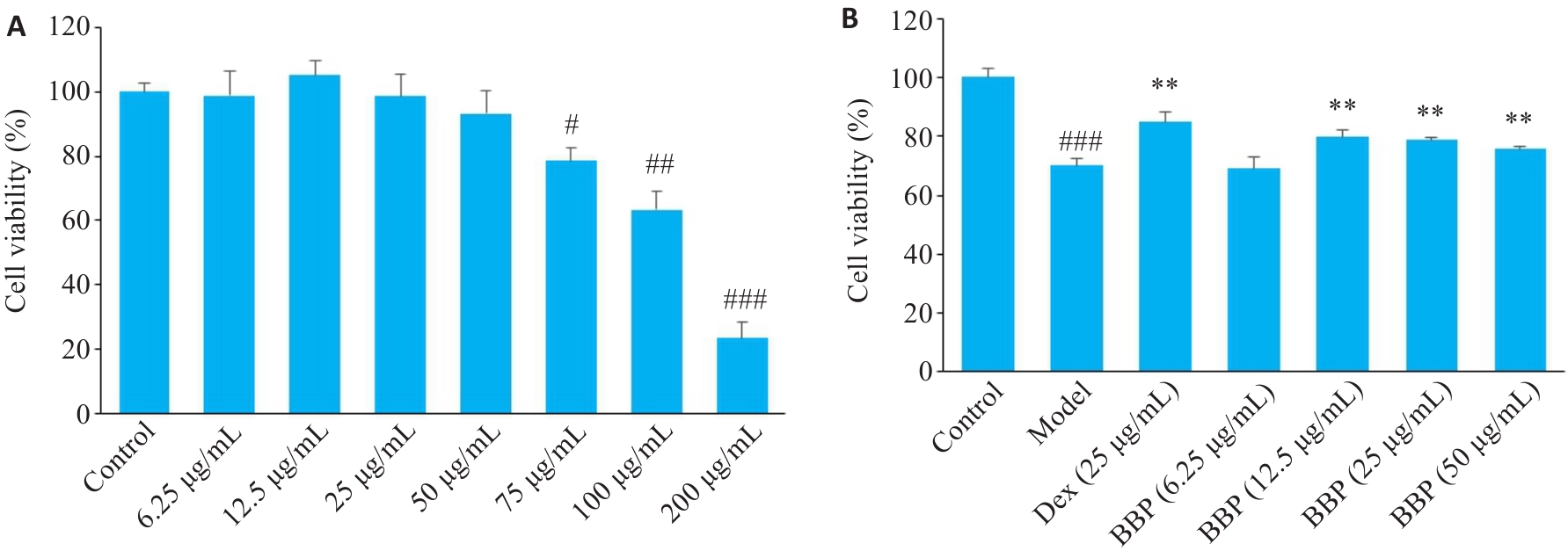
图1 CCK-8法检测结果
Fig.1 Results of CCK-8 assay. A: Cell viability assessed by CCK-8 assay in RAW 264.7 cells treated with different concentrations of BBP. B: Cell viability detected by CCK-8 assay of LPS-induced RAW 264.7 cells treated with different concentrations of BBP. Data are presented as Mean±SD (n=3). #P<0.05, ##P<0.01, ###P<0.001 vs Control group; **P<0.01 vs Model group.
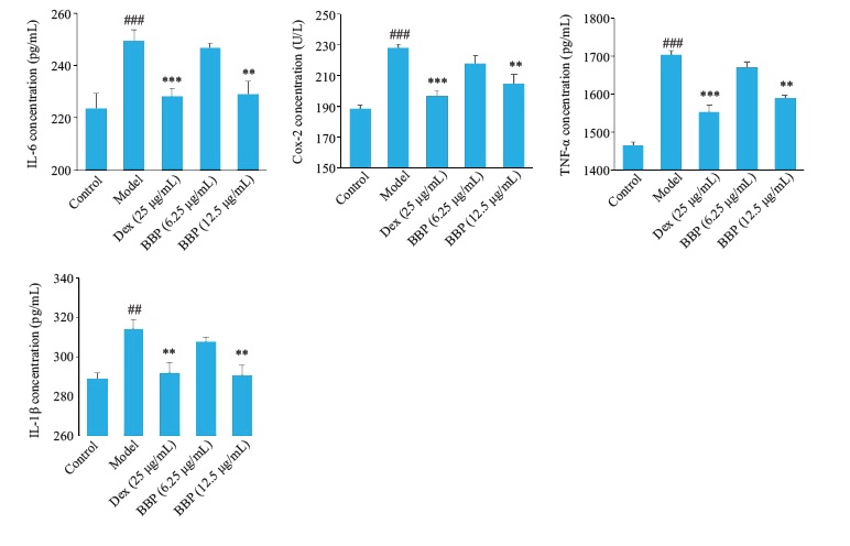
图2 BBP对LPS诱导的RAW264.7细胞炎症因子水平的影响
Fig.2 Effect of BBP on inflammatory cytokine levels in LPSinducedRAW264.7 cells Mean±SD, n=3). ##P<0.01, ###P<0.001 vs Control group; **P<0.010.01, ***P<0.010.001 vs Model group.
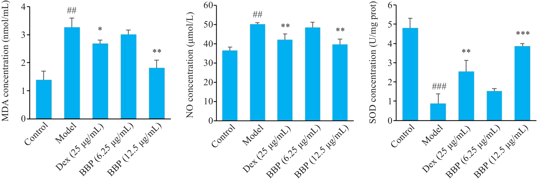
图3 BBP对LPS诱导RAW264.7细胞中MDA、SOD和NO水平的影响
Fig.3 Effect of BBP on MDA, SOD and NO in LPS-induced RAW264.7 cells (Mean±SD, n=3). ##P<0.01, ###P<0.001 vs Control group; *P<0.05, **P<0.01, ***P<0.001 vs Model group.
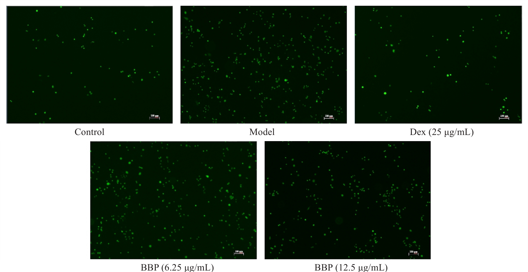
图4 各组细胞ROS的表达
Fig.4 Effect of BBP pretreatment on ROS in RAW264.7 cells assessed using DCFH-DA fluorescent probe (Scale bar=100 μm).
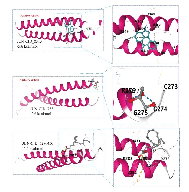
图5 qPCR检测LPS诱导的RAW264.7细胞中Keap-1、HO-1、Nrf-2、NF-κB、TNF-α、IκB-α和IL-1β的mRNA表达水平
Fig.5 qPCR for detecting mRNA expressions of the Keap-1, HO-1, Nrf-2,NF-κB, TNF-α, IκB-α and IL-1β in LPS-induced RAW264.7 cells (Mean±SD, n=3). ##P<0.01, ###P<0.001 vs Control group; **P<0.010.01, ***P<0.010.001 vsModel group.
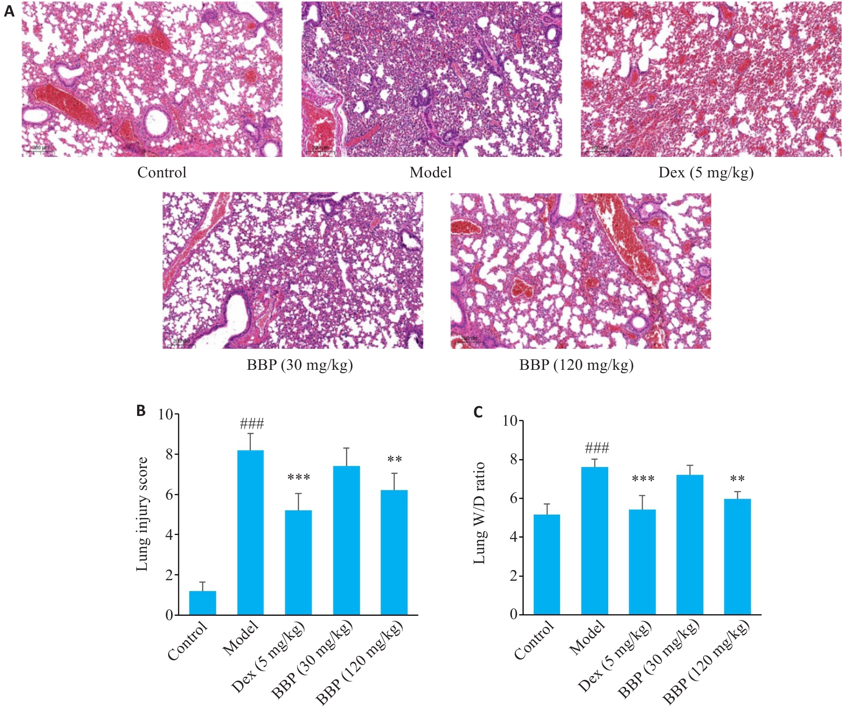
图6 BBP对LPS诱导的ALI小鼠肺组织病理学变化的影响
Fig.6 Effects of BBP on LPS-induced histopathological changes in lung tissues of ALI mice. A: HE staining for examining histological alterations in mouse lung tissues (Scale bar=200 μm). B: Lung injury score. C: Lung wet-to-dry ratio. Data are presented as Mean±SD (n=6). ###P<0.001 vs Control group; **P<0.01, ***P<0.001 vs Model group.
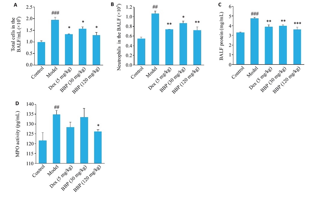
图7 BBP对LPS诱导的ALI小鼠BALF中总细胞数、中性粒细胞数、蛋白水平及肺组织中MPO水平的影响
Fig.7 Effects of BBP on changes cell counts in BALF from LPSinducedALI mice. A: Number of total cells in the BALF. B: Number ofneutrophils in the BALF. C: Levels of BALF protein. D: MPO activitydetected using corresponding assay kits in the lung tissue. Data arepresented as Mean±SD, n=6). ##P<0.01, ###P<0.001 vs Control group; *P<0.05, **P<0.01, ***P<0.001 vs Model group.
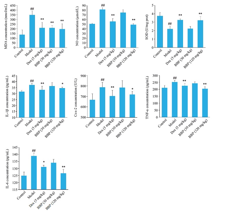
图8 BBP对LPS诱导的ALI小鼠肺组织中炎症因子和氧化因子水平的影响
Fig.8 Effects of BBP on levels of inflammatory factors (IL-1β, COX-2,TNF‑α and IL-6) and oxidative stress indicators (NO, SOD andMDA) in lung tissues of LPS-induced ALI mice. Data are presentedas Mean±SD, n=6). ##P<0.01 vs control group; *P<0.05, **P<0.01 vsmodel group.
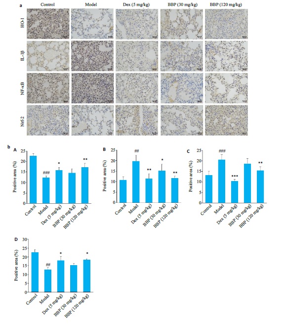
图9 BBP对ALI小鼠肺组织中Nrf-2、HO-1、IL-1β 和 NF-κB(p65)表达水平的影响
Fig.9 Effects of BBP on expression levels of Nrf-2, HO-1, IL-1β, and NF-κB (p65) in lung tissues of mice with ALI. (a) IHC results of Nrf-2, HO-1,IL-1β and NF-κB (p65) in lung tissues of mice in each group (Scale bar=50 μm). (b) Proportion of positive cells for (A) HO-1, (B) NF-κB (p65),(C) IL-1β, and (D) Nrf-2 in lung tissues of mice in each group (%). Dataare presented as Mean±SD (n=6). ##P<0.01, ###P<0.001 vs Control group; *P<0.05, **P<0.01 vs Model group.
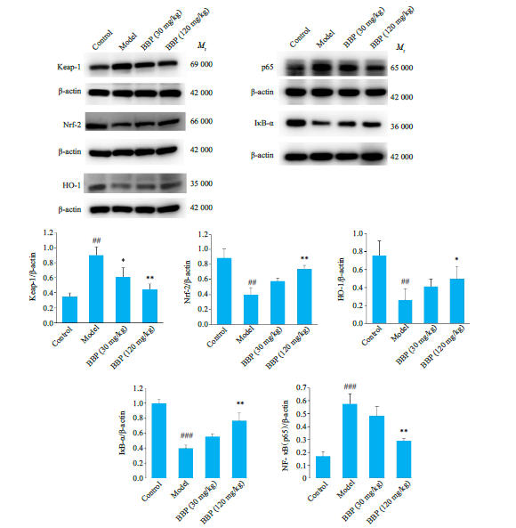
图10 BBP对LPS诱导的ALI小鼠肺组织中Keap-1、Nrf-2、HO-1、IκB-α和NF- κB(p65)蛋白水平的影响
Fig.10 Effect of BBP on expressions of Keap-1, Nrf2, HO-1, IκB-α and NF‑κB (p65) in mouse lung tissues. Data are presented as Mean±SD (n=6). ##P<0.01, ###P<0.001 vs Control group; *P<0.05, **P<0.01 vs Model group.
| [1] | Zheng JJ, Li Y, Kong XB, et al. Exploring immune-related pathogenesis in lung injury: Providing new insights Into ALI/ARDS[J]. Biomed Pharmacother, 2024, 175: 116773. doi:10.1016/j.biopha.2024.116773 |
| [2] | Evans CE. Hypoxia-inducible factor signaling in inflammatory lung injury and repair[J]. Cells, 2022, 11(2): 183. doi:10.3390/cells11020183 |
| [3] | Su YC, Lucas R, Fulton DJR, et al. Mechanisms of pulmonary endothelial barrier dysfunction in acute lung injury and acute respiratory distress syndrome[J]. Chin Med J Pulm Crit Care Med, 2024, 2(2): 80-7. doi:10.1016/j.pccm.2024.04.002 |
| [4] | Yin ZH, Song RZ, Yu T, et al. Natural compounds regulate macrophage polarization and alleviate inflammation against ALI/ARDS[J]. Biomolecules, 2025, 15(2): 192. doi:10.3390/biom15020192 |
| [5] | Liu HQ, Dong JL, Xu CL, et al. Acute lung injury: pathogenesis and treatment[J]. J Transl Med, 2025, 23(1): 926. doi:10.1186/s12967-025-06994-2 |
| [6] | Cai YJ, Shang LR, Zhou FY, et al. Macrophage pyroptosis and its crucial role in ALI/ARDS[J]. Front Immunol, 2025, 16: 1530849. doi:10.3389/fimmu.2025.1530849 |
| [7] | Wang J, Peng XY, Yuan N, et al. Interplay between pulmonary epithelial stem cells and innate immune cells contribute to the repair and regeneration of ALI/ARDS[J]. Transl Res, 2024, 272. 111-25. doi:10.1016/j.trsl.2024.05.012 |
| [8] | Liu C, Xiao K, Xie LX. Progress in preclinical studies of macrophage autophagy in the regulation of ALI/ARDS[J]. Front Immunol, 2022, 13: 922702. doi:10.3389/fimmu.2022.922702 |
| [9] | Qian J, Liu KJ, Zhong CH, et al. Sivelestat sodium alleviated sepsis-induced acute lung injury by inhibiting TGF‑β/Smad signaling pathways through upregulating microRNA-744-5p[J]. J DisThorac, 2024, 16(10): 6616-33. doi:10.21037/jtd-24-65 |
| [10] | Peinado-Acevedo JS, Rivera-Bustamante T, Rivera J, et al. Balancing inflammation and adverse effects of glucocorticoids in clinical practice[J]. Rev Colomb De Reumatología, 2024, 31(4): 498-510. doi:10.1016/j.rcreu.2023.08.004 |
| [11] | 李新月, 苏芳芳, 蒋 超, 等.中药熊胆功效演进及成分研究概述[J]. 中国中药杂志, 2022, 47(18): 4846-53. |
| [12] | Wu JS, Fang S, Li WK, et al. Metabolomics research on the hepatoprotective effect of cultured bear bile powder in α-naphthylisothiocyanate-induced cholestatic mice[J]. J Chromatogr B, 2020, 1153: 122269. doi:10.1016/j.jchromb.2020.122269 |
| [13] | Zhu H, Wang GR, Bai YY, et al. Natural bear bile powder suppresses neuroinflammation in lipopolysaccharide-treated mice via regulating TGR5/AKT/NF-κB signaling pathway[J]. J Ethnopharmacol, 2022, 289: 115063. doi:10.1016/j.jep.2022.115193 |
| [14] | 贾贵阳, 倪元萍, 王梦茹, 等. 熊胆粉及熊去氧胆酸对肝癌癌前病变的作用[J].中国实验 动物学报, 2024, 32(11): 1424-32. doi:10.3969/j.issn.1005-4847.2024.11.007 |
| [15] | Chen HW, Shen AL, Liu LY, et al. Bear bile powder inhibits growth of hepatocellular carcinoma via suppressing STAT3 signaling pathway in mice[J]. Chin J Integr Med, 2020, 26(5): 370-4. doi:10.1007/s11655-019-3010-1 |
| [16] | Cui N, Zhang WS, Su FZ, et al. Metabolomics and lipidomics study unveils the impact of tauroursodeoxycholic acid on hyperlipidemic mice[J]. Molecules, 2023, 28: 6352-65. doi:10.3390/molecules28176352 |
| [17] | Chen XL, Cai K, Zhang W, et al. Bear bile powder ameliorates type 2 diabetes via modulation of metabolic profiles, gut microbiota, and metabolites[J]. Front Pharmacol, 2023, 13: 1090955. doi:10.3389/fphar.2022.1090955 |
| [18] | 朴仁范. 朝药学[M]. 延边大学出版社, 2013: 86-7. |
| [19] | 元持常. 东医四象新编:东医四象新编剂方[M]. 京城: 文友社, 1929: 17. |
| [20] | Hu CL, Li JL, Tan YS, et al. Tanreqing injection attenuates macrophage activation and the inflammatory response via the lncRNA-SNHG1/HMGB1 axis in lipopolysaccharide-induced acute lung injury[J]. Front Immunol, 2022, 13: 820718. doi:10.3389/fimmu.2022.820718 |
| [21] | 岳 斌, 张 明, 武可泗, 等. HPLC-ELSD测定蛇胆川贝口服液等药中西贝素的含量[J].中 成药, 2007, (3): 474-6. doi:10.3969/j.issn.1001-1528.2007.03.056 |
| [22] | Cheng L, Tian HL, Lei HY, et al. Bear bile powder ameliorates LPS-induced acute lung injury by inhibiting CD14 pathway and improving intestinal flora: exploration of "Fei (lung)-Dachang (large intestine) interaction" theory[J]. Chin J Integr Med, 2025, 31(9): 821-9. doi:10.1007/s11655-024-3556-4 |
| [23] | Yang SY, Chen KJ, Yu JK, et al. Inhibition of cathepsin L ameliorates inflammation through the A20/NF‑κB pathway in endotoxin-induced acute lung injury[J]. iScience, 2024, 27(11): 111024. doi:10.1016/j.isci.2024.111024 |
| [24] | Liu SX, Liu JC, Wang YH, et al. Oxidative stress: signaling pathways, biological functions, and disease[J]. MedComm, 2025, 6(7): e70268. doi:10.1002/mco2.70268 |
| [25] | Sharif O, Bolshakov VN, Raines S, et al. Transcriptional profiling of the LPS induced NF‑κB response in macrophages[J]. BMC Immunol, 2007, 8(1): 1. doi:10.1186/1471-2172-8-1 |
| [26] | Bellezza I, Giambanco I, Minelli A, et al. Nrf2-Keap1 signaling in oxidative and reductive stress[J]. Biochim Biophys Acta BBA Mol Cell Res, 2018, 1865(5): 721-33. doi:10.1016/j.bbamcr.2018.02.010 |
| [27] | Wardyn JD, Ponsford AH, Sanderson CM. Dissecting molecular cross-talk between Nrf2 and NF‑κB response pathways[J]. Biochem Soc Trans, 2015, 43(4): 621-6. doi:10.1042/bst20150014 |
| [28] | Ahmed SMU, Luo L, Namani A, et al. Nrf2 signaling pathway: Pivotal roles in inflammation[J]. Biochim Biophys Acta BBA Mol Basis Dis, 2017, 1863(2): 585-97. doi:10.1016/j.bbadis.2016.11.005 |
| [29] | Song LY, Li MJ, Li JY, et al. Synthesis and evaluation of lansiumamide B analogues to ameliorate acute lung injury by reducing oxidative stress and inflammation via Keap1/Nrf2/NF-κB pathway[J]. Eur J Med Chem, 2025, 299: 118052. doi:10.1016/j.ejmech.2025.118052 |
| [30] | Patangrao Renushe A, Kumar Banothu A, Kumar Bharani K, et al. Vincamine, an active constituent of Vinca rosea ameliorates experimentally induced acute lung injury in Swiss albino mice through modulation of Nrf-2/NF‑κB signaling cascade[J]. Int Immunopharmacol, 2022, 108: 108773. doi:10.1016/j.intimp.2022.108773 |
| [31] | Niri P, Saha A, Polopalli S, et al. β‑Caryophyllene attenuates oxidative stress and inflammatory response in LPS induced acute lung injury by targeting ACE2/MasR and Nrf2/HO-1/NF‑κB axis[J]. Biochem Biophys Res Commun, 2025, 746: 151286. doi:10.1016/j.bbrc.2024.151286 |
| [32] | González-Bosch C, Boorman E, Zunszain PA, et al. Short-chain fatty acids as modulators of redox signaling in health and disease[J]. Redox Biol, 2021, 47: 102165. doi:10.1016/j.redox.2021.102165 |
| [33] | Milivojac T, Grabež M, Amidžić L, et al. Ursodeoxycholic and chenodeoxycholic bile acids alleviate endotoxininduced acute lung injury in rats by modulating aquaporin expression and pathways associated with apoptosis and inflammation[J]. Frontiers Pharmacol, 2025, 16: 1484292-311. doi:10.3389/fphar.2025.1484292 |
| [34] | Gong ZG, Mao W, Ren PP, et al. Taurochenodeoxycholic acid ameliorates the Staphylococcus aureus infection-induced acute lung injury through toll-like receptor 2 in mice[J]. Int Immunopharmacol, 2024, 142: 113228. doi:10.1016/j.intimp.2024.113228 |
| [35] | He XD, Shi JY, Bu LN, et al. Ursodeoxycholic acid alleviates fat embolism syndrome-induced acute lung injury by inhibiting the p38 MAPK/NF‑κB signalling pathway through FXR[J]. Pharmacol Biochem, 2024, 230: 116574. doi:10.1016/j.bcp.2024.116574 |
| [36] | Sipos A, Kerekes É, Szeőcs D, et al. Ursodeoxycholic acid prompts glycolytic dominance,reductive stress and epithelial-to-mesenchymal transition in ovarian cancer cells through NRF2 activation[J]. Cell Death Discov, 2025, 11: 134. doi:10.1038/s41420-025-02398-9 |
| [1] | 祝金超, 邵宁宁, 黄泽彧, 刘燕青, 孟祥艳, 刘子泉, 孙予杰, 董津睿, 樊毫军. 薤白超细粉通过抑制铁死亡并调控NF-κB/STAT3信号轴缓解脂多糖诱导的小鼠急性肺损伤[J]. 南方医科大学学报, 2026, 46(5): 1006-1017. |
| [2] | 范正媛, 沈子涵, 李亚, 沈婷婷, 李高峰, 李素云. 补肺益肾方对香烟烟雾提取物诱导的人支气管上皮细胞损伤的保护作用及其机制[J]. 南方医科大学学报, 2025, 45(7): 1372-1379. |
| [3] | 孙亚磊, 罗萌, 郭长胜, 高静, 苏凯奇, 陈立典, 冯晓东. 穗花杉双黄酮通过抑制细胞焦亡减轻小鼠急性肺损伤[J]. 南方医科大学学报, 2025, 45(4): 692-701. |
| [4] | 姜一凡, 李小荣, 耿嘉逸, 陈永锋, 唐碧, 康品方. 槲皮素通过抑制HMGB1/RAGE/NF-κB信号通路减轻糖尿病引起的大鼠肾脏损伤[J]. 南方医科大学学报, 2024, 44(9): 1769-1775. |
| [5] | 张珊苑, 蔡巧燕, 祁江晗, 殷恺馨, 何晨晨, 高铸烨, 张铃, 褚剑锋. 清心解瘀颗粒抗动脉粥样硬化的药效学及调控机制[J]. 南方医科大学学报, 2024, 44(8): 1518-1528. |
| [6] | 凌旭光, 徐雯雯, 庞观来, 洪旭星, 刘凤芹, 李 洋. 茶多酚通过抑制NLRP3炎症小体改善脓毒症小鼠的急性肺损伤[J]. 南方医科大学学报, 2024, 44(2): 381-386. |
| [7] | 王丽娅, 田美惠, 李 蓉, 吴 越, 王莎莎, 吕 恒, 刘忠义, 于 影. 乙醛脱氢酶2改善急性肺损伤小鼠的肺内皮屏障及维持线粒体动力学平衡[J]. 南方医科大学学报, 2023, 43(8): 1388-1395. |
| [8] | 房尚萍, 袁 冉, 孙任珂, 马同军. 敲除S1PR3可缓解小鼠的急性肺损伤:基于抑制MAPK信号通路[J]. 南方医科大学学报, 2022, 42(12): 1815-1821. |
| [9] | 陈加宝, 刘 华. 2,3,5,4'-四羟基二苯乙烯-2-O-β-D-葡萄糖苷可减轻脂多糖诱导的大鼠急性肺损伤[J]. 南方医科大学学报, 2021, 41(7): 1101-1106. |
| [10] | 韩 冰, 陈梦婷, 杨传铭, 李晓红. 右美托咪定预处理通过抑制NLRP3炎性小体激活减轻大鼠肠缺血再灌注诱导的急性肺损伤[J]. 南方医科大学学报, 2021, 41(12): 1857-1863. |
| [11] | 谢璨灿, 吴双华, 李峥嵘, 黄 冰, 曾维忠. 电针刺激通过JAK1/STAT3通路减轻脓毒症大鼠的急性肺损伤[J]. 南方医科大学学报, 2020, 40(11): 1662-1667. |
| [12] | 杨 倩,高培宇,母迷迷,陶象男,何 晶,吴凤娇,郭术俊,钱中清,宋传旺. 急性肺损伤小鼠肺泡巨噬细胞吞噬功能降低[J]. 南方医科大学学报, 2020, 40(03): 376-381. |
| [13] | 谭继翔,何进,秦文熠,赵林. 槲皮素通过抑制TLR4/NF-κB通路缓解脂多糖诱导的急性肾损伤[J]. 南方医科大学学报, 2019, 39(05): 598-. |
| [14] | 鲍权,洪小杨,唐靖,王刚,陈曦,秦再生. 改良兔体外循环急性肺损伤模型的建立[J]. 南方医科大学学报, 2017, 37(06): 797-. |
| [15] | 赵燕,程黎,宋志新,邓欣雨,何婧,邓旺,王导新. 白介素17对急性肺损伤时肺水肿液清除的影响[J]. 南方医科大学学报, 2017, 37(04): 494-. |
| 阅读次数 | ||||||
|
全文 |
|
|||||
|
摘要 |
|
|||||
