
南方医科大学学报 ›› 2026, Vol. 46 ›› Issue (10): 2405-2414.doi: 10.12122/j.issn.1673-4254.2026.10.10
• • 上一篇
何梦茹1( ), 金串1, 余亦璇3, 王静娴1, 童旭1, 陈沐岩3, 宋宏伟2, 王颖2, 周莉2, 王钧1, 王洪巨1, 李妙男1()
收稿日期:2026-03-20
出版日期:2026-10-20
发布日期:2026-10-26
通讯作者:
李妙男
E-mail:3021997475@qq.com;13855265385@163.com
作者简介:何梦茹,在读硕士研究生,E-mail: 3021997475@qq.com
基金资助:
Mengru HE1(), Chuan JIN1, Yixuan YU3, Jingxian WANG1, Xu TONG1, Muyan CHEN3, Hongwei SONG2, Ying WANG2, Li ZHOU2, Jun WANG1, Hongju WANG1, Miaonan LI1()
Received:2026-03-20
Online:2026-10-20
Published:2026-10-26
Contact:
Miaonan LI
E-mail:3021997475@qq.com;13855265385@163.com
摘要:
目的 探讨心脏磁共振(CMR)指标心肌整体纵向应变(GLS)与外周血炎症指标中性粒细胞/淋巴细胞比值(NLR)联合对首发急性心肌梗死(AMI)患者经皮冠状动脉介入治疗(PCI)术后发生主要不良心血管事件(MACEs)的预测价值。 方法 本研究为一项单中心、前瞻性队列研究。纳入2022年6月~2025年7月于蚌埠医学院第一附属医院首次确诊为AMI的患者134例,其中男性99例,女性35例,年龄33~83(60.19±11.7)岁,出院后对其进行中位时间为538 d的门诊或电话随访,根据是否发生MACEs分为2组:非MACEs组(n=103),MACEs组(n=31)。 结果 基线资料显示MACEs组患者年龄、白细胞计数、中性粒细胞计数、NLR、PLR、NT-pro BNP明显高于非MACEs组(P<0.05),单核细胞计数明显低于非MACEs组(P<0.05)。MACEs组患者Gensini评分、前壁心肌梗死的发生率及左前降支(LAD)作为罪犯血管的比例显著高于非MACEs组(P<0.05),右冠状动脉(RCA)作为罪犯血管的比例及胸痛TIMI低危比例明显低于非MACEs组(P<0.05)。CMR结果分析表明,MACEs组患者的LGE、左室收缩末期容积指数、矛盾运动及室壁瘤发生率均显著高于非MACEs组(P<0.05),而左室射血分数、梗死节段径向、周向、纵向及整体径向、周向、纵向的应变值均显著低于非MACEs组(P<0.05)。多因素Logistic回归结果显示NLR、GLS是MACEs的独立预测因素(P<0.05)。受试者工作特征(ROC)曲线分析结果显示GLS及NLR对首发AMI患者PCI术后是否发生MACEs有一定预测能力,曲线下面积(AUC)分别为0.825、0.820;二者联合的AUC为0.921,高于单一指标(P<0.05)。 结论 心脏磁共振GLS与外周血NLR均与首发AMI患者PCI术后预后密切相关,二者联合应用能够更好地预测首发AMI患者的预后。
何梦茹, 金串, 余亦璇, 王静娴, 童旭, 陈沐岩, 宋宏伟, 王颖, 周莉, 王钧, 王洪巨, 李妙男. 基于心肌整体纵向应变和外周血中性粒细胞与淋巴细胞比值精准预测首发急性心肌梗死患者经皮冠状动脉介入治疗术后发生主要不良心血管事件风险:一项单中心前瞻性队列研究[J]. 南方医科大学学报, 2026, 46(10): 2405-2414.
Mengru HE, Chuan JIN, Yixuan YU, Jingxian WANG, Xu TONG, Muyan CHEN, Hongwei SONG, Ying WANG, Li ZHOU, Jun WANG, Hongju WANG, Miaonan LI. Myocardium global longitudinal strain and peripheral blood neutrophil-to-lymphocyte ratio for predicting MACE risks following percutaneous coronary intervention for first-time acute myocardial infarction: a prospective cohort study[J]. Journal of Southern Medical University, 2026, 46(10): 2405-2414.
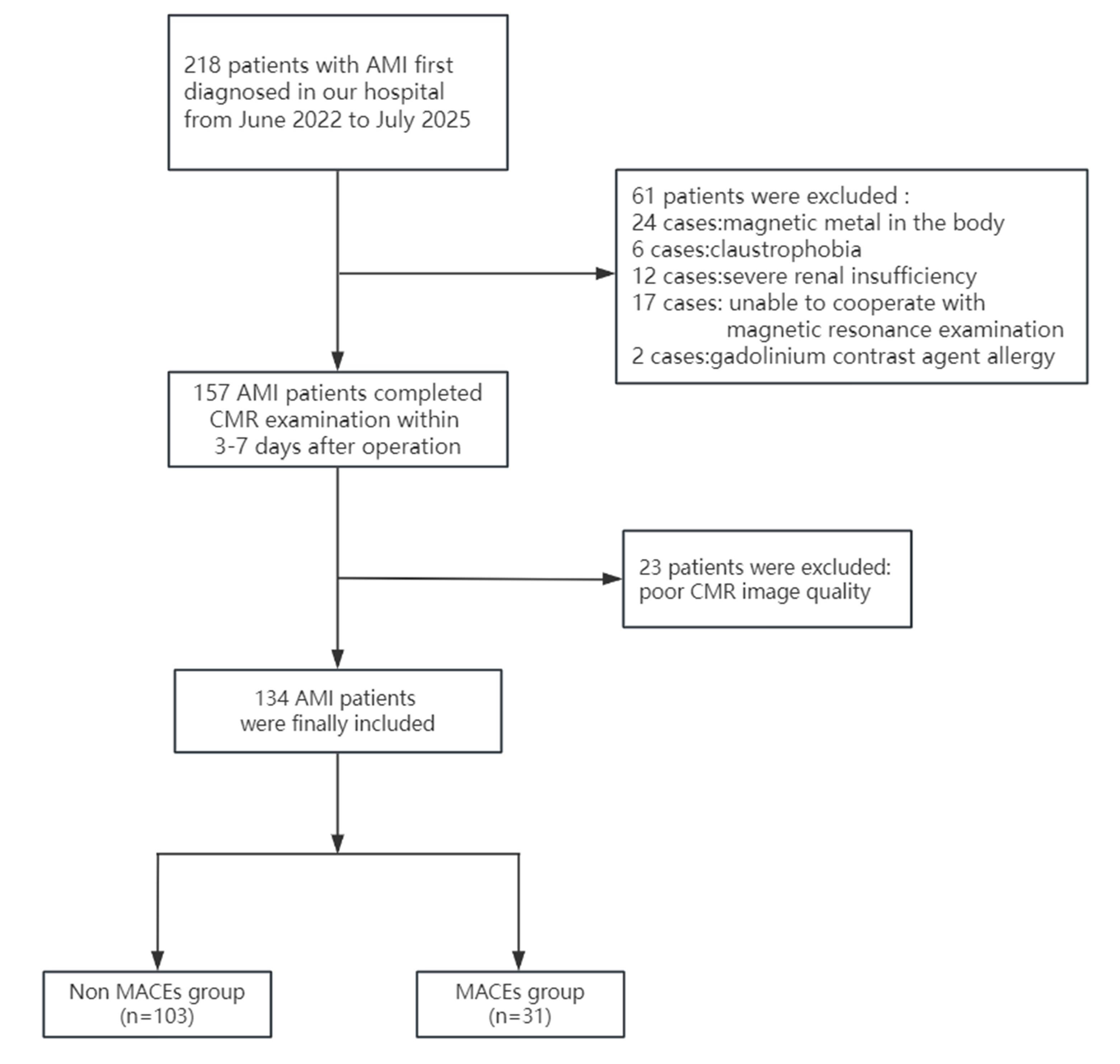
图1 患者入组流程图和分组结果
Fig.1 Flow chart of patient enrollment and grouping.
| Characteristics | Non-MACEs group (n=103) | MACEs group (n=31) | P |
|---|---|---|---|
| Age (year) | 59±11 | 64±12 | 0.041 |
| Male [n (%)] | 79 (76.7%) | 20 (64.5%) | 0.176 |
| BMI (kg/m2) | 25.0±3.7 | 25.0±3.2 | 0.921 |
| Hypertension [n (%)] | 61 (59.2%) | 15 (48.4%) | 0.286 |
| Diabetes [n (%)] | 25 (24.3%) | 9 (29.0%) | 0.593 |
| AF [n (%)] | 4 (3.9%) | 4 (12.9%) | 0.083 |
| Previous stroke [n (%)] | 8 (7.8%) | 4 (12.9%) | 0.472 |
| Previous smoking [n (%)] | 59 (57.3%) | 17 (54.8%) | 0.810 |
| WBC (109/L) | 9.69 (8.04, 11.47) | 11.47 (10.29, 13.49) | <0.001 |
| Neutrophil (109/L) | 7.37 (5.71, 9.22) | 9.56 (8.55, 11.66) | <0.001 |
| Lymphocyte (109/L) | 0.48 (0.36, 0.67) | 0.48 (0.27, 0.61) | 0.364 |
| Monocyte (109/L) | 1.43 (1.13, 2.01) | 1.02 (0.88, 1.39) | <0.001 |
| Platelet (109/L) | 205 (168, 240) | 214 (171, 254) | 0.388 |
| NLR | 5.3 (3.2, 7.0) | 9.2 (7.4, 12.4) | <0.001 |
| PLR | 142 (93, 182) | 173 (142, 254) | <0.001 |
| FBG (mmol/L) | 6.5 (5.4, 8.2) | 6.3 (5.1, 8.8) | 0.959 |
| UA (µmol/L) | 315 (259, 381) | 334 (260, 394) | 0.513 |
| SCr (µmol/L) | 62 (51, 74) | 66 (51, 82) | 0.593 |
| TC (mmol/L) | 4.35 (3.73, 5.25) | 4.80 (4.13, 5.53) | 0.110 |
| TG (mmol/L) | 1.52 (1.02, 2.09) | 1.32 (0.89, 2.08) | 0.754 |
| LDL-C (mmol/L) | 2.77 (2.07, 3.38) | 2.93 (2.40, 3.66) | 0.297 |
| HDL-C (mmol/L) | 1.05 (0.89, 1.22) | 1.13 (0.98, 1.39) | 0.129 |
| CK-MB(U/L) | 40 (20, 100) | 38 (20, 141) | 0.829 |
| cTn I (μg/L) | 11 (3, 22) | 8 (1, 47) | 0.827 |
| NT-proBNP(pg/mL) | 258 (61, 697) | 1,030 (123, 2,330) | 0.001 |
表1 两组AMI患者基线特征
Tab.1 Baseline characteristics of the two groups of AMI patients
| Characteristics | Non-MACEs group (n=103) | MACEs group (n=31) | P |
|---|---|---|---|
| Age (year) | 59±11 | 64±12 | 0.041 |
| Male [n (%)] | 79 (76.7%) | 20 (64.5%) | 0.176 |
| BMI (kg/m2) | 25.0±3.7 | 25.0±3.2 | 0.921 |
| Hypertension [n (%)] | 61 (59.2%) | 15 (48.4%) | 0.286 |
| Diabetes [n (%)] | 25 (24.3%) | 9 (29.0%) | 0.593 |
| AF [n (%)] | 4 (3.9%) | 4 (12.9%) | 0.083 |
| Previous stroke [n (%)] | 8 (7.8%) | 4 (12.9%) | 0.472 |
| Previous smoking [n (%)] | 59 (57.3%) | 17 (54.8%) | 0.810 |
| WBC (109/L) | 9.69 (8.04, 11.47) | 11.47 (10.29, 13.49) | <0.001 |
| Neutrophil (109/L) | 7.37 (5.71, 9.22) | 9.56 (8.55, 11.66) | <0.001 |
| Lymphocyte (109/L) | 0.48 (0.36, 0.67) | 0.48 (0.27, 0.61) | 0.364 |
| Monocyte (109/L) | 1.43 (1.13, 2.01) | 1.02 (0.88, 1.39) | <0.001 |
| Platelet (109/L) | 205 (168, 240) | 214 (171, 254) | 0.388 |
| NLR | 5.3 (3.2, 7.0) | 9.2 (7.4, 12.4) | <0.001 |
| PLR | 142 (93, 182) | 173 (142, 254) | <0.001 |
| FBG (mmol/L) | 6.5 (5.4, 8.2) | 6.3 (5.1, 8.8) | 0.959 |
| UA (µmol/L) | 315 (259, 381) | 334 (260, 394) | 0.513 |
| SCr (µmol/L) | 62 (51, 74) | 66 (51, 82) | 0.593 |
| TC (mmol/L) | 4.35 (3.73, 5.25) | 4.80 (4.13, 5.53) | 0.110 |
| TG (mmol/L) | 1.52 (1.02, 2.09) | 1.32 (0.89, 2.08) | 0.754 |
| LDL-C (mmol/L) | 2.77 (2.07, 3.38) | 2.93 (2.40, 3.66) | 0.297 |
| HDL-C (mmol/L) | 1.05 (0.89, 1.22) | 1.13 (0.98, 1.39) | 0.129 |
| CK-MB(U/L) | 40 (20, 100) | 38 (20, 141) | 0.829 |
| cTn I (μg/L) | 11 (3, 22) | 8 (1, 47) | 0.827 |
| NT-proBNP(pg/mL) | 258 (61, 697) | 1,030 (123, 2,330) | 0.001 |
| Characteristics | Non-MACEs group (n=103) | MACEs group (n=31) | P |
|---|---|---|---|
| Gensini score | 72 (46, 102) | 88 (75, 166) | 0.004 |
| Number of diseased vessels [n (%)] | 0.205 | ||
| One vessel disease | 26 (25.2%) | 4 (12.9%) | 0.148 |
| Two vessel disease | 25 (24.3%) | 6 (19.4%) | 0.569 |
| Three vessel disease | 52 (50.5%) | 21 (67.7%) | 0.091 |
| Anterior myocardial infarction | 47 (45.6%) | 27 (87.1%) | <0.001 |
| Culprit vessel [n (%)] | <0.001 | ||
| LAD | 44 (42.7%) | 24 (77.4%) | <0.001 |
| LCX | 15 (14.6%) | 4 (12.9%) | 0.815 |
| RCA | 44 (42.7%) | 3 (9.7%) | <0.001 |
| Chest pain TIMI classification [n (%)] | 0.011 | ||
| Low risk | 60 (58.3%) | 9 (29.0%) | 0.004 |
| Intermediate risk | 31 (30.1%) | 14 (45.2%) | 0.119 |
| High risk | 12 (11.7%) | 8 (25.8%) | 0.099 |
表2 两组AMI患者冠脉病变情况的比较
Tab.2 Comparison of coronary artery lesions between the two groups of AMI patients
| Characteristics | Non-MACEs group (n=103) | MACEs group (n=31) | P |
|---|---|---|---|
| Gensini score | 72 (46, 102) | 88 (75, 166) | 0.004 |
| Number of diseased vessels [n (%)] | 0.205 | ||
| One vessel disease | 26 (25.2%) | 4 (12.9%) | 0.148 |
| Two vessel disease | 25 (24.3%) | 6 (19.4%) | 0.569 |
| Three vessel disease | 52 (50.5%) | 21 (67.7%) | 0.091 |
| Anterior myocardial infarction | 47 (45.6%) | 27 (87.1%) | <0.001 |
| Culprit vessel [n (%)] | <0.001 | ||
| LAD | 44 (42.7%) | 24 (77.4%) | <0.001 |
| LCX | 15 (14.6%) | 4 (12.9%) | 0.815 |
| RCA | 44 (42.7%) | 3 (9.7%) | <0.001 |
| Chest pain TIMI classification [n (%)] | 0.011 | ||
| Low risk | 60 (58.3%) | 9 (29.0%) | 0.004 |
| Intermediate risk | 31 (30.1%) | 14 (45.2%) | 0.119 |
| High risk | 12 (11.7%) | 8 (25.8%) | 0.099 |
| Characteristics | Non-MACEs group (n=103) | MACEs group (n=31) | P |
|---|---|---|---|
| Myocardial mass (g) | 101 (85, 117) | 100 (87, 120) | 0.667 |
| LGE (%) | 16 (12, 25) | 22 (17, 33) | 0.011 |
| LVEDV/BSA (mL/m2 ) | 72 (61, 84) | 72 (61, 95) | 0.478 |
| LVESV/BSA (mL/m2 ) | 37 (28, 45) | 41 (35, 60) | 0.017 |
| LVEF (%) | 49 ± 10 | 40±11 | <0.001 |
| Reverse motion [n (%)] | 75 (72.8%) | 28 (90.3%) | 0.043 |
| MVO [n (%)] | 57 (55.3%) | 23 (74.2%) | 0.061 |
| MVO volume (mL) | 2.87 (1.03, 4.70) | 2.08 (0.92, 5.47) | 0.698 |
| Ventricular aneurysm [n (%)] | 16 (15.5%) | 16 (51.6%) | <0.001 |
| Myocardial strain in infarct segment [n (%)] | |||
| Radial strain | 16 (13, 22) | 12 (11, 17) | 0.003 |
| Circumferential strain | 11.5 (9.4, 14.1) | 10.1 (9.0, 11.4) | 0.018 |
| Longitudinal strain | 11.2 (9.0, 14.3) | 7.7 (6.7, 9.5) | <0.001 |
| Global myocardial strain | |||
| Radial strain | 26 (21, 32) | 24 (19, 26) | 0.018 |
| Circumferential strain | 15.11±3.10 | 13.57±2.77 | 0.011 |
| Longitudinal strain | 11.6 (9.3, 14.4) | 8.3 (6.8, 9.4) | <0.001 |
表3 两组AMI患者CMR参数的比较
Tab.3 Comparison of cardiac magnetic resonance (CMR) parameters between the two groups of AMI patients
| Characteristics | Non-MACEs group (n=103) | MACEs group (n=31) | P |
|---|---|---|---|
| Myocardial mass (g) | 101 (85, 117) | 100 (87, 120) | 0.667 |
| LGE (%) | 16 (12, 25) | 22 (17, 33) | 0.011 |
| LVEDV/BSA (mL/m2 ) | 72 (61, 84) | 72 (61, 95) | 0.478 |
| LVESV/BSA (mL/m2 ) | 37 (28, 45) | 41 (35, 60) | 0.017 |
| LVEF (%) | 49 ± 10 | 40±11 | <0.001 |
| Reverse motion [n (%)] | 75 (72.8%) | 28 (90.3%) | 0.043 |
| MVO [n (%)] | 57 (55.3%) | 23 (74.2%) | 0.061 |
| MVO volume (mL) | 2.87 (1.03, 4.70) | 2.08 (0.92, 5.47) | 0.698 |
| Ventricular aneurysm [n (%)] | 16 (15.5%) | 16 (51.6%) | <0.001 |
| Myocardial strain in infarct segment [n (%)] | |||
| Radial strain | 16 (13, 22) | 12 (11, 17) | 0.003 |
| Circumferential strain | 11.5 (9.4, 14.1) | 10.1 (9.0, 11.4) | 0.018 |
| Longitudinal strain | 11.2 (9.0, 14.3) | 7.7 (6.7, 9.5) | <0.001 |
| Global myocardial strain | |||
| Radial strain | 26 (21, 32) | 24 (19, 26) | 0.018 |
| Circumferential strain | 15.11±3.10 | 13.57±2.77 | 0.011 |
| Longitudinal strain | 11.6 (9.3, 14.4) | 8.3 (6.8, 9.4) | <0.001 |
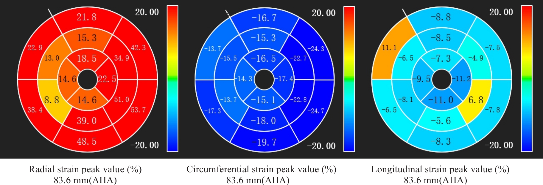
图2 1例71岁女性患者, 前壁心肌梗死, 整体纵向应变显著下降伴矛盾运动, PCI术后17个月发生MACEs
Fig.2 A 71-year-old female patient with anterior myocardial infarction showing significantly reduced global longitudinal strain with paradoxical motion. She experienced a major adverse cardiovascular events (MACEs) 17 months after percutaneous coronary intervention (PCI).
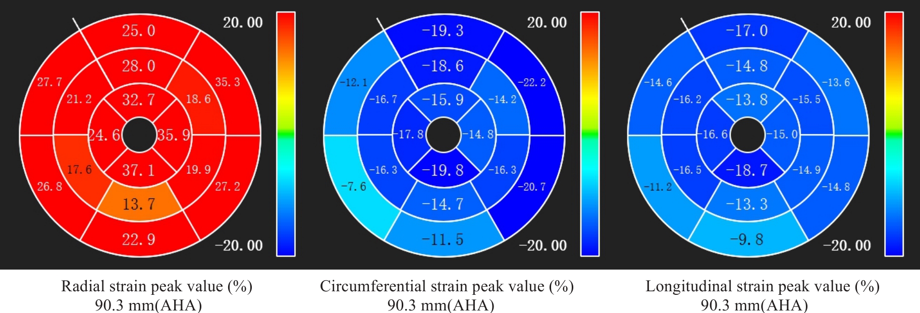
图3 1例70岁男性患者, 下壁心肌梗死, 整体纵向应变未见明显下降, PCI术后22个月未出现MACEs
Fig.3 A 70-year-old male patient with inferior wall myocardial infarction showing no significant decrease in global longitudinal strain. He remained free of MACEs 22 months after PCI.
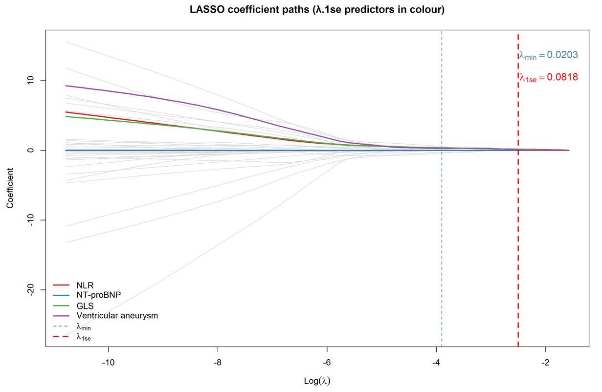
图4 基于 LASSO 回归的变量筛选结果
Fig.4 Based on the variable screening results of LASSO regression (λ.1se criterion), 4 indicators were selected: NLR, GLS, NT proBNP, and ventricular aneurysm (n=134).
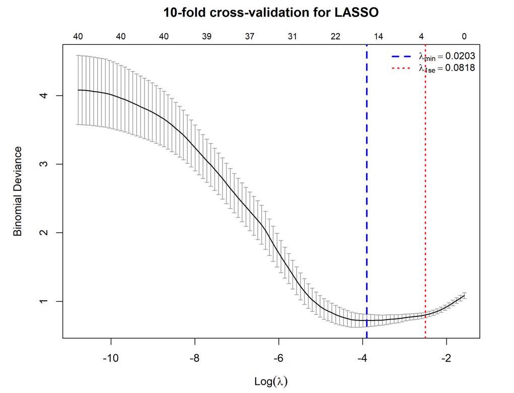
图5 LASSO 回归的 10 折交叉验证曲线
Fig.5 10-fold cross validation curve of LASSO regression (n=134).
| Index | β | SE | Wald | P | OR | 95% CI |
|---|---|---|---|---|---|---|
| NLR | 0.371 | 0.096 | 15.037 | <0.001 | 1.449 | 1.201-1.747 |
| GLS | -0.475 | 0.132 | 12.946 | <0.001 | 0.622 | 0.480-0.805 |
| NT-proBNP | 0.020 | 0.015 | 1.732 | 0.188 | 1.020* | 0.990-1.051 |
| Ventricular aneurysm | 0.563 | 1.282 | 0.870 | 0.351 | 1.755 | 0.538-5.726 |
表4 多因素logistic回归分析MACEs影响因素
Tab.4 Multivariate logistic regression analysis of factors affecting post-PCI MACEs
| Index | β | SE | Wald | P | OR | 95% CI |
|---|---|---|---|---|---|---|
| NLR | 0.371 | 0.096 | 15.037 | <0.001 | 1.449 | 1.201-1.747 |
| GLS | -0.475 | 0.132 | 12.946 | <0.001 | 0.622 | 0.480-0.805 |
| NT-proBNP | 0.020 | 0.015 | 1.732 | 0.188 | 1.020* | 0.990-1.051 |
| Ventricular aneurysm | 0.563 | 1.282 | 0.870 | 0.351 | 1.755 | 0.538-5.726 |
| Index | β | SE | Wald | P | OR | 95% CI |
|---|---|---|---|---|---|---|
| NLR | 0.388 | 0.092 | 17.946 | <0.001 | 1.475 | 1.232-1.765 |
| GLS | -0.531 | 0.129 | 16.804 | <0.001 | 0.588 | 0.456-0.758 |
| Constant | 1.160 | 1.224 | 0.898 | - | 3.190 | - |
表5 GLS与NLR预测MACEs的二元logistic回归分析
Tab.5 Binary logistic regression analysis of GLS and NLR for predicting MACEs
| Index | β | SE | Wald | P | OR | 95% CI |
|---|---|---|---|---|---|---|
| NLR | 0.388 | 0.092 | 17.946 | <0.001 | 1.475 | 1.232-1.765 |
| GLS | -0.531 | 0.129 | 16.804 | <0.001 | 0.588 | 0.456-0.758 |
| Constant | 1.160 | 1.224 | 0.898 | - | 3.190 | - |
| Parameter | Cutoff value | Sensitivity(%) | Specificity(%) | AUC | 95% CI | Youden index |
|---|---|---|---|---|---|---|
| NLR | 7.223 | 0.774 | 0.777 | 0.820 | 0.735-0.905 | 0.551 |
| GLS | -9.65 | 0.839 | 0.718 | 0.825 | 0.740-0.910 | 0.557 |
| Joint prediction | 0.303 | 0.839 | 0.883 | 0.911 | 0.858-0.965 | 0.722 |
表6 预测MACEs的风险模型
Tab.6 Predictive model for the risk of MACEs
| Parameter | Cutoff value | Sensitivity(%) | Specificity(%) | AUC | 95% CI | Youden index |
|---|---|---|---|---|---|---|
| NLR | 7.223 | 0.774 | 0.777 | 0.820 | 0.735-0.905 | 0.551 |
| GLS | -9.65 | 0.839 | 0.718 | 0.825 | 0.740-0.910 | 0.557 |
| Joint prediction | 0.303 | 0.839 | 0.883 | 0.911 | 0.858-0.965 | 0.722 |
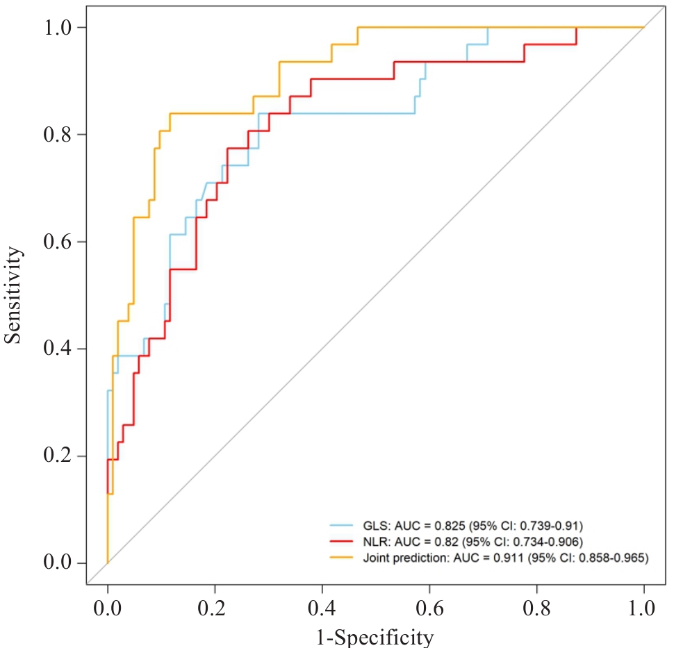
图6 NLR、GLS及联合诊断对MACEs预测价值的ROC曲线
Fig.6 ROC curve of MACEs predicted by NLR, GLS and their combination (n=134).
| [1] | 国家心血管病中心. 中国心血管健康与疾病报告2024[M]. 北京: 中国协和医科大学出版社, 2025. |
| [2] | 中华医学会心血管病学分会, 中华心血管病杂志编辑委员会. 急性ST段抬高型心肌梗死诊断和治疗指南 [J]. 中华心血管病杂志, 2010, (8): 675-690. |
| [3] | 张丽伟, 赵 赫, 曾辰华. 血浆C型凝集素样受体2水平与急性ST段抬高型心肌梗死患者经皮冠状动脉介入术后发生主要不良心血管事件的关系[J]. 中华实用诊断与治疗杂志, 2020, 34(12): 1239-43. |
| [4] | Yang J, Yin P, Li MX, et al. Long-term cardiovascular and noncardiovascular mortality after acute coronary syndrome a nationwide competing-risks analysis of 1.56 million patients[J]. JACC, 2026, 87(12): 1511-22. doi:10.1016/j.jacc.2025.12.064 |
| [5] | Yang XY, Wen L, Sun M, et al. Prediction of cardiac deterioration in acute heart failure patients: Evaluation of the efficacy of single laboratory indicator models versus comprehensive models[J]. Medicine, 2024, 103(44): e40266. doi:10.1097/md.0000000000040266 |
| [6] | von Knobelsdorff-Brenkenhoff F, Schulz-Menger J. Cardiovascular magnetic resonance in the guidelines of the European Society of Cardiology: a comprehensive summary and update[J]. J Cardiovasc Magn Reson, 2023, 25(1): 42. doi:10.1186/s12968-023-00950-z |
| [7] | Zhang Y, Ko SM. The imaging spectrum of myocardial infarction and its associated complications: a contemporary pictorial review of computed tomography and magnetic resonance imaging[J]. Diagn Interv Radiol, 2026. DOI:10.4274/dir.2026.263576 . |
| [8] | Pan JL, Ng SM, Neubauer S, et al. Phenotyping heart failure by cardiac magnetic resonance imaging of cardiac macro- and microscopic structure: state of the art review[J]. Eur Heart J Cardiovasc Imaging, 2023, 24(10): 1302-17. doi:10.1093/ehjci/jead124 |
| [9] | Thomas KE, Fotaki A, Botnar RM, et al. Imaging methods: magnetic resonance imaging[J]. Circ Cardiovasc Imaging, 2023, 16(1): e014068. doi:10.1161/circimaging.122.014068 |
| [10] | Ren Y, Li S, Chen YC, et al. Prognostic value of left ventricular strain determined using CMR-feature tracking in patients with alcoholic cardiomyopathy[J]. Int J Cardiol, 2025, 435: 133391. doi:10.1016/j.ijcard.2025.133391 |
| [11] | Frișan AC, Lazăr MA, Șoșdean R, et al. Myocardial work analysis in ST-elevation myocardial infarction: insights into left ventricular ejection fraction: a pilot study[J]. Life, 2025, 15(3): 338. doi:10.3390/life15030338 |
| [12] | Alhawamdeh N, Bani Hani DA, Alshraideh JA, et al. Novel inflammatory markers predict cardiovascular events after percutaneous coronary intervention in patients with acute coronary syndrome[J]. Heart Lung, 2026, 75: 111-6. doi:10.1016/j.hrtlng.2025.09.014 |
| [13] | 张 慧, 王 黎, 上官海娟, 等. 基于心脏磁共振评估的冠状动脉微血管阻塞对急性ST段抬高型心肌梗死患者急诊经皮冠状动脉介入治疗术后预后的临床研究[J]. 中国介入心脏病学杂志, 2025, 33(6): 327-33. |
| [14] | Hu YN, Liu YS, Wang YP, et al. Dual-pathway model of IL-6 and HSP22/HSP27 dynamics predicts post-PCI MACE in STEMI patients[J]. BMC Cardiovasc Disord, 2025, 25(1): 863. doi:10.1186/s12872-025-05304-4 |
| [15] | Thygesen K, Alpert JS, Jaffe AS, et al. Fourth universal definition of myocardial infarction (2018)[J]. Circulation, 2018, 138(20): e618-51. doi:10.1161/cir.0000000000000632 |
| [16] | Byrne RA, Rossello X, Coughlan JJ, et al. 2023 ESC guidelines for the management of acute coronary syndromes[J]. Eur Heart J, 2023, 44(38): 3720-826. |
| [17] | Norman S, Koshy A, Biswas S, et al. A systematic comparison of ACC/AHA, ESC and JCS guidelines on intravascular imaging: level of evidence and clinical use scenarios[J]. Heart Lung Circ, 2024, 33: S549-50. doi:10.1016/j.hlc.2024.06.927 |
| [18] | Tudela Martínez JI, Alcaraz Pérez P, Martínez Encarnación L, et al. Cardiac magnetic resonance in adults: an updated review of the diagnostic approach to major heart diseases[J]. J Clin Med, 2025, 14(20): 7323. doi:10.3390/jcm14207323 |
| [19] | Thrane PG, Olesen KKW, Thim T, et al. Temporal trends in cardiovascular events after primary percutaneous coronary intervention for ST-segment elevation myocardial infarction[J]. JACC Adv, 2025, 4(3): 101614. doi:10.1016/j.jacadv.2025.101614 |
| [20] | 杨彦杰, 王 鸽, 严 研, 等. 低风险急性ST段抬高型心肌梗死患者早期出院的可行性分析[J]. 中国心血管病研究, 2023, 21(8): 680-3. doi:10.3969/j.issn.1672-5301.2023.08.002 |
| [21] | 杜晓彬, 王方方, 杨耀峰. 血清S100A4、ANGPTK4、CX3CL1水平联合检测对急性心肌梗死患者PCI术后MACE发生的预测价值[J]. 黑龙江医药科学, 2024, 47(4): 41-4. |
| [22] | Peles I, Barrett O, Cafri C, et al. Predictors of adverse outcome in high-risk percutaneous coronary interventions patients[J]. Can J Cardiol, 2023, 39(10): 1369-79. doi:10.1016/j.cjca.2023.04.021 |
| [23] | Chen YF, Jiang M, Xia C, et al. A novel deep learning system for STEMI prognostic prediction from multi-sequence cardiac magnetic resonance[J]. Sci Bull, 2025, 70(24): 4241-52. doi:10.1016/j.scib.2025.11.027 |
| [24] | Wang CP, Zhou L, Liang Y, et al. Interactions of ST-elevation myocardial infarction, age, and sex and the risk of major adverse cardiovascular events among Chinese adults: a secondary analysis of a single-centre prospective cohort[J]. BMJ Open, 2022, 12(7): e058494. doi:10.1136/bmjopen-2021-058494 |
| [25] | 李洋洋, 梁力晖, 陈柏林, 等. 急性冠脉综合征患者经皮冠状动脉介入术后院内MACE发作风险预测模型构建[J]. 心脏杂志, 2022, 34(5): 531-6. |
| [26] | 郭智超, 范春雨. 全身免疫炎症指数对急性心肌梗死病人PCI术后院内不良预后的预测价值[J]. 中西医结合心脑血管病杂志, 2024, 22(24): 4530-3. |
| [27] | Fang CY, Chen ZF, Zhang J, et al. The predictive value of neutrophil-lymphocyte ratio combined with the Global Registry of Acute Coronary Events score for inhospital adverse cardiovascular events in patients with acute ST-elevation myocardial infarction[J]. J Res Med Sci, 2024, 29: 41. doi:10.4103/jrms.jrms_485_22 |
| [28] | Matter MA, Paneni F, Libby P, et al. Inflammation in acute myocardial infarction: the good, the bad and the ugly[J]. Eur Heart J, 2024, 45(2): 89-103. doi:10.1093/eurheartj/ehad486 |
| [29] | Mornar Jelavić M, Babić Z, Babić D, et al. Ten-year outcomes after acute stemi treated with primary pci - the role of acute biomarkers and other parameters in predicting clinical severity and prognosis[J]. Acta Clin Croat, 2024, 63(3/4): 733-44. |
| [30] | Zhang C, Zeng S, Ji WJ, et al. Synergistic role of circulating CD14++CD16+ monocytes and fibrinogen in predicting the cardiovascular events after myocardial infarction[J]. Clin Cardiol, 2023, 46(5): 521-8. doi:10.1002/clc.24005 |
| [31] | Yang F, Yu M, Yuan YF, et al. A new comprehensive parameter combining residual C-reactive protein and neutrophil risk predicting adverse events in Chinese patients after percutaneous coronary intervention: a multi-center prospective cohort study[J]. Chin Med J, 2026, 139(1): 139-41. doi:10.1097/cm9.0000000000003885 |
| [32] | Helali J, Ramesh K, Brown J, et al. Late gadolinium enhancement on cardiac MRI: a systematic review and meta-analysis of prognosis across cardiomyopathies[J]. Int J Cardiol, 2025, 419: 132711. doi:10.1016/j.ijcard.2024.132711 |
| [33] | Troger F, Pamminger M, Poskaite P, et al. Clinical impact of persistent microvascular obstruction in CMR after reperfused STEMI[J]. Circ Cardiovasc Imag, 2025, 18(6): e017645. doi:10.1161/circimaging.124.017645 |
| [34] | Li M, Wang J, Ding S, et al. Cardiac magnetic resonance imaging-derived pathophysiology and prognosis of diabetes mellitus with acute myocardial infarction after revascularization: a prospective cohort study. Ann Med. 2024, 56(1): 2399751. doi:10.1080/07853890.2024.2399751 |
| [35] | Guglielmo M, Fusini L, Baessato F, et al. PROGnostic RolE of strain measurements in stress cardiac MRI in predicting major adverse cardiac events[J]. Int J Cardiol, 2024, 412: 132337. doi:10.1016/j.ijcard.2024.132337 |
| [36] | Jering KS, Claggett BL, Braunwald E, et al. NT-proBNP in the early convalescent phase after high-risk myocardial infarction is associated with adverse cardiovascular outcomes: the PARADISE-MI trial[J]. J Card Fail, 2026, 32(3): 586-95. doi:10.1016/j.cardfail.2025.03.018 |
| [37] | Charkiewicz-Szeremeta K, Sawicka-Śmiarowska E, Dubatówka M, et al. Predictors of long-term prognosis focused on kidney function in patients with chronic coronary syndrome[J]. Diseases, 2026, 14(2): 78. doi:10.3390/diseases14020078 |
| [38] | Feng YL, Zhuo YF, Cheng HJ. Predictive value of circulating cardiomyocyte-specific cell-free DNA levels for heart failure risk after acute ST-segment elevation myocardial infarction[J]. Postepy Kardiol Interwencyjnej, 2025, 21(4): 565-76. doi:10.5114/aic.2025.156807 |
| [39] | Zornitzki L, Barlev Y, Gefen S, et al. Left ventricular global longitudinal strain in patients with coronary microvascular dysfunction and preserved ejection fraction: a single-center study[J]. Pol Heart J, 2026, 84(2): 208-16. doi:10.33963/v.phj.110652 |
| [40] | Shereef AS, Abdelmajeed MG, Alshair MH, et al. Coronary slow flow and its correlation with reduced left ventricle global longitudinal strain: a case–control study[J]. Echo Res Pract, 2024, 11(1): 2. doi:10.1186/s44156-023-00037-6 |
| [41] | Ersbøll M, Valeur N, Mogensen UM, et al. Global left ventricular longitudinal strain is closely associated with increased neurohormonal activation after acute myocardial infarction in patients with both reduced and preserved ejection fraction: a two-dimensional speckle tracking study[J]. Eur J Heart Fail, 2012, 14(10): 1121-9. doi:10.1093/eurjhf/hfs107 |
| [42] | Kolenda Zloić S, Martins da Fonseca J, Ayogu CI, et al. Prognostic impact of left ventricular strain by feature tracking in acute myocardial infarction treated with PCI: a meta-analysis[J]. Clin Imaging, 2025, 124: 110514. doi:10.1016/j.clinimag.2025.110514 |
| [43] | Li XB, Yu C, Liu XW, et al. A prediction model based on systemic immune-inflammatory index combined with other predictors for major adverse cardiovascular events in acute myocardial infarction patients[J]. J Inflamm Res, 2024, 17: 1211-25. doi:10.2147/jir.s443153 |
| [44] | Aghezzaf S, Coisne A, Bauters C, et al. Feasibility and prognostic significance of ventricular-arterial coupling after myocardial infarction: the RIGID-MI cohort[J]. Eur Heart J Cardiovasc Imaging, 2024, 25(5): 668-77. doi:10.1093/ehjci/jead342 |
| [45] | Jigoranu RA, Mitu O, Oancea AF, et al. Diagnostic and severity assessment of coronary artery disease using ApoB/ApoA-I ratio: insights from a statin-treated eastern European cohort[J]. Medicina, 2026, 62(2): 297. doi:10.3390/medicina62020297 |
| [46] | Zhang Y, Luo R, Sun XC, et al. The relationship between Gensini score and rates of 30-day mortality in acute coronary syndrome patients in China[J]. BMC Cardiovasc Disord, 2025, 26(1): 3. doi:10.1186/s12872-025-05303-5 |
| [47] | 李萌玫, 孙洪巧, 万雪梅, 等. 急性心肌梗死急诊PCI术后罪犯血管部位对心力衰竭及心肌标志物的影响[J]. 临床心血管病杂志, 2025, 41(10): 774-80. |
| [48] | Vidal-Calés P, Bujak K, Rinaldi R, et al. Long-term prognostic impact of the left anterior descending coronary artery as the STEMI-related culprit vessel: subanalysis of the EXAMINATION-EXTEND trial[J]. REC Interv Cardiol, 2025, 7(2): 99-108. |
| [1] | 黄凌霄, 徐海喆, 黄凌燕, 姚新波, 周开元, 高勇占. 面向边界模糊与异质性区域的半监督MRI图像分割双网络模型[J]. 南方医科大学学报, 2026, 46(8): 1956-1966. |
| [2] | 胡欣怡, 王圩健, 李慧, 陈宗正, 周忻, 袁俊菲, 陈亮. 急性ST段抬高型心肌梗死患者直接经皮冠状动脉介入治疗后30天主要不良心血管事件的列线图预测模型:基于血清GSDMD蛋白表达水平[J]. 南方医科大学学报, 2026, 46(3): 550-558. |
| [3] | 马振岩, 阿鑫, 赵蕾, 张洪博, 刘科, 赵依晴, 钱赓. 急性ST段抬高型心肌梗死经皮冠状动脉介入术后左心室不良重构的新型风险预测模型:基于心脏磁共振的多中心前瞻性研究[J]. 南方医科大学学报, 2025, 45(4): 669-683. |
| [4] | 刘科, 马振岩, 付磊, 张丽萍, 阿鑫, 肖少波, 张震, 张洪博, 赵蕾, 钱赓. 心脏磁共振成像整体纵向应变对急性ST段抬高型心肌梗死后左心室重构的预测价值:403例前瞻性研究[J]. 南方医科大学学报, 2024, 44(6): 1033-1039. |
| [5] | 崔佳宁, 刘文佳, 闫 非, 赵亚男, 陈伟杰, 罗春材, 张兴华, 李 涛. 心脏磁共振成像对急性ST段抬高型心肌梗死后左心室不良重构的预测价值[J]. 南方医科大学学报, 2024, 44(3): 553-562. |
| [6] | 王燕妮, 黄霞, 陈福恒, 高圆圆, 崔向荣, 严琴, 井宣. 急性心肌梗死患者血清中 BIN1 水平与 Killip 分级之间的关系[J]. 南方医科大学学报, 2024, 44(12): 2388-2395. |
| [7] | 崔佳宁, 李润霞, 刘学谦, 赵亚男, 张兴华, 刘清波, 李 涛. 心脏磁共振特征追踪技术可评估心肌淀粉样变性患者的心脏功能[J]. 南方医科大学学报, 2023, 43(4): 660-666. |
| [8] | 邓钰莹, 陈华锋, 李功辉, 陈立桁, 傅 强. 慢性肾脏病对肌钙蛋白诊断急性心肌梗死准确性的影响[J]. 南方医科大学学报, 2023, 43(2): 300-307. |
| [9] | 苏小凤, 韩继明, 高莹卉, 何子君, 赵 哲, 林俊岭, 郭静静, 陈开兵, 高 燕, 刘 霖. 老年阻塞性睡眠呼吸暂停与代谢综合征各组分的相关性及其对远期不良心血管事件发生风险的影响[J]. 南方医科大学学报, 2021, 41(11): 1592-1599. |
| [10] | 李红兵, 赵茂军, 徐英生. COVID-19普通型与危重型患者的生化分析及中性粒细胞/淋巴细胞比值的临床应用[J]. 南方医科大学学报, 2020, 40(07): 965-971. |
| [11] | 侯季秋,陈雅丽,马 迪,王 超,靳会会,安 莹,赵海滨. 从CXCL12/CXCR4轴探析空瓶诱导的慢性情绪应激对急性心肌梗死大鼠炎性因子的影响[J]. 南方医科大学学报, 2020, 40(05): 624-631. |
| [12] | 夏欣田,温敏勇,詹少锋,何 靖,陈伟焘. 中性粒细胞/淋巴细胞比值可作为重型COVID-19的预警信号[J]. 南方医科大学学报, 2020, 40(03): 333-336. |
| [13] | 朱建,杨燕,胡司淦,李辉,张恒. Connexin 43在急性冠脉综合征患者外周血单核细胞中的表达[J]. 南方医科大学学报, 2019, 39(04): 471-. |
| [14] | 李慧娣,向定成,张金霞,段天兵,龙锋,李爱敏. 急性心肌梗死早期脑钠肽浓度的动态演变规律及其对心力衰竭的诊断价值[J]. 南方医科大学学报, 2018, 38(01): 112-. |
| [15] | 姜大庆,谷天祥,徐兆发,刘爽,李学渊. 心肌球源性心肌干细胞联合心室肌细胞外基质治疗大鼠急性心肌梗死效果[J]. 南方医科大学学报, 2016, 36(10): 1316-. |
| 阅读次数 | ||||||
|
全文 |
|
|||||
|
摘要 |
|
|||||
