
南方医科大学学报 ›› 2026, Vol. 46 ›› Issue (7): 1553-1562.doi: 10.12122/j.issn.1673-4254.2026.07.09
• • 上一篇
木尼拉·麦合木提1( ), 黄利思2, 李家楷1, 蔡如龙1, 冯棋1, 林于迪1, 黄泽泓1, 伍思佳1, Umar Muhammad1, 方航1()
收稿日期:2026-01-22
出版日期:2026-07-20
发布日期:2026-07-20
通讯作者:
方航
E-mail:1814370730@qq.com;fanghang@ smu.edu.cn
作者简介:木尼拉·麦合木提,硕士,E-mail:1814370730@qq.com
基金资助:
Maihemuti Munila1(), Lisi HUANG2, Jiakai LI1, Rulong CAI1, Qi FENG1, Yudi LIN1, Zehong HUANG1, Sijia WU1, Muhammad Umar1, Hang FANG1()
Received:2026-01-22
Online:2026-07-20
Published:2026-07-20
Contact:
Hang FANG
E-mail:1814370730@qq.com;fanghang@ smu.edu.cn
摘要:
目的 探讨耐甲氧西林金黄色葡萄球菌(MRSA)诱导的小鼠膝关节假体周围感染(PJI)模型中三级淋巴结构(TLS)的结构与功能特征及其与局部骨溶解的关系。 方法 构建MRSA诱导的小鼠膝关节PJI模型,将C57BL/6J小鼠随机分为假体组(PBS)和PJI组(1×104 CFU MRSA USA300),观察60 d。检测外周血白细胞计数(WBC)、红细胞沉降率(ESR)及C反应蛋白(CRP)等炎症指标;HE染色评估假体周围组织TLS形成及成熟程度;多重免疫荧光分析T/B细胞分区(CD3/B220)、滤泡树突状细胞(CD21)及TLS功能活性(Ki67、PNAd);TRAP染色进行骨形态计量学分析,评估破骨细胞数量(N.Oc/BS)。 结果 术后21d,PJI组外周血炎症指标显著高于假体组(P<0.0001)。组织学显示,PJI组假体周围形成大量成熟TLS,截面积达17212.10±7624.28 μm²,显著高于假体组(P<0.01)。多重免疫荧光证实PJI组呈现“T细胞环绕B细胞”的成熟TLS典型空间结构,并伴有FDC网络;TLS内存在大量Ki67阳性增殖细胞,边缘及实质内可见PNAd阳性高内皮微静脉。TRAP分析显示,PJI组单位骨表面破骨细胞数量为(28.75±4.13)/mm,显著高于假体组的(1.63±0.74)/mm(P<0.0001)。 结论 MRSA慢性感染可在小鼠假体周围诱导形成具备典型结构分区及功能活性的成熟TLS,并伴随显著骨溶解。
木尼拉·麦合木提, 黄利思, 李家楷, 蔡如龙, 冯棋, 林于迪, 黄泽泓, 伍思佳, Umar Muhammad, 方航. 小鼠MRSA慢性假体周围感染诱导成熟三级淋巴结构形成及骨溶解[J]. 南方医科大学学报, 2026, 46(7): 1553-1562.
Maihemuti Munila, Lisi HUANG, Jiakai LI, Rulong CAI, Qi FENG, Yudi LIN, Zehong HUANG, Sijia WU, Muhammad Umar, Hang FANG. Chronic MRSA periprosthetic joint infection induces formation of mature tertiary lymphoid structures and osteolysis in mice[J]. Journal of Southern Medical University, 2026, 46(7): 1553-1562.
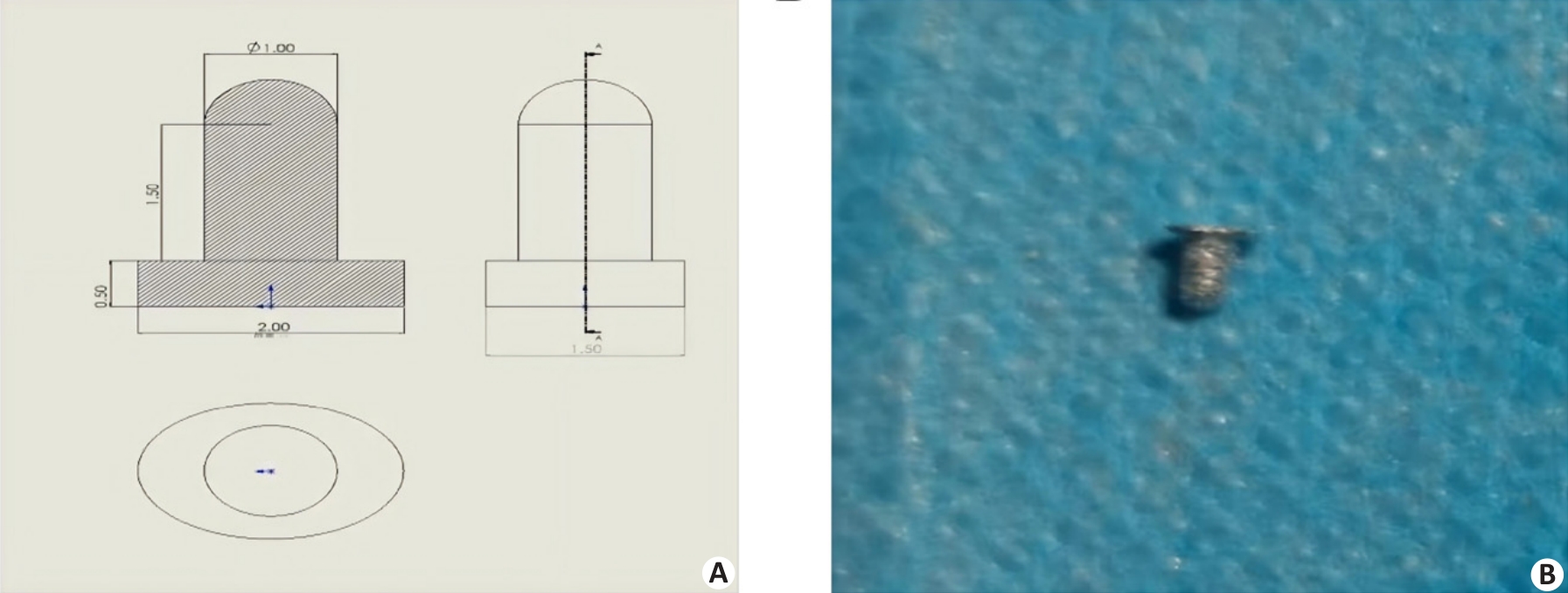
图1 小鼠膝关节微型假体的设计与制备
Fig.1 Design and manufacturing of the murine knee prosthesis. A: The micro-prosthesis model designed via CAD software, tailored to the distal femoral anatomy of C57BL/6J mice. B: Macroscopic view of the final Ti6Al4V implant produced by selective laser melting.
| Powder Ti Al V O N C H Fe Zn P Cu Ag | |
|---|---|
| Ti6Al4V powder | - 5.5-6.5 3.5-4.5 <0.13 <0.03 <0.08 <0.012 <0.25 - - - - |
表1 Ti6Al4V粉末的化学成分
Tab.1 Chemical composition of Ti6Al4V powder (wt%)
| Powder Ti Al V O N C H Fe Zn P Cu Ag | |
|---|---|
| Ti6Al4V powder | - 5.5-6.5 3.5-4.5 <0.13 <0.03 <0.08 <0.012 <0.25 - - - - |
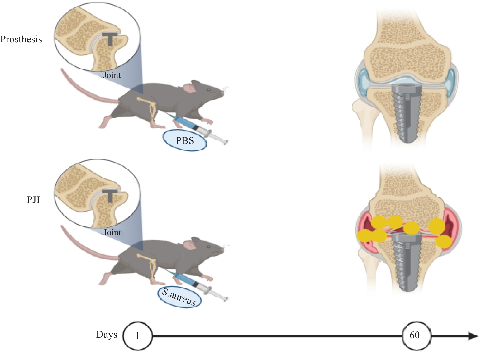
图2 小鼠膝关节PJI模型构建流程示意图
Fig.2 Schematic diagram of construction workflow for establishing mouse models of chronic knee periprosthetic joint infection (PJI) with methicillin-resistant Staphylococcus aureus (MRSA). Left: Experimental grouping and modeling procedure. Right: Schematic illustration of the periprosthetic microenvironment in the presence of infection (indicated by the red box).
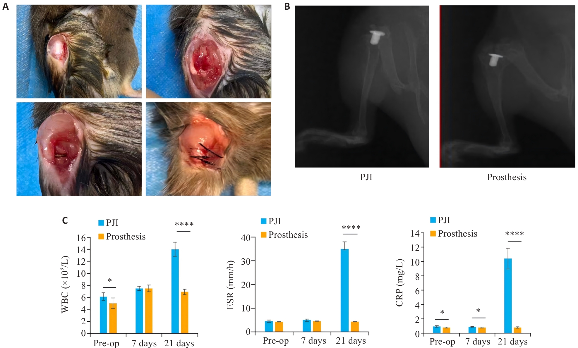
图3 小鼠膝关节PJI模型建立及术后评价
Fig.3 Establishment and postoperative evaluation of the mouse knee PJI model. A: Surgical procedures of the PJI model. B: X-ray images of mouse knee joints at 28 days post-surgery. C: Quantitative analysis of peripheral blood WBC, ESR, and CRP levels. Data are presented as Mean±SD, n=6. *P<0.05, ****P<0.0001.
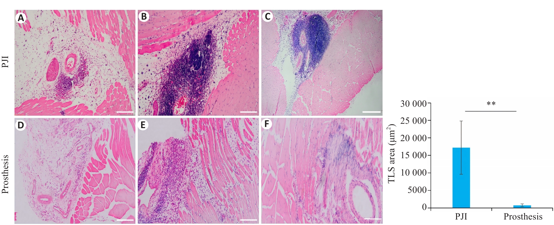
图4 小鼠膝关节假体周围软组织HE染色及TLS定量分析
Fig.4 HE staining and quantitative analysis of tertiary lymphoid structures (TLS) in periprosthetic soft tissues of mouse knee joints (n=6). A-C: HE staining showing massive dense inflammatory cell infiltration and formation of typical lymphoid follicle-like aggregate structures in PJI group. D-F: HE staining showing only sparse inflammatory cells in the prosthesis (control) group. Scale bar=100 μm. Data are presented as Mean±SD. **P<0.01.
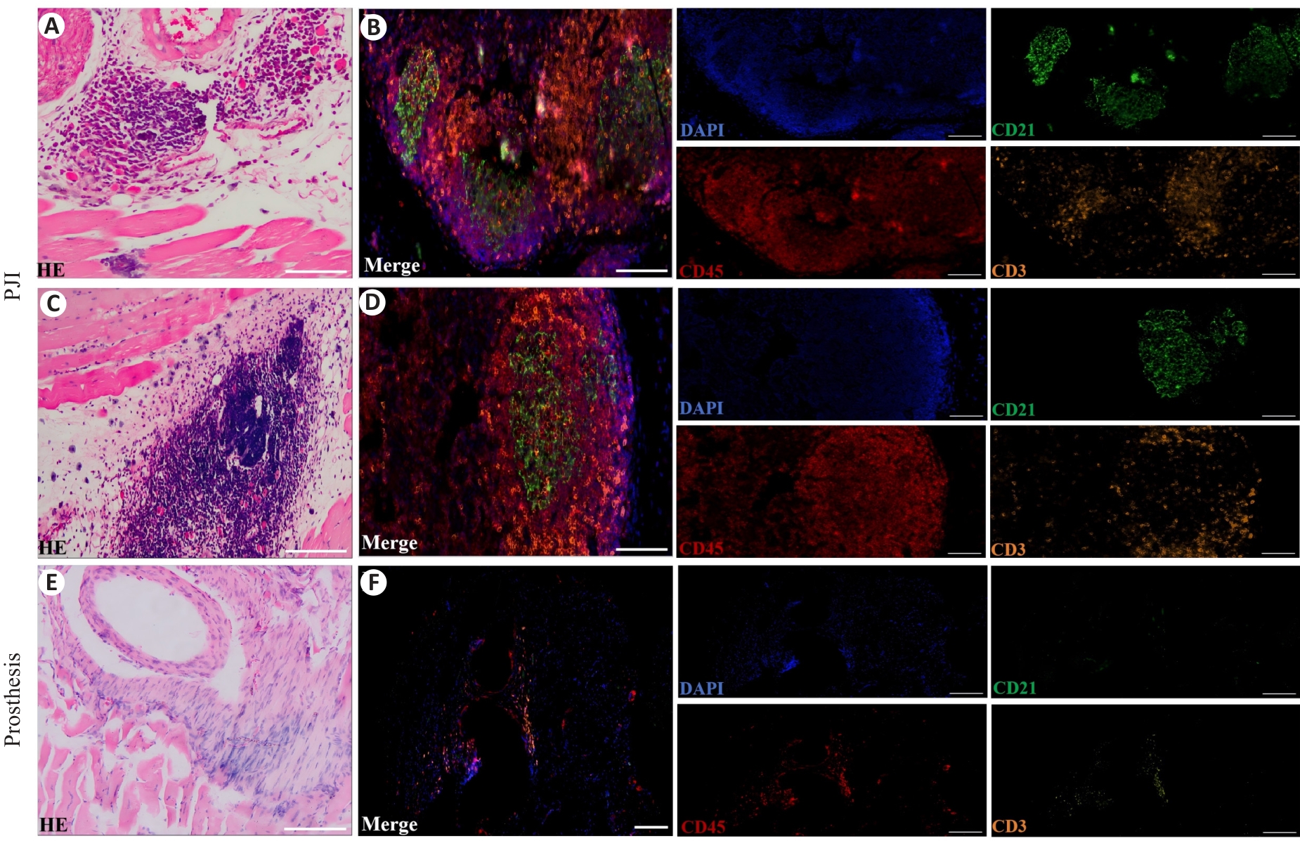
图5 小鼠假体周围组织免疫细胞HE染色及mIF染色
Fig.5 HE staining and multiplex immunofluorescence (mIF) staining of periprosthetic tissues. A-D: HE staining and merged mIF images in PJI group (CD21, green; CD45, red; CD3, yellow). E, F: HE staining and merged mIF image in prosthesis group (Scale bar=50 μm).
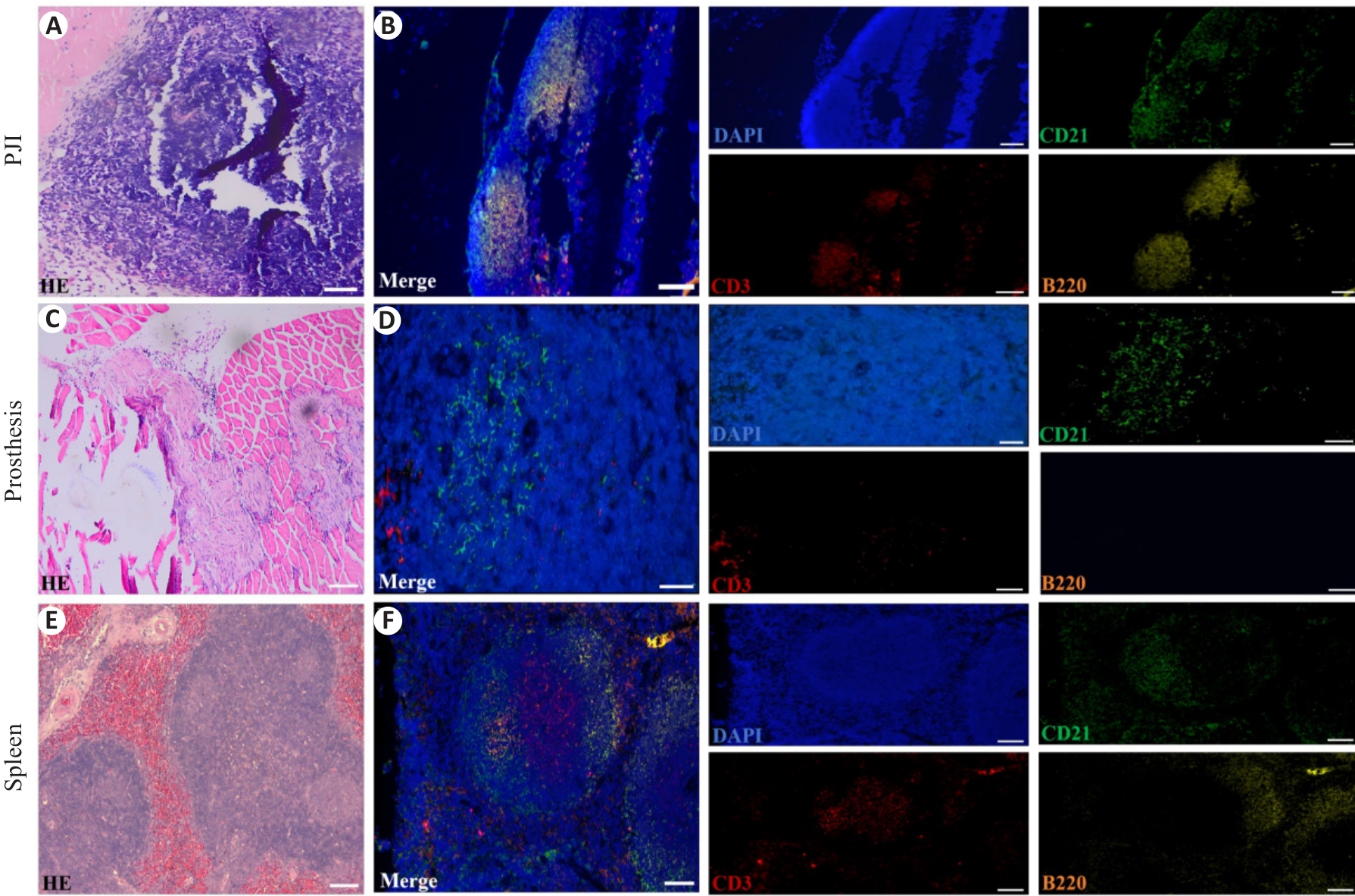
图6 小鼠假体周围组织中T细胞、B细胞及FDC的组织学特征与空间分布
Fig.6 Histological features and spatial organization of T cells, B cells, and follicular dendritic cells (FDCs) in periprosthetic tissues of the mice, using spleen tissue as the positive control. A, C, E: Representative HE staining images of the PJI group (A), prosthesis group (C), and the spleen (E). B, D, F: Corresponding merged mIF images showing immune cell distribution in PJI group (B), prosthesis group (D), and the spleen (F). The markers are colored as follows: CD3 (red), B220 (yellow), CD21 (green), and nuclei are counterstained with DAPI (blue) (Scale bar=50 μm).
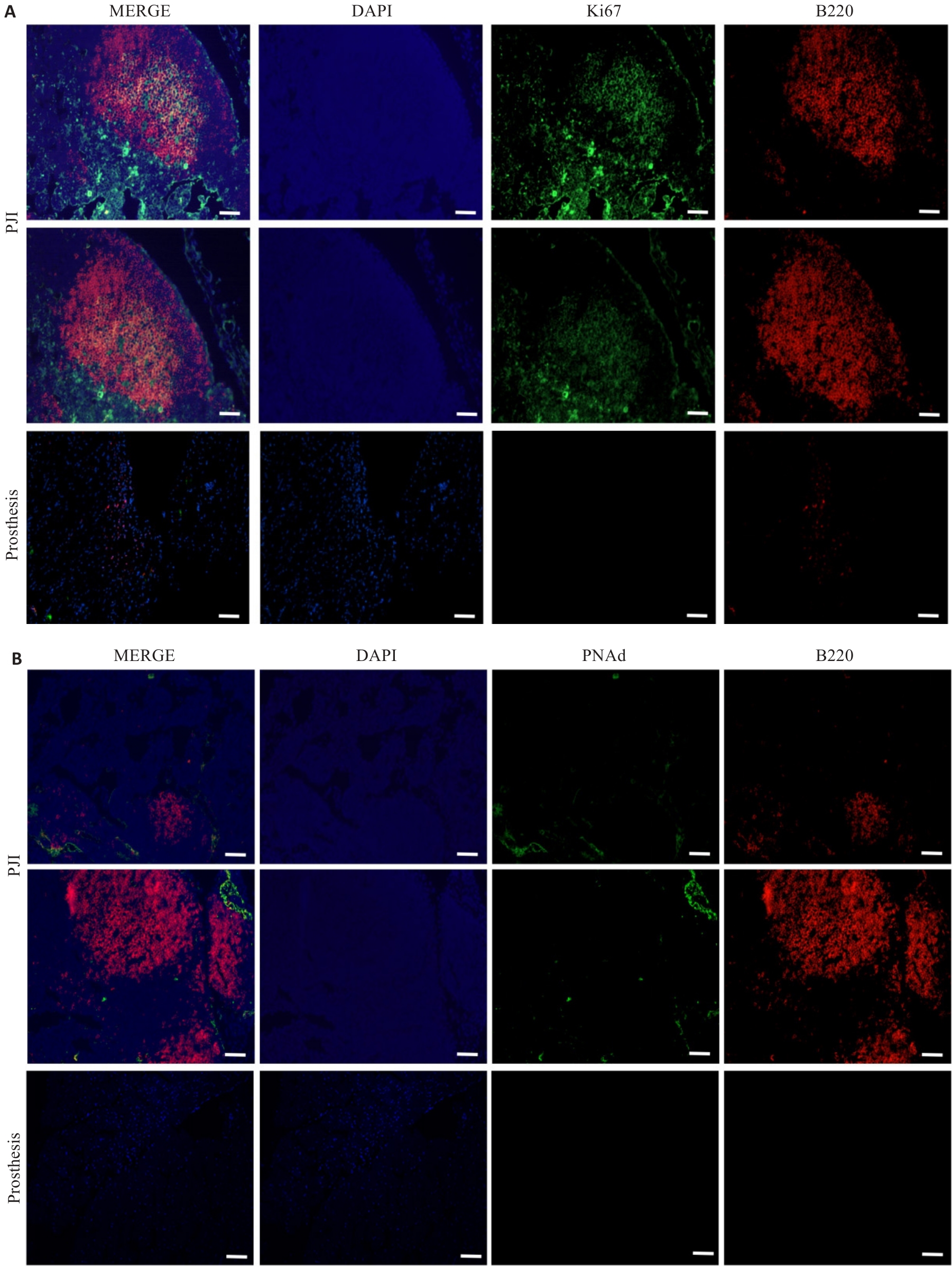
图7 小鼠假体周围组织中Ki67与PNAd的表达特征
Fig.7 Expression of Ki67 and PNAd in periprosthetic tissues of the mice (Scale bar=50 μm). A: Representative mIF images of Ki67 and B220 in PJI group (top two rows) and prosthesis group (bottom row). B: Representative mIF images of PNAd and B220 in PJI group (top two rows) and prosthesis group (bottom row). The markers are colored as follows: Ki67 or PNAd (green), B220 (red), and nuclei are counterstained with DAPI (blue).
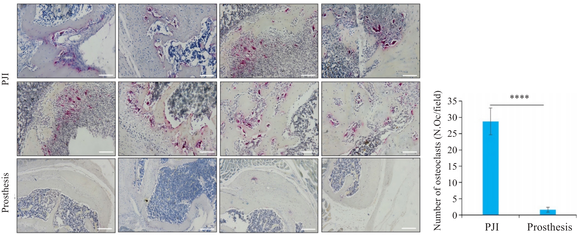
图8 小鼠假体周围骨组织TRAP染色及破骨细胞定量分析
Fig.8 TRAP staining and quantitative analysis of osteoclasts in periprosthetic bone tissues of the mice in PJI and prosthesis groups (Scale bar=100 μm). Data are presented as Mean±SD (n=6). ****P<0.0001.
| [1] | Borens O, Yusuf E, Trampuz A. Surgical site infections (SSIs): risk factors and prevention strategies[M]//European Instructional Lectures. Berlin, Heidelberg: Springer, 2013: 15-24. doi:10.1007/978-3-642-36149-4_2 |
| [2] | Kurtz SM, Lau E, Watson H, et al. Economic burden of periprosthetic joint infection in the United States[J]. J Arthroplasty, 2012, 27(8 ): 61-5.e1. doi:10.1016/j.arth.2012.02.022 |
| [3] | Costerton JW, Stewart PS, Greenberg EP. Bacterial biofilms: a common cause of persistent infections[J]. Science, 1999, 284(5418): 1318-22. doi:10.1126/science.284.5418.1318 |
| [4] | Foster TJ. Immune evasion by staphylococci[J]. Nat Rev Microbiol, 2005, 3(12): 948-58. doi:10.1038/nrmicro1289 |
| [5] | de Mesy Bentley KL, Trombetta R, Nishitani K, et al. Evidence of Staphylococcus aureus deformation, proliferation, and migration in canaliculi of live cortical bone in murine models of osteomyelitis[J]. J Bone Miner Res, 2017, 32(5): 985-90. doi:10.1002/jbmr.3055 |
| [6] | Visperas A, Santana D, Klika AK, et al. Current treatments for biofilm-associated periprosthetic joint infection and new potential strategies[J]. J Orthop Res, 2022, 40(7): 1477-91. doi:10.1002/jor.25345 |
| [7] | Muthukrishnan G, Masters EA, Daiss JL, et al. Mechanisms of immune evasion and bone tissue colonization that make Staphylococcus aureus the primary pathogen in osteomyelitis[J]. Curr Osteoporos Rep, 2019, 17(6): 395-404. doi:10.1007/s11914-019-00548-4 |
| [8] | Tang HD, Zhu MZ, Qiao J, et al. Lymphotoxin signalling in tertiary lymphoid structures and immunotherapy[J]. Cell Mol Immunol, 2017, 14(10): 809-18. doi:10.1038/cmi.2017.13 |
| [9] | Mueller CG, Nayar S, Gardner D, et al. Cellular and vascular components of tertiary lymphoid structures[M]//Tertiary Lymphoid Structures. New York, NY: Springer New York, 2018: 17-30. doi:10.1007/978-1-4939-8709-2_2 |
| [10] | Dieu-Nosjean MC, Goc J, Giraldo NA, et al. Tertiary lymphoid structures in cancer and beyond[J]. Trends Immunol, 2014, 35(11): 571-80. doi:10.1016/j.it.2014.09.006 |
| [11] | Song IH, Heo SH, Bang WS, et al. Predictive value of tertiary lymphoid structures assessed by high endothelial venule counts in the neoadjuvant setting of triple-negative breast cancer[J]. Cancer Res Treat, 2017, 49(2): 399-407. doi:10.4143/crt.2016.215 |
| [12] | Wang XQ, Sadovskaya I, Leterme D, et al. A comparative study of antibodies against proteins extracted from staphylococcal biofilm for the diagnosis of orthopedic prosthesis-related infections in an animal model and in humans[J]. Diagn Microbiol Infect Dis, 2013, 75(2): 124-9. doi:10.1016/j.diagmicrobio.2012.10.013 |
| [13] | Carli AV, Ross FP, Bhimani SJ, et al. Developing a clinically representative model of periprosthetic joint infection[J]. J Bone Joint Surg Am, 2016, 98(19): 1666-76. doi:10.2106/jbjs.15.01432 |
| [14] | Inzana JA, Schwarz EM, Kates SL, et al. A novel murine model of established Staphylococcal bone infection in the presence of a fracture fixation plate to study therapies utilizing antibiotic-laden spacers after revision surgery[J]. Bone, 2015, 72: 128-36. doi:10.1016/j.bone.2014.11.019 |
| [15] | Taniguchi N, Fujibayashi S, Takemoto M, et al. Effect of pore size on bone ingrowth into porous titanium implants fabricated by additive manufacturing: an in vivo experiment[J]. Mater Sci Eng C Mater Biol Appl, 2016, 59: 690-701. doi:10.1016/j.msec.2015.10.069 |
| [16] | Gbejuade HO, Lovering AM, Webb JC. The role of microbial biofilms in prosthetic joint infections[J]. Acta Orthop, 2015, 86(2): 147-58. doi:10.3109/17453674.2014.966290 |
| [17] | Chen ZT, Klein T, Murray RZ, et al. Osteoimmunomodulation for the development of advanced bone biomaterials[J]. Mater Today, 2016, 19(6): 304-21. doi:10.1016/j.mattod.2015.11.004 |
| [18] | Heim CE, Vidlak D, Kielian T. Interleukin-10 production by myeloid-derived suppressor cells contributes to bacterial persistence during Staphylococcus aureus orthopedic biofilm infection[J]. J Leukoc Biol, 2015, 98(6): 1003-13. doi:10.1189/jlb.4vma0315-125rr |
| [19] | Buckley CD, Barone F, Nayar S, et al. Stromal cells in chronic inflammation and tertiary lymphoid organ formation[J]. Annu Rev Immunol, 2015, 33: 715-45. doi:10.1146/annurev-immunol-032713-120252 |
| [20] | Pitzalis C, Jones GW, Bombardieri M, et al. Ectopic lymphoid-like structures in infection, cancer and autoimmunity[J]. Nat Rev Immunol, 2014, 14(7): 447-62. doi:10.1038/nri3700 |
| [21] | Cao J, Xu F, Lin SH, et al. IL-27 controls sepsis-induced impairment of lung antibacterial host defence[J]. Thorax, 2014, 69(10): 926-37. doi:10.1136/thoraxjnl-2014-205777 |
| [22] | Ruddle NH. Lymphoid neo-organogenesis: lymphotoxin's role in inflammation and development[J]. Immunol Res, 1999, 19(2/3): 119-25. doi:10.1007/bf02786481 |
| [23] | Denton AE, Linterman MA. Stromal networking: cellular connections in the germinal centre[J]. Curr Opin Immunol, 2017, 45: 103-11. doi:10.1016/j.coi.2017.03.001 |
| [24] | Aloisi F, Pujol-Borrell R. Lymphoid Neogenesis in chronic inflammatory diseases[J]. Nat Rev Immunol, 2006, 6(3): 205-17. doi:10.1038/nri1786 |
| [25] | Rangel-Moreno J, Carragher DM, de la Luz Garcia-Hernandez M, et al. The development of inducible Bronchus-associated lymphoid tissue depends on IL-17[J]. Nat Immunol, 2011, 12(7): 639-46. doi:10.1038/ni.2053 |
| [26] | Moyron-Quiroz JE, Rangel-Moreno J, Kusser K, et al. Role of inducible Bronchus associated lymphoid tissue (iBALT) in respiratory immunity[J]. Nat Med, 2004, 10(9): 927-34. doi:10.1038/nm1091 |
| [27] | Heesters BA, Myers RC, Carroll MC. Follicular dendritic cells: dynamic antigen libraries[J]. Nat Rev Immunol, 2014, 14(7): 495-504. doi:10.1038/nri3689 |
| [28] | Krimpenfort LT, Degn SE, Heesters BA. The follicular dendritic cell: At the germinal center of autoimmunity?[J]. Cell Rep, 2024, 43(3): 113869. doi:10.1016/j.celrep.2024.113869 |
| [29] | Kranich J, Krautler NJ. How follicular dendritic cells shape the B-cell antigenome[J]. Front Immunol, 2016, 7: 225. doi:10.3389/fimmu.2016.00225 |
| [30] | Dapunt U, Maurer S, Giese T, et al. The macrophage inflammatory proteins MIP1α (CCL3) and MIP2α (CXCL2) in implant-associated osteomyelitis: linking inflammation to bone degradation[J]. Mediators Inflamm, 2014, 2014: 728619. doi:10.1155/2014/728619 |
| [31] | Teillaud JL, Dieu-Nosjean MC. Tertiary lymphoid structures: an anti-tumor school for adaptive immune cells and an antibody factory to fight cancer?[J]. Front Immunol, 2017, 8: 830. doi:10.3389/fimmu.2017.00830 |
| [32] | Scherr TD, Heim CE, Morrison JM, et al. Hiding in plain sight: interplay between staphylococcal biofilms and host immunity[J]. Front Immunol, 2014, 5: 37. doi:10.3389/fimmu.2014.00037 |
| [33] | Titorencu I, Jinga VV, Constantinescu E, et al. Proliferation, differentiation and characterization of osteoblasts from human BM mesenchymal cells[J]. Cytotherapy, 2007, 9(7): 682-96. doi:10.1080/14653240701561329 |
| [34] | Meednu N, Zhang HW, Owen T, et al. Production of RANKL by memory B cells: a link between B cells and bone erosion in rheumatoid arthritis[J]. Arthritis Rheumatol, 2016, 68(4): 805-16. doi:10.1002/art.39489 |
| [35] | Takayanagi H. Osteoimmunology: shared mechanisms and crosstalk between the immune and bone systems[J]. Nat Rev Immunol, 2007, 7(4): 292-304. doi:10.1038/nri2062 |
| [36] | Okamoto K, Nakashima T, Shinohara M, et al. Osteoimmunology: the conceptual framework unifying the immune and skeletal systems[J]. Physiol Rev, 2017, 97(4): 1295-349. doi:10.1152/physrev.00036.2016 |
| [37] | Sato K, Suematsu A, Okamoto K, et al. Th17 functions as an osteoclastogenic helper T cell subset that links T cell activation and bone destruction[J]. J Exp Med, 2006, 203(12): 2673-82. doi:10.1084/jem.20061775 |
| [38] | Marriott I. Osteoblast responses to bacterial pathogens: a previously unappreciated role for bone-forming cells in host defense and disease progression[J]. Immunol Res, 2004, 30(3): 291-308. doi:10.1385/ir:30:3:291 |
| [1] | 蒋露, 赵敏, 刘星, 杨浩, 牟韵竹. 外用二甲双胍联合Q开关Nd:YAG激光通过调控CREB/MITF通路改善黄褐斑小鼠色素沉着[J]. 南方医科大学学报, 2026, 46(5): 1149-1156. |
| [2] | 王泽安, 刘丽丽, 施美, 侯勇, 董莉莉, 张国梁. 益气解毒方通过抑制NF-κB通路的过度激活改善小鼠肝癌免疫微环境[J]. 南方医科大学学报, 2026, 46(3): 629-637. |
| [3] | 张宏博, 闫梦宇, 张建东, 孙培旺, 汪蕊, 郭园园. 吡非尼酮抑制调节性T细胞延缓小鼠膀胱癌进展[J]. 南方医科大学学报, 2025, 45(7): 1513-1518. |
| [4] | 庆顺杰, 沈智勇. 过表达己糖激酶2通过激活JAK/STAT途径促进结直肠癌细胞的增殖、迁移和侵袭并调节肿瘤免疫微环境[J]. 南方医科大学学报, 2025, 45(3): 542-553. |
| [5] | 张芡, 刘博文, 雷丽, 王晔, 张馨月, 毛樟坤, 唐鹏, 张金梅, 杨佳宜, 彭彦茜, 刘泽. 丝氨酸蛋白酶抑制剂E1过表达通过诱导M2型巨噬细胞极化促进三阴性乳腺癌细胞增殖与紫杉醇耐药[J]. 南方医科大学学报, 2025, 45(12): 2551-2560. |
| [6] | 贺松其, 刘洋, 秦梦晨, 何春雨, 江稳滔, 王一钦, 谭思蕊, 孙海燕, 孙海涛. 中医药调控糖酵解重塑肿瘤免疫微环境的研究进展[J]. 南方医科大学学报, 2025, 45(10): 2277-2284. |
| [7] | 何欣容, 熊斯丽, 朱真如, 孙景苑, 曹传辉, 王惠. UBE2T通过调节性T细胞诱导肝细胞癌的放疗抵抗[J]. 南方医科大学学报, 2024, 44(6): 1149-1158. |
| [8] | 高志强, 林 洁, 洪 鹏, 胡再宏, 董军君, 石秦林, 田小毛, 刘 丰, 魏光辉. 基于高通量 RNA 测序分析 Wilms 瘤中关键基因对预后及免疫应答的影响[J]. 南方医科大学学报, 2024, 44(4): 727-738. |
| [9] | 陈守峰, 张舒超, 樊伟林, 孙 巍, 刘贝贝, 刘建民, 郭园园. 吡非尼酮联合PD-L1抑制剂抑制小鼠异位膀胱肿瘤的生长[J]. 南方医科大学学报, 2024, 44(2): 210-216. |
| [10] | 姚倚钠, 刘 佳, 周想军, 刘泽宇, 邱士珍, 何颖政, 周雪琼. TTC9A在泛癌中的表达水平与多种癌症的预后和免疫微环境相关[J]. 南方医科大学学报, 2024, 44(1): 70-82. |
| [11] | 赵爱月, 苏云霞, 傅德强. MiR-4772通过调控卵巢癌免疫相关基因改变肿瘤免疫微环境[J]. 南方医科大学学报, 2022, 42(11): 1638-1645. |
| [12] | 刘见荣, 沈卫星, 程海波, 范旻旻, 肖 君, 徐长亮, 谭佳妮, 赖岳阳, 余成涛, 孙东东, 李 柳. 参白解毒方显著抑制小鼠结直肠腺瘤的形成及癌变: 基于PTEN/PI3K/AKT通路[J]. 南方医科大学学报, 2022, 42(10): 1452-1461. |
| [13] | 徐小惠, 李 锐, 曾 欣, 王 欣, 薛炳乾, 黄道超, 黄 轶. NDUFA13失活在诱导小鼠自发性肝炎病理进程中的作用及机制[J]. 南方医科大学学报, 2021, 41(1): 55-63. |
| [14] | 刘严友葓,徐虹铃,赖 楠,杨子科,康世均 . 过表达IL-12的恶性黑色素瘤细胞在肿瘤免疫微环境重建过程中抑制T细胞表面PD-1的表达[J]. 南方医科大学学报, 2020, 40(06): 856-863. |
| [15] | 林芳波,刘鑫,谢婧雯,罗静,奉夏露,侯德仁. SORL1基因敲除小鼠可作为散发性阿尔茨海默病模型[J]. 南方医科大学学报, 2018, 38(03): 289-. |
| 阅读次数 | ||||||
|
全文 |
|
|||||
|
摘要 |
|
|||||
