Journal of Southern Medical University ›› 2024, Vol. 44 ›› Issue (8): 1589-1598.doi: 10.12122/j.issn.1673-4254.2024.08.18
Previous Articles Next Articles
Linyu XIAO1( ), Ting DUAN2, Yongsheng XIA4, Yue CHEN1, Yang SUN1, Yibo XU6, Lei XU1, Xingzhou YAN1, Jianguo HU3,5()
Received:2024-06-03
Online:2024-08-20
Published:2024-09-06
Contact:
Jianguo HU
E-mail:xiaolinyu1123@163.com;jghu9200@bbmc.edu.cn
Supported by:Linyu XIAO, Ting DUAN, Yongsheng XIA, Yue CHEN, Yang SUN, Yibo XU, Lei XU, Xingzhou YAN, Jianguo HU. Linarin inhibits microglia activation-mediated neuroinflammation and neuronal apoptosis in mouse spinal cord injury by inhibiting the TLR4/NF-κB pathway[J]. Journal of Southern Medical University, 2024, 44(8): 1589-1598.
Add to citation manager EndNote|Ris|BibTeX
URL: https://www.j-smu.com/EN/10.12122/j.issn.1673-4254.2024.08.18
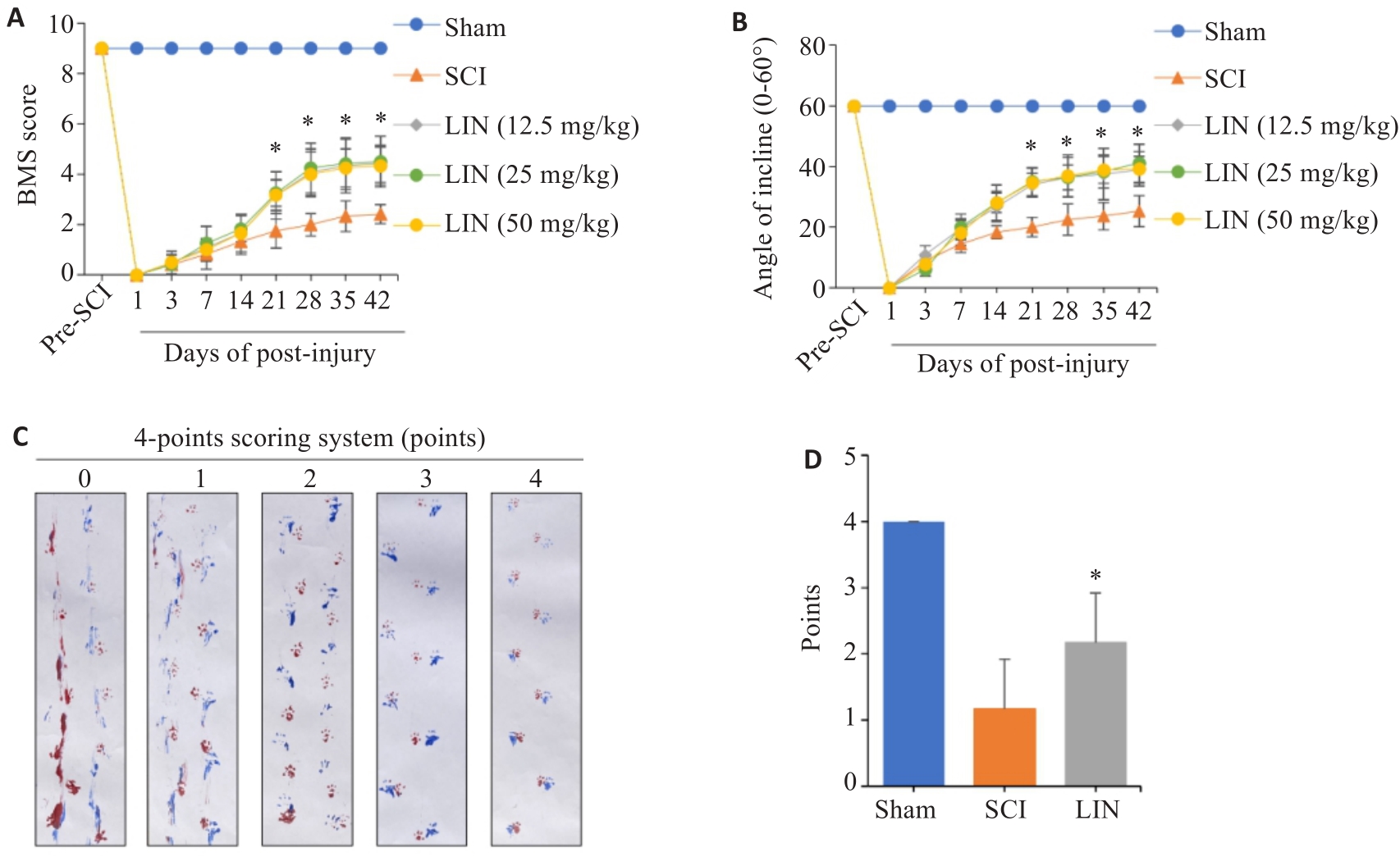
Fig.1 Linarin (LIN) treatment improves motor function in SCI mice. A: BMS score. B: Inclined plate experiment. C, D: Footprint score. *P<0.05 vs SCI group.
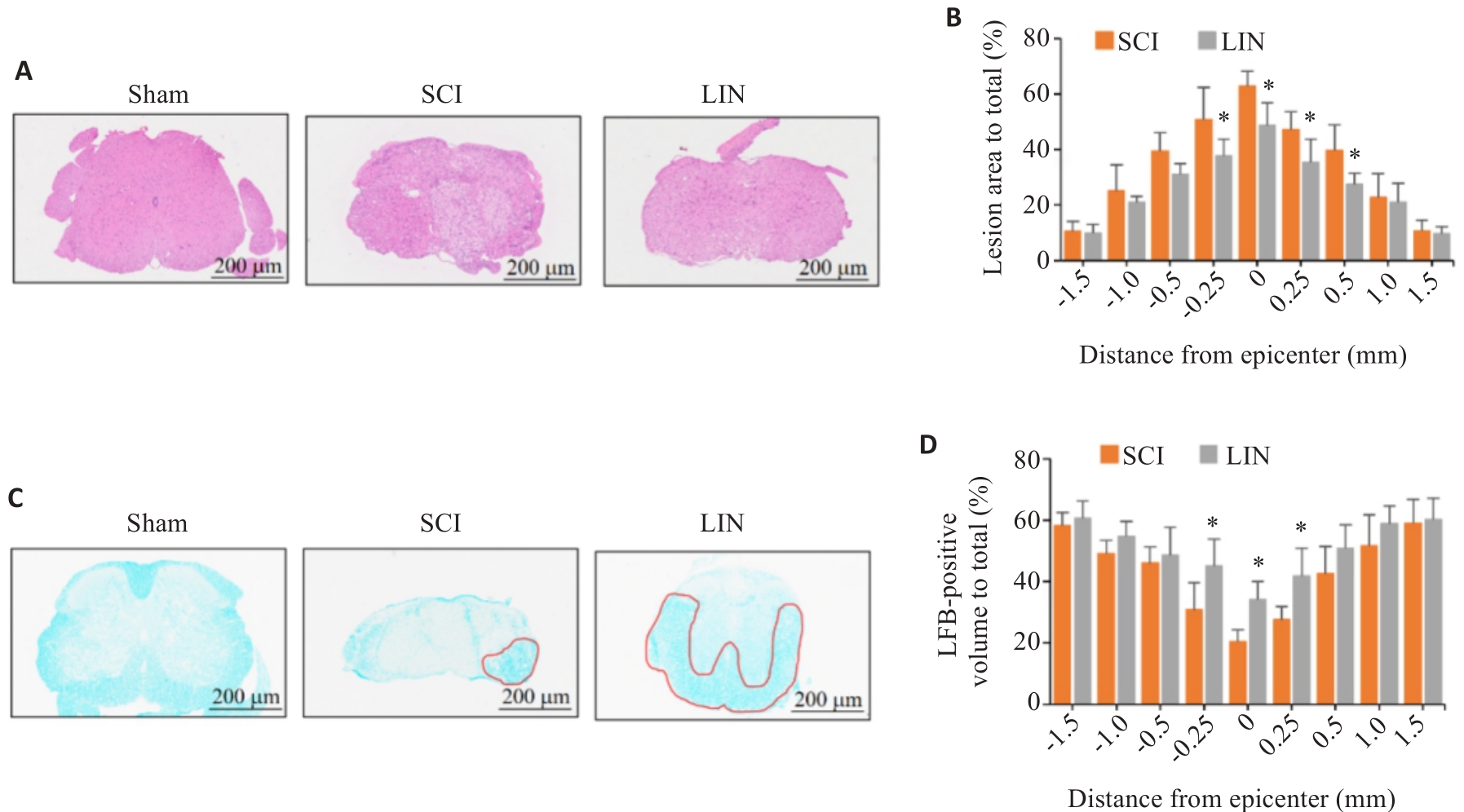
Fig.2 LIN treatment improves spinal cord histopathology in SCI mice. A: HE staining of a cross section at the center of spinal cord injury. B: Quantitative analysis of spinal cord injury area in LIN treatment group and SCI group. C: LFB staining of a cross section at the center of spinal cord injury. D: Quantitative analysis of myelination area in LIN treatment group and SCI group. *P<0.05 vs SCI group.
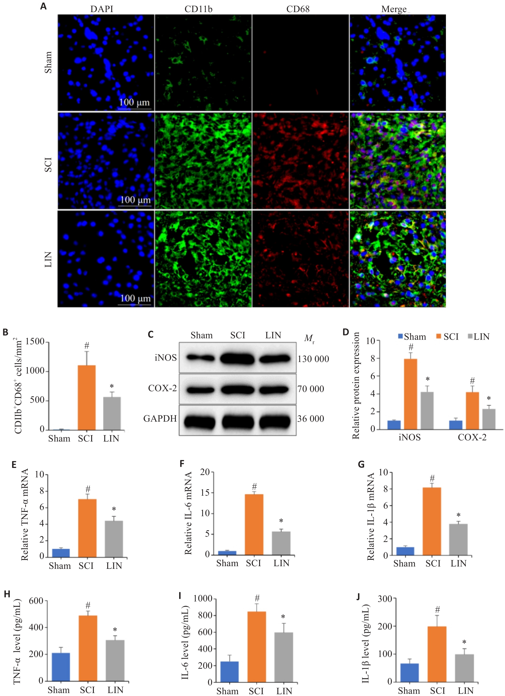
Fig3 LIN treatment inhibits microglia activation and inflammatory factor secretion in mouse spinal cord tissues. A: Microglia marker (CD11b) and activation marker (CD68) detected by immunofluorescence assay. B: Statistical graphs of the number of CD11b+CD68+ cells. C, D: Protein expression of iNOS and COX-2 detected by Western blotting and quantitative analyses. E-G: RT-qPCR for detecting relative expression levels of TNF-α, IL-6 and IL-1β mRNA. H-J: ELISA for detecting protein expression levels of TNF-α, IL-6 and IL-1β. #P<0.05 vs Sham group, *P<0.05 vs SCI group.
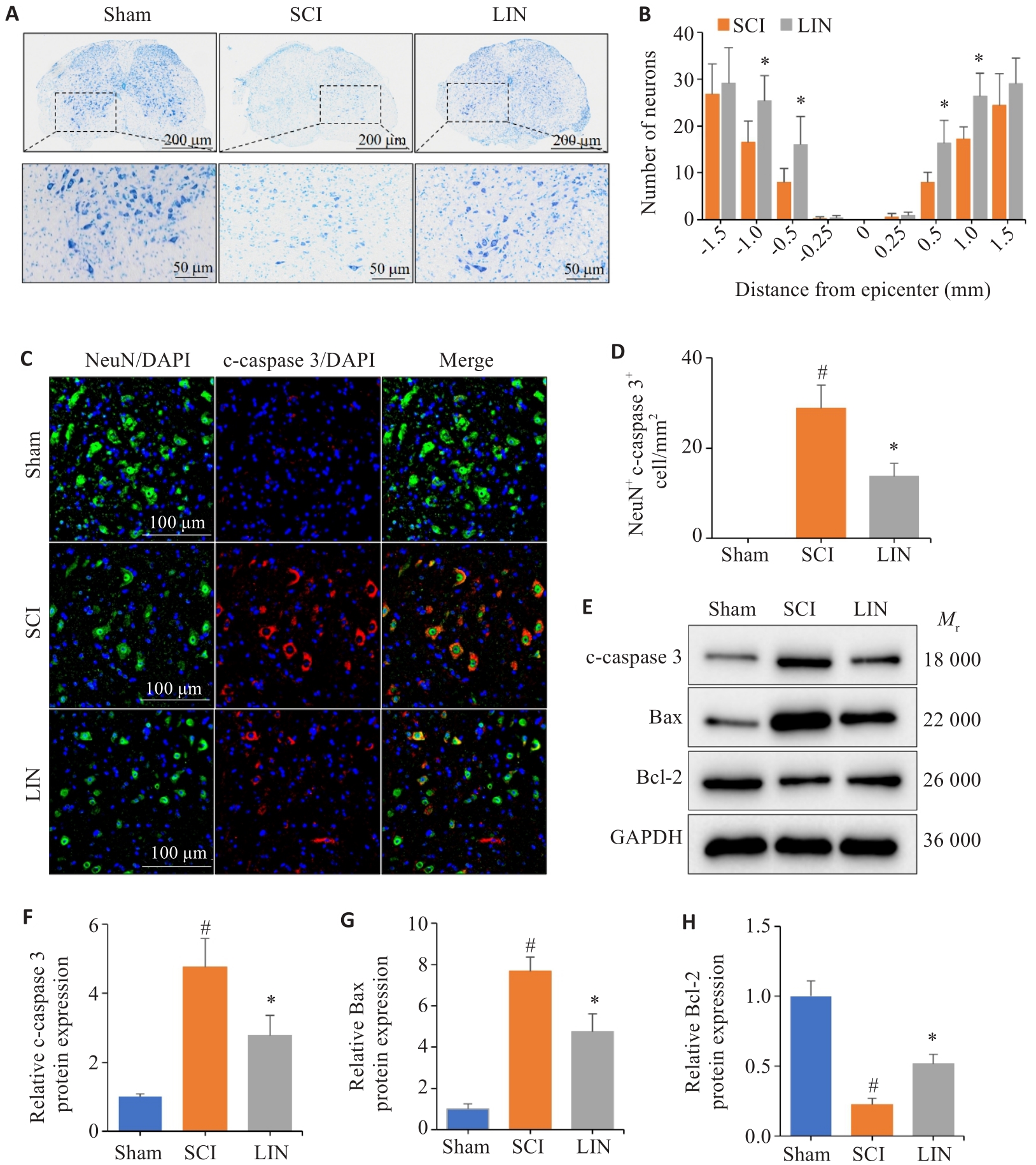
Fig.4 LIN treatment reduces neuronal apoptosis in mouse spinal cord after SCI. A: Nissl staining of the center of injury. B: Number of residual motor neurons in the anterior horn of the spinal cord in LIN-treated group and SCI group. C: Immunofluorescence detection of neuronal cell marker NeuN and apoptotic protein cleaved-caspase 3 (c-caspase 3). D: Statistical graph of the number of NeuN+c-caspase 3+ cells. E-H: Statistical graph of c-caspase 3, Bax and Bcl-2 protein expressions detected by Western blotting. #P<0.05 vs Sham group, *P<0.05 vs SCI group.
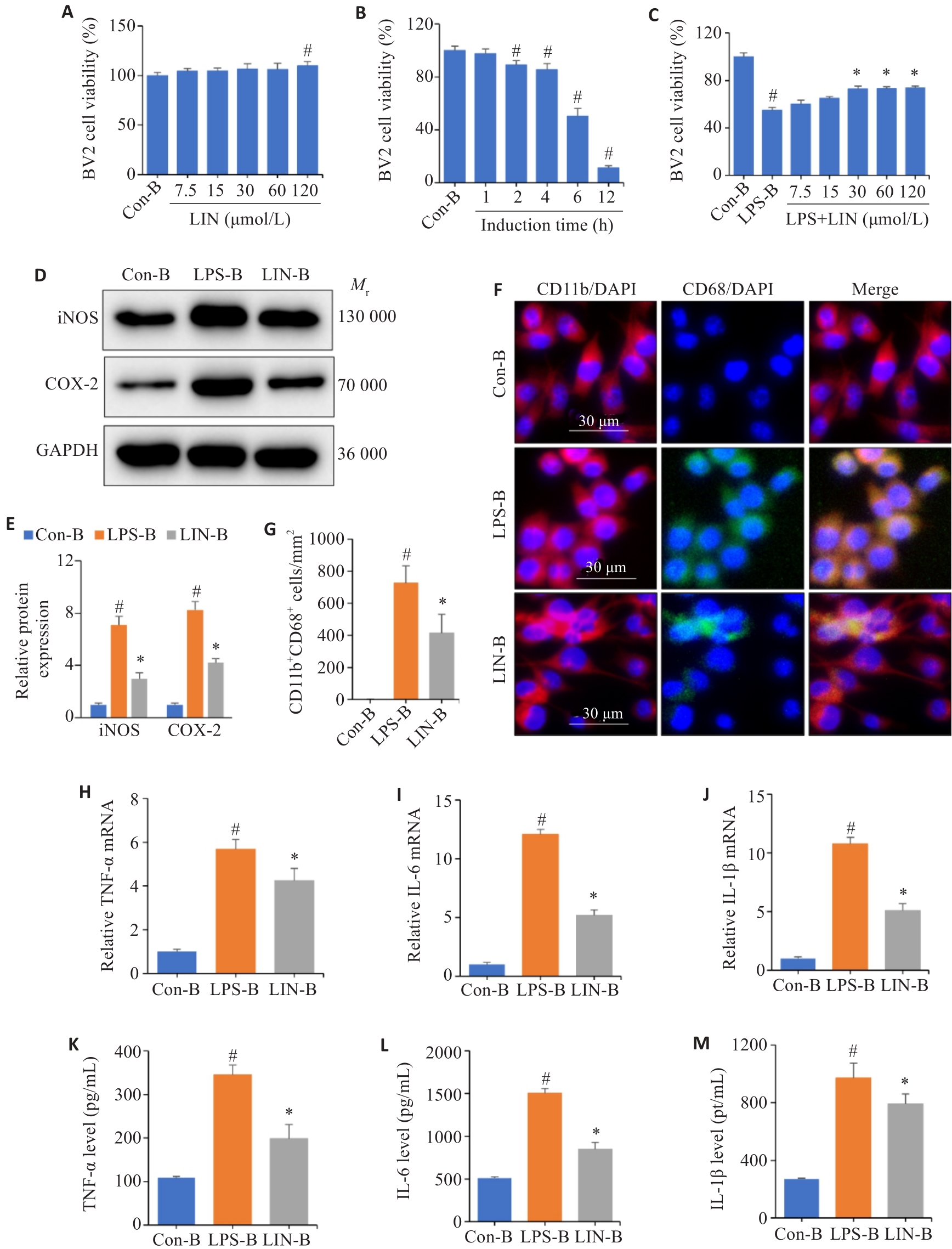
Fig.5 LIN inhibits microglia activation and secretion of inflammatory factors in BV2 cells. A: Cytotoxicity assay of LIN in BV2 cells. B: Optimal time determination of LPS-induced BV2 cell injury. C: Effect of LIN on LPS-induced BV2 cell viability. D, E: Statistical graphs of protein expression and quantitative analysis of iNOS and COX-2 detected by Western blotting. F: Immunofluorescence detection of microglia marker (CD11b) and activation marker (CD68) in BV2 cells. G: Statistical graphs of the number of CD11b+CD68+ cells. H-J: Relative mRNA expression levels of TNF-α, IL-6 and IL-1β detected by RT-qPCR. K-M: Protein expression levels of TNF-α, IL-6 and IL-1β detected by ELISA. #P<0.05 vs Con-B group, *P<0.05 vs LPS-B group.
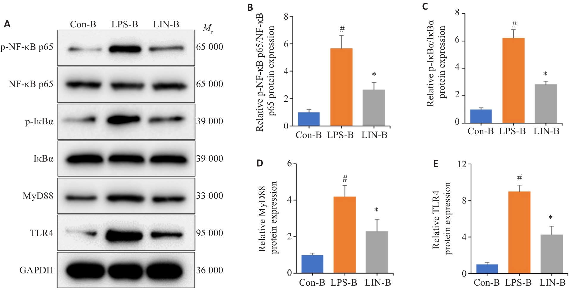
Fig6 LIN inhibits the TLR4/NF‑κB signaling pathway. A-E: Statistical graphs of the expression and quantitative analysis of TLR4/NF-κB pathway proteins after LIN intervention detected by Western blotting. #P<0.05 vs Con-B group, *P<0.05 vs LPS-B group.

Fig.7 Protective effect of LIN agaisnt inflammation-mediated neuronal apoptosis. A: CCK8 detection of LIN toxicity on HT22 cells. B-E: Statistical graphs of cleaved caspase-3 (c-caspase 3), Bax and Bcl-2 protein expression and quantitative analysis in HT22 cells detected by Western blotting. #P<0.05 vs Con-H group, *P<0.05 vs LPS-H group.
| 1 | Lin FX, Pan QL, Gu HY, et al. The role of resveratrol on spinal cord injury: from bench to bedside[J]. Mol Neurobiol, 2024, 61(1): 104-19. |
| 2 | Hu X, Xu W, Ren YL, et al. Spinal cord injury: molecular mechanisms and therapeutic interventions[J]. Signal Transduct Target Ther, 2023, 8(1): 245. |
| 3 | Xiao SN, Zhang Y, Liu ZH, et al. Alpinetin inhibits neuroinflammation and neuronal apoptosis via targeting the JAK2/STAT3 signaling pathway in spinal cord injury[J]. CNS Neurosci Ther, 2023, 29(4): 1094-108. |
| 4 | Benedetti B, Weidenhammer A, Reisinger M, et al. Spinal cord injury and loss of cortical inhibition[J]. Int J Mol Sci, 2022, 23(10): 5622. |
| 5 | Lv B, Zhang X, Yuan JS, et al. Biomaterial-supported MSC transplantation enhances cell-cell communication for spinal cord injury[J]. Stem Cell Res Ther, 2021, 12(1): 36. |
| 6 | Mottaghipisheh J, Taghrir H, Boveiri Dehsheikh A, et al. Linarin, a glycosylated flavonoid, with potential therapeutic attributes: a comprehensive review[J]. Pharmaceuticals, 2021, 14(11): 1104. |
| 7 | Qi WH, Chen YL, Sun SB, et al. Inhibiting TLR4 signaling by linarin for preventing inflammatory response in osteoarthritis[J]. Aging, 2021, 13(4): 5369-82. |
| 8 | Kim B, Lee JH, Seo MJ, et al. Linarin down-regulates phagocytosis, pro-inflammatory cytokine production, and activation marker expression in RAW264.7 macrophages[J]. Food Sci Biotechnol, 2016, 25(5): 1437-42. |
| 9 | Pan HY, Zhang JH, Wang YY, et al. Linarin improves the dyskinesia recovery in Alzheimer's disease zebrafish by inhibiting the acetylcholinesterase activity[J]. Life Sci, 2019, 222: 112-6. |
| 10 | Lou HY, Fan PH, Perez RG, et al. Neuroprotective effects of linarin through activation of the PI3K/Akt pathway in amyloid-β-induced neuronal cell death[J]. Bioorg Med Chem, 2011, 19(13): 4021-7. |
| 11 | Zhang YQ, Gao SH, Xia SN, et al. Linarin ameliorates ischemia-reperfusion injury by the inhibition of endoplasmic reticulum stress targeting AKR1B1[J]. Brain Res Bull, 2024, 207: 110868. |
| 12 | Xie C, Yue SQ, Li XZ, et al. A linarin derivative protects against ischemia-induced neuronal injury in mice by promoting cerebral blood flow recovery via KDELR-dependent CSPG4 activation[J]. Oxid Med Cell Longev, 2022, 2022: 6434086. |
| 13 | Chen YQ, Wang SN, Shi YJ, et al. CRID3, a blocker of apoptosis associated speck like protein containing a card, ameliorates murine spinal cord injury by improving local immune microenvironment[J]. J Neuroinflammation, 2020, 17(1): 255. |
| 14 | Han X, Wu YC, Meng M, et al. Linarin prevents LPS-induced acute lung injury by suppressing oxidative stress and inflammation via inhibition of TXNIP/NLRP3 and NF‑κB pathways[J]. Int J Mol Med, 2018, 42(3): 1460-72. |
| 15 | Basso DM, Fisher LC, Anderson AJ, et al. Basso Mouse Scale for locomotion detects differences in recovery after spinal cord injury in five common mouse strains[J]. J Neurotrauma, 2006, 23(5): 635-59. |
| 16 | Metz GA, Merkler D, Dietz V, et al. Efficient testing of motor function in spinal cord injured rats[J]. Brain Res, 2000, 883(2): 165-77. |
| 17 | Liu ZY, Yao XQ, Jiang WS, et al. Advanced oxidation protein products induce microglia-mediated neuroinflammation via MAPKs-NF‑κB signaling pathway and pyroptosis after secondary spinal cord injury[J]. J Neuroinflammation, 2020, 17(1): 90. |
| 18 | Al-Sammarraie N, Mahmood M, Ray SK. Neuroprotective role of Noggin in spinal cord injury[J]. Neural Regen Res, 2023, 18(3): 492-6. |
| 19 | Moura MVN, Mesquita da Conceição Bahia G, Gonçalves Correa M, et al. Neuroprotective effects of crude extracts, compounds, and isolated molecules obtained from plants in the central nervous system injuries: a systematic review[J]. Front Neurosci, 2023, 17: 1249685. |
| 20 | 钱永帅, 余惠凡, 张 艳, 等. 蒙花苷药理作用研究进展[J]. 中药药理与临床, 2023, 39(10): 124-8. |
| 21 | Yu Q, Li X, Cao X. Linarin could protect myocardial tissue from the injury of Ischemia-reperfusion through activating Nrf-2[J]. Biomed Pharmacother, 2017, 90: 1-7. |
| 22 | Zhuang ZJ, Shan CW, Li B, et al. Linarin enriched extract attenuates liver injury and inflammation induced by high-fat high-cholesterol diet in rats[J]. Evid Based Complement Alternat Med, 2017, 2017: 4701570. |
| 23 | Ankeny DP, Popovich PG. Mechanisms and implications of adaptive immune responses after traumatic spinal cord injury[J]. Neuroscience, 2009, 158(3): 1112-21. |
| 24 | Deng JH, Meng FQ, Zhang KX, et al. Emerging roles of microglia depletion in the treatment of spinal cord injury[J]. Cells, 2022, 11(12): 1871. |
| 25 | Li LL, Lan Y, Wang FQ, et al. Linarin protects against CCl4-induced acute liver injury via activating autophagy and inhibiting the inflammatory response: involving the TLR4/MAPK/Nrf2 pathway[J]. Drug Des Devel Ther, 2023, 17: 3589-604. |
| 26 | Jin CN, Liu JY, Jin RY, et al. Linarin ameliorates dextran sulfate sodium-induced colitis in C57BL/6J mice via the improvement of intestinal barrier, suppression of inflammatory responses and mo-dulation of gut microbiota[J]. Food Funct, 2022, 13(20): 10574-86. |
| 27 | Lu YC, Yeh WC, Ohashi PS. LPS/TLR4 signal transduction pathway[J]. Cytokine, 2008, 42(2): 145-51. |
| 28 | Takeda K, Akira S. TLR signaling pathways[J]. Semin Immunol, 2004, 16(1): 3-9. |
| 29 | Azam S, Jakaria M, Kim IS, et al. Regulation of toll-like receptor (TLR) signaling pathway by polyphenols in the treatment of age-linked neurodegenerative diseases: focus on TLR4 signaling[J]. Front Immunol, 2019, 10: 1000. |
| 30 | Huang Y, Lin J, Chen XW, et al. Pannexin-1 contributes to the apoptosis of spinal neurocytes in spinal cord injury[J]. Front Physiol, 2021, 12: 656647. |
| 31 | Kim HN, McCrea MR, Li SX. Advances in molecular therapies for targeting pathophysiology in spinal cord injury[J]. Expert Opin Ther Targets, 2023, 27(3): 171-87. |
| 32 | Wang S, Smith GM, Selzer ME, et al. Emerging molecular therapeutic targets for spinal cord injury[J]. Expert Opin Ther Targets, 2019, 23(9): 787-803. |
| [1] | Xiaoyu CHANG, Hanwen ZHANG, Hongting CAO, Ling HOU, Xin MENG, Hong TAO, Yan LUO, Guanghua LI. Heat stress affects expression levels of circadian clock gene Bmal1 and cyclins in rat thoracic aortic endothelial cells [J]. Journal of Southern Medical University, 2025, 45(7): 1353-1362. |
| [2] | Yujia YANG, Lifang YANG, Yaling WU, Zhaoda DUAN, Chunze YU, Chunyun WU, Jianyun YU, Li YANG. Cannabidiol inhibits neuronal endoplasmic reticulum stress and apoptosis in rats with multiple concussions by regulating the PERK-eIF2α-ATF4-CHOP pathway [J]. Journal of Southern Medical University, 2025, 45(6): 1240-1250. |
| [3] | Guanglü HE, Wanyu CHU, Yan LI, Xin SHENG, Hao LUO, Aiping XU, Mingjie BIAN, Huanhuan ZHANG, Mengya WANG, Chao ZHENG. Orexin-A promotes motor function recovery of rats with spinal cord injury by regulating ionotropic glutamate receptors [J]. Journal of Southern Medical University, 2025, 45(5): 1023-1030. |
| [4] | Xiaotao LIANG, Yifan XIONG, Xueqi LIU, Xiaoshan LIANG, Xiaoyu ZHU, Wei XIE. Huoxue Shufeng Granule alleviates central sensitization in chronic migraine mice via TLR4/NF-κB inflammatory pathway [J]. Journal of Southern Medical University, 2025, 45(5): 986-994. |
| [5] | Yang YANG, Kai WANG, Jianxiu LIU, Zhimo ZHOU, Wen JIA, Simou WU, Jinxing LI, Fang HE, Ruyue CHENG. Early life Bifidobacterium bifidum BD-1 intervention alleviates hyperactivity of juvenile female rats with attention deficit hyperactivity disorder [J]. Journal of Southern Medical University, 2025, 45(4): 702-710. |
| [6] | Yue CHEN, Linyu XIAO, Lü REN, Xue SONG, Jing LI, Jianguo HU. Monotropein improves motor function of mice with spinal cord injury by inhibiting the PI3K/AKT signaling pathway to suppress neuronal apoptosis [J]. Journal of Southern Medical University, 2025, 45(4): 774-784. |
| [7] | Fei CHU, Xiaohua CHEN, Bowen SONG, Jingjing YANG, Lugen ZUO. Moslosooflavone ameliorates dextran sulfate sodium-induced colitis in mice by suppressing intestinal epithelium apoptosis via inhibiting the PI3K/AKT signaling pathway [J]. Journal of Southern Medical University, 2025, 45(4): 819-828. |
| [8] | Yi ZHANG, Yu SHEN, Zhiqiang WAN, Song TAO, Yakui LIU, Shuanhu WANG. High expression of CDKN3 promotes migration and invasion of gastric cancer cells by regulating the p53/NF-κB signaling pathway and inhibiting cell apoptosis [J]. Journal of Southern Medical University, 2025, 45(4): 853-861. |
| [9] | Di CHEN, Ying LÜ, Yixin GUO, Yirong ZHANG, Ruixuan WANG, Xiaoruo ZHOU, Yuxin CHEN, Xiaohui WU. Dihydroartemisinin enhances doxorubicin-induced apoptosis of triple negative breast cancer cells by negatively regulating the STAT3/HIF-1α pathway [J]. Journal of Southern Medical University, 2025, 45(2): 254-260. |
| [10] | Yu BIN, Ziwen LI, Suwei ZUO, Sinuo SUN, Min LI, Jiayin SONG, Xu LIN, Gang XUE, Jingfang WU. High expression of apolipoprotein C1 promotes proliferation and inhibits apoptosis of papillary thyroid carcinoma cells by activating the JAK2/STAT3 signaling pathway [J]. Journal of Southern Medical University, 2025, 45(2): 359-370. |
| [11] | Shuo LIU, Jing LI, Xingwang WU. Swertiamarin ameliorates 2,4,6-trinitrobenzenesulfonic acid-induced colitis in mice by inhibiting intestinal epithelial cell apoptosis [J]. Journal of Southern Medical University, 2024, 44(8): 1545-1552. |
| [12] | Xiaofan CONG, Teng CHEN, Shuo LI, Yuanyuan WANG, Longyun ZHOU, Xiaolong LI, Pei ZHANG, Xiaojin SUN, Surong ZHAO. Dihydroartemisinin enhances sensitivity of nasopharyngeal carcinoma HNE1/DDP cells to cisplatin-induced apoptosis by promoting ROS production [J]. Journal of Southern Medical University, 2024, 44(8): 1553-1560. |
| [13] | Mengdong ZHENG, Yan LIU, Jiaojiao LIU, Qiaozhen KANG, Ting WANG. Effect of deletion of protein 4.1R on proliferation, apoptosis and glycolysis of hepatocyte HL-7702 cells [J]. Journal of Southern Medical University, 2024, 44(7): 1355-1360. |
| [14] | Yuanguo WANG, Peng ZHANG. Ferroptosis suppressor genes are highly expressed in esophageal cancer to inhibit tumor cell ferroptosis [J]. Journal of Southern Medical University, 2024, 44(7): 1389-1396. |
| [15] | Zhijun REN, Jianxin DIAO, Yiting WANG. Xionggui Decoction alleviates heart failure in mice with myocardial infarction by inhibiting oxidative stress-induced cardiomyocyte apoptosis [J]. Journal of Southern Medical University, 2024, 44(7): 1416-1424. |
| Viewed | ||||||
|
Full text |
|
|||||
|
Abstract |
|
|||||