
Journal of Southern Medical University ›› 2026, Vol. 46 ›› Issue (9): 2023-2034.doi: 10.12122/j.issn.1673-4254.2026.09.03
Lixuan YE1( ), Yuxin LIAO2, Qiong LIU2, Jia ZHANG2, Lei GAO2, Zhaoyang DONG1()
Received:2026-01-20
Online:2026-09-20
Published:2026-09-30
Contact:
Zhaoyang DONG
E-mail:2669556676@qq.com;021092@ gzucm.edu.cn
Supported by:Lixuan YE, Yuxin LIAO, Qiong LIU, Jia ZHANG, Lei GAO, Zhaoyang DONG. Xiaoyao San alleviates abnormal α‑syn aggregation and neuronal injury in a Caenorhabditis elegans model of Parkinson disease via the SKN-1 antioxidative pathway[J]. Journal of Southern Medical University, 2026, 46(9): 2023-2034.
Add to citation manager EndNote|Ris|BibTeX
URL: https://www.j-smu.com/EN/10.12122/j.issn.1673-4254.2026.09.03
| Time (min) | Flow (mL/min) | % B |
|---|---|---|
| 0.00 | 0.30 | 5.0 |
| 2.00 | 0.30 | 5.0 |
| 18.00 | 0.30 | 95.0 |
| 20.00 | 0.30 | 95.0 |
Tab.1 Chromatography gradient program
| Time (min) | Flow (mL/min) | % B |
|---|---|---|
| 0.00 | 0.30 | 5.0 |
| 2.00 | 0.30 | 5.0 |
| 18.00 | 0.30 | 95.0 |
| 20.00 | 0.30 | 95.0 |
| Gene | Forward primer (5'-3') | Reverse primer (5'-3') |
|---|---|---|
| Dat-1 | TTGGTGCCTACAGACGATCC | GCTGTTCCGTCTTCTGACCA |
| Cat-2 | ACTGAAGAACCTGCGGTACG | CCGATGGTGATTGCTCCTGC |
| Gst-4 | GATGCTCGTGCTCTTGCTGA | GGAGTCGTTGGCTTCAGCTT |
| Ced-9 | TGCTCAGGACTTGCCATCAC | GCGTGAAATAGTCGACCACA |
| Sod-3 | ATCTACTGCTCGCACTGCTT | ACGTAGGTGGCATGATGCTT |
| β-actin | ACGACGAGTCCGGCCCATCC | GAAAGCTGGTGGTGACGATGGTT |
Tab.2 Primer sequences of target genes
| Gene | Forward primer (5'-3') | Reverse primer (5'-3') |
|---|---|---|
| Dat-1 | TTGGTGCCTACAGACGATCC | GCTGTTCCGTCTTCTGACCA |
| Cat-2 | ACTGAAGAACCTGCGGTACG | CCGATGGTGATTGCTCCTGC |
| Gst-4 | GATGCTCGTGCTCTTGCTGA | GGAGTCGTTGGCTTCAGCTT |
| Ced-9 | TGCTCAGGACTTGCCATCAC | GCGTGAAATAGTCGACCACA |
| Sod-3 | ATCTACTGCTCGCACTGCTT | ACGTAGGTGGCATGATGCTT |
| β-actin | ACGACGAGTCCGGCCCATCC | GAAAGCTGGTGGTGACGATGGTT |
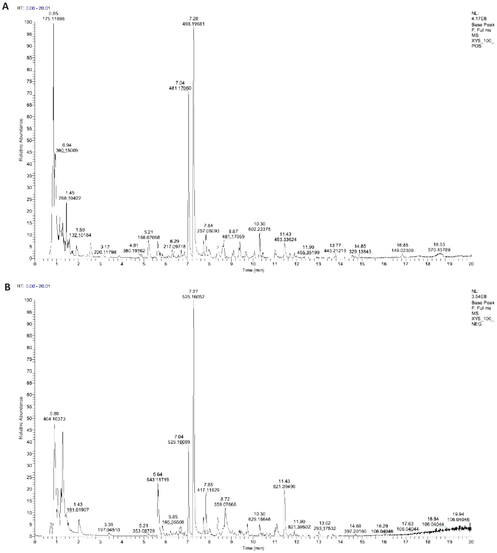
Fig.1 UHPLC-HRMS total ion chromatogram (TIC) of Xiaoyao San (XYS). A: Positive ion mode (POS). B: Negative ion mode (NEG).
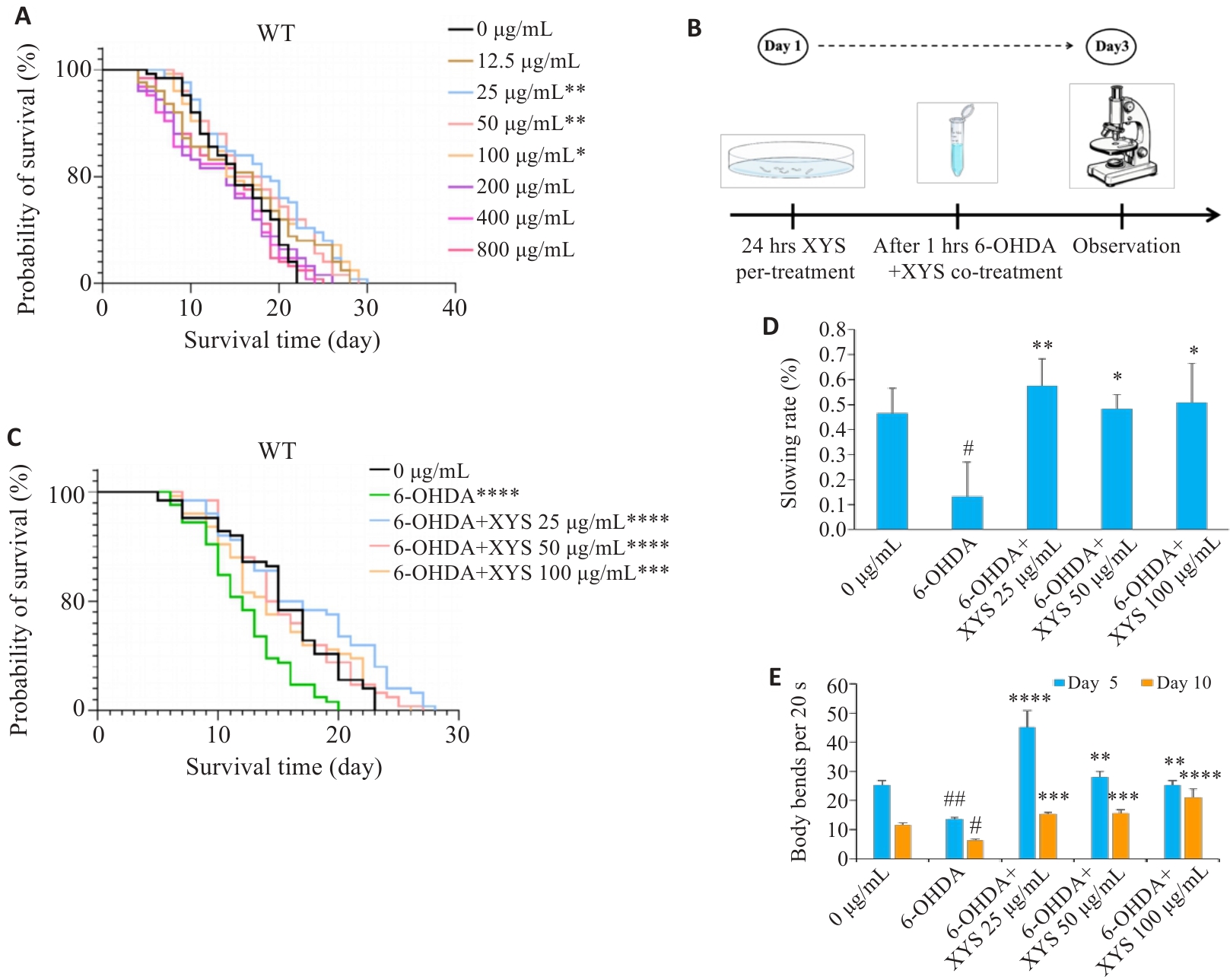
Fig.2 XYS extends lifespan and improves locomotor activity of C. elegans PD models. A: Survival curves of C. elegans exposed to different concentrations of XYS. B: Schematic diagram of the experimental design for combined 6-OHDA and XYS treatment. C: Survival curves of 6-OHDA-induced PD model worms with or without XYS treatment. D: Quantification of the basal slowing response. E: Quantification of body bends within 20 s on days 5 and 10 post-treatment. Data represent Mean±SD from at least 3 independent biological replicates (n=50 for lifespan assays and n=30 for other assays). #P<0.05, ##P<0.01 vs control group; *P<0.05, **P<0.01, ***P<0.001, ****P<0.0001 vs model group.
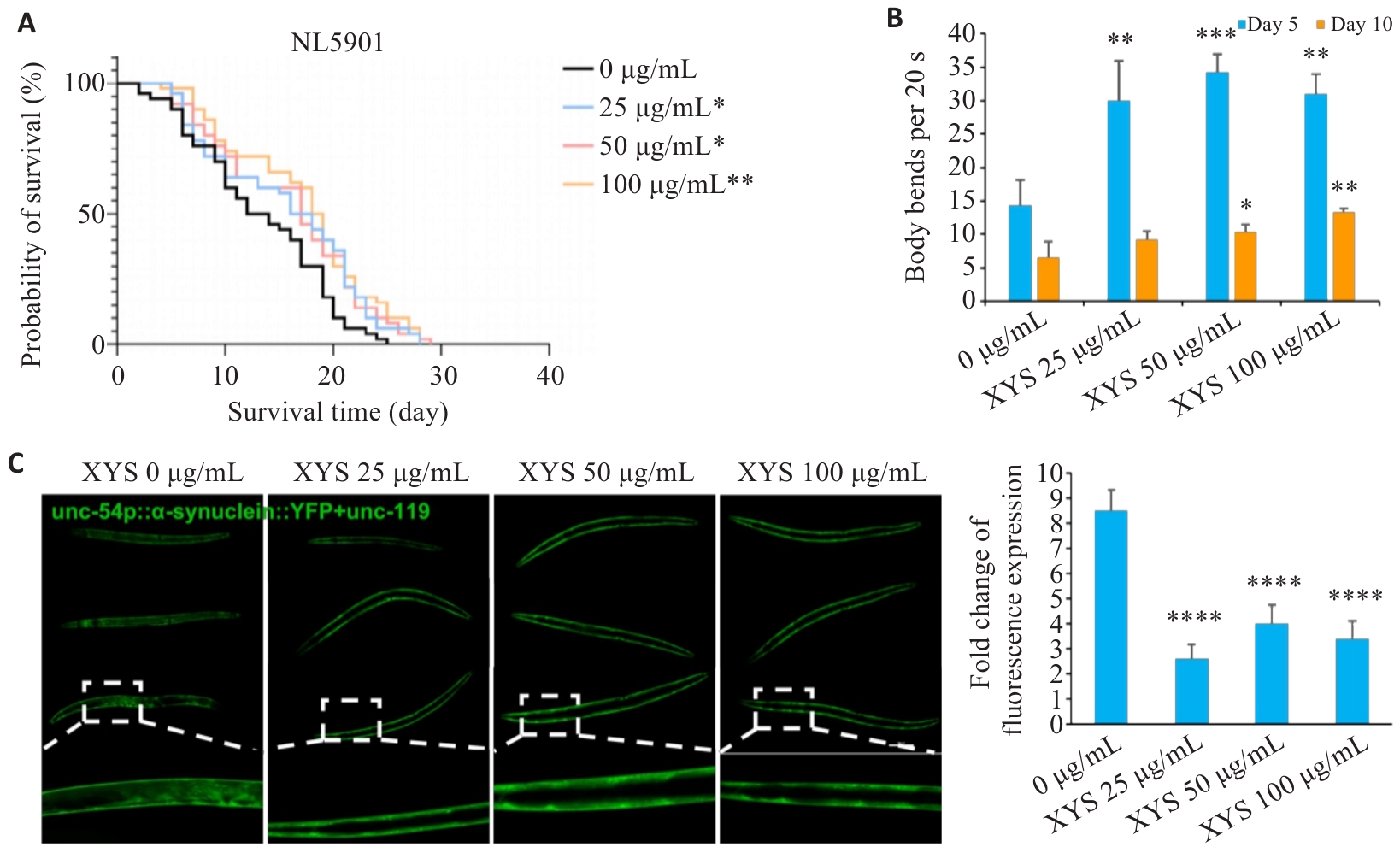
Fig.3 XYS extends lifespan and reduces α-syn aggregation in NL5901 worms. A: Survival curves of NL5901 worms with or without XYS treatment. B: Quantification of body bends within 20 s on days 5 and 10 post-treatment. C: Representative images of α‑syn::YFP fluorescence in body wall muscle cells and quantification of fluorescence intensity (Scale bar=100 μm). Data represent Mean±SD from at least 3 independent biological replicates (n=50 for lifespan assays and n=30 for other assays). *P<0.05, **P<0.01, ***P<0.001, ****P<0.0001 vs control group.
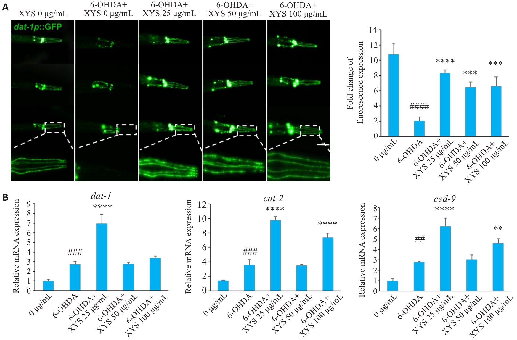
Fig.4 XYS protects against 6-OHDA-induced dopaminergic neurodegeneration in C. elegans. A: Representative GFP images of dopaminergic neurons with or without XYS treatment and quantification of fluorescence intensity (Scale bar=100 μm). B: Relative mRNA expression levels of dat-1, cat-2, and ced-9. Data represent Mean±SD from at least three independent biological replicates (n=30 for fluorescence analysis and n=3 for qPCR analysis). ##P<0.01, ###P<0.001, ####P<0.0001 vs control group; **P<0.01, ***P<0.001, ****P<0.0001 vs model group.
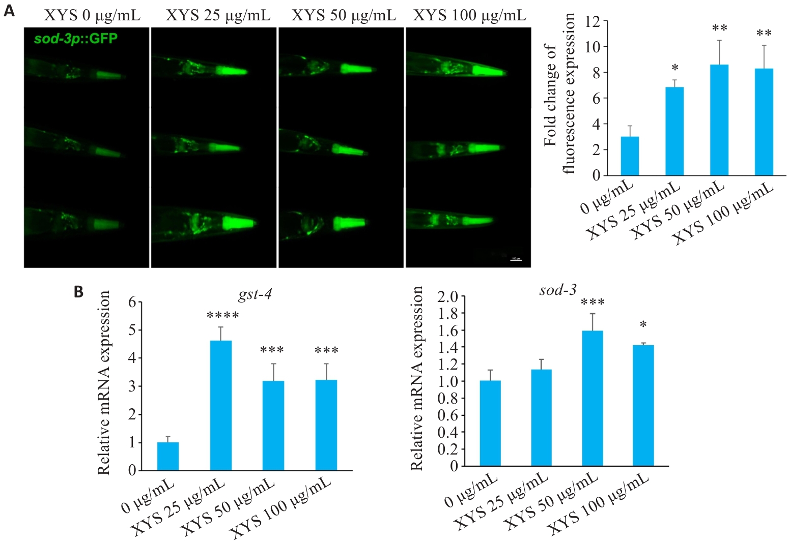
Fig.5 XYS significantly enhances antioxidant capacity in C. elegans. A: Representative fluorescence images of sod-3::GFP worms and quantification of sod-3::GFP fluorescence intensity (Scale bar=100 μm). B: Relative mRNA expression levels of sod-3 and gst-4. Data represent Mean±SD from at least 3 independent biological replicates (n=30 for fluorescence analysis and n=3 for qPCR analysis). *P<0.05, **P<0.01, ***P<0.001, ****P<0.0001 vs control group.
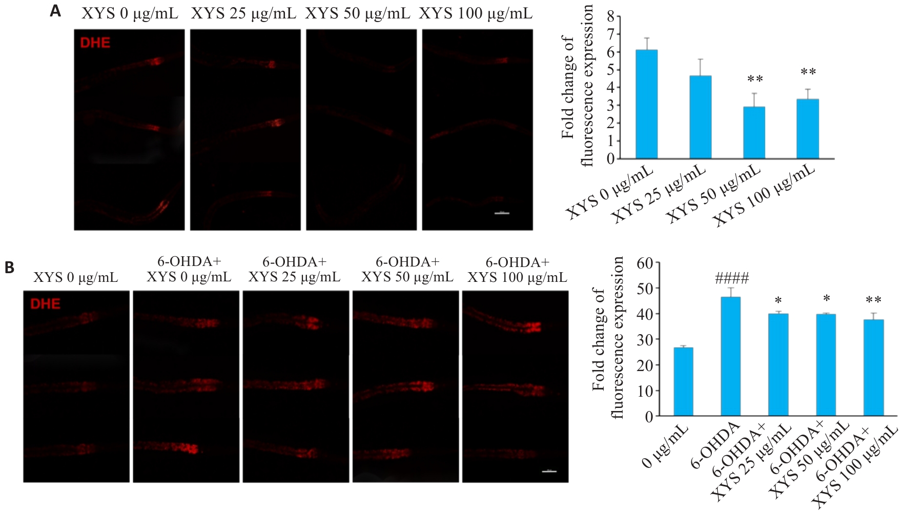
Fig.6 XYS significantly reduces oxidative stress levels in different C. elegans models of PD. A: Representative DHE staining images of NL5901 worms with or without XYS treatment and quantification of DHE fluorescence intensity. B: Representative DHE staining images of 6-OHDA-treated N2 worms with or without XYS treatment and quantification of DHE fluorescence intensity. Scale bar=100 μm. Data represent Mean±SD from at least 3 independent biological replicates (n=30). ####P<0.0001 vs control group; *P<0.05, **P<0.01 vs model group.
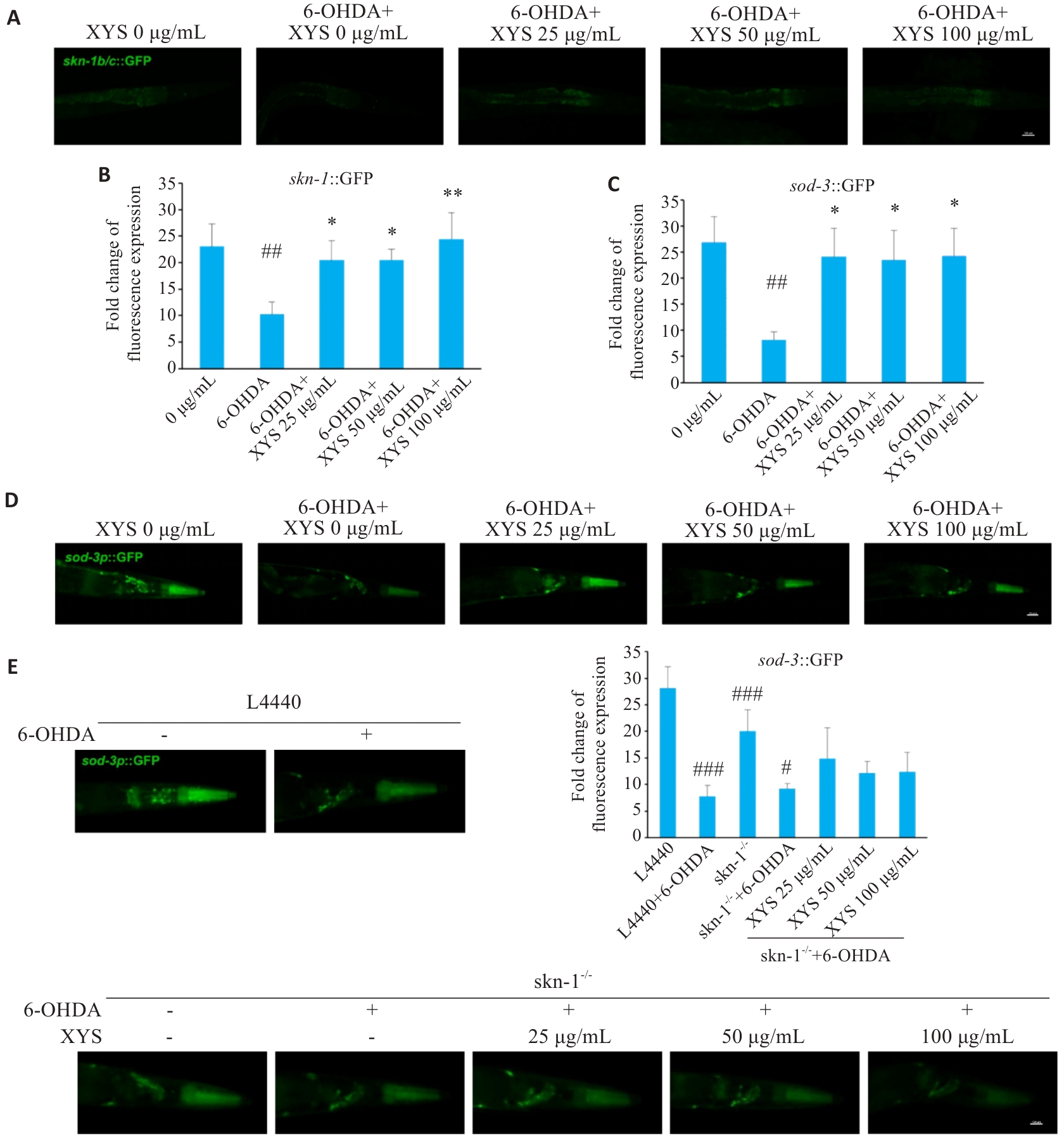
Fig.7 XYS improves antioxidant responses in PD C. elegans models through upregulation of SKN-1. A: Representative fluorescence images of LD1 worms. B: Quantification of fluorescence intensity in LD1 worms. C: Quantification of fluorescence intensity in CF1553 worms. D: Representative fluorescence images of CF1553 worms. E: Representative fluorescence images of skn-1 RNAi-treated worms and quantification of fluorescence intensity. Scale bar=100 μm. Data represent Mean±SD from at least 3 independent biological replicates (n=30). ##P<0.01, ###P<0.001 vs control group; #P<0.05 vs skn-1 RNAi group; *P<0.05, **P<0.01 vs model group.
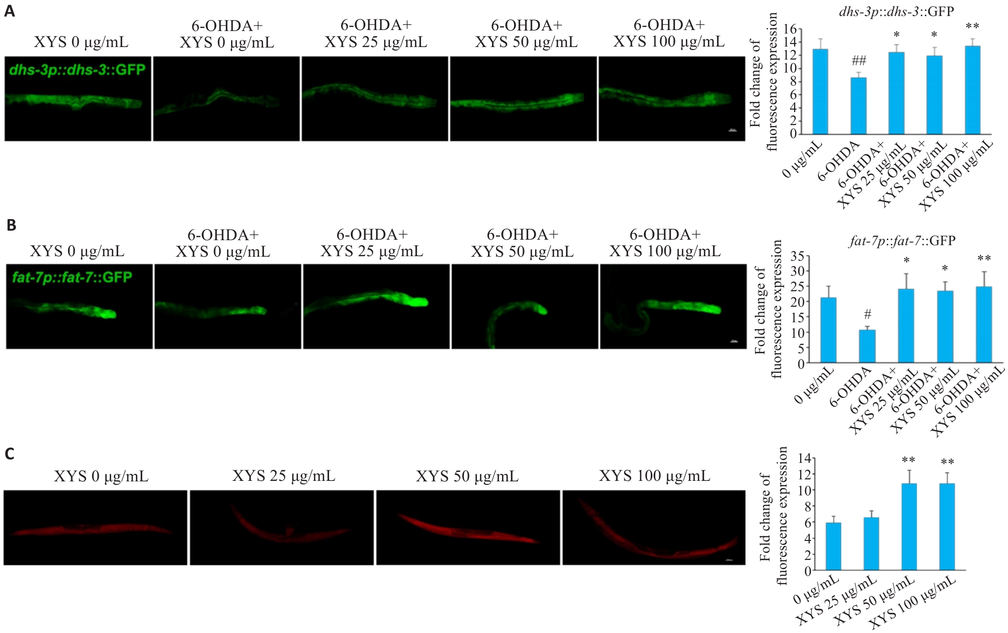
Fig.8 XYS ameliorates lipid metabolic dysfunction in PD C. elegans models. A: Representative fluorescence images of LIU104 worms and quantification of fluorescence intensity. B: Representative fluorescence images of DMS303 worms and quantification of fluorescence intensity. C: Representative Nile Red staining images of NL5901 worms and quantification of fluorescence intensity. Scale bar=100 μm. Data represent Mean±SD from at least 3 independent biological replicates (n=30). #P<0.05, ##P<0.01 vs control group; *P<0.05, **P<0.01 vs model group.
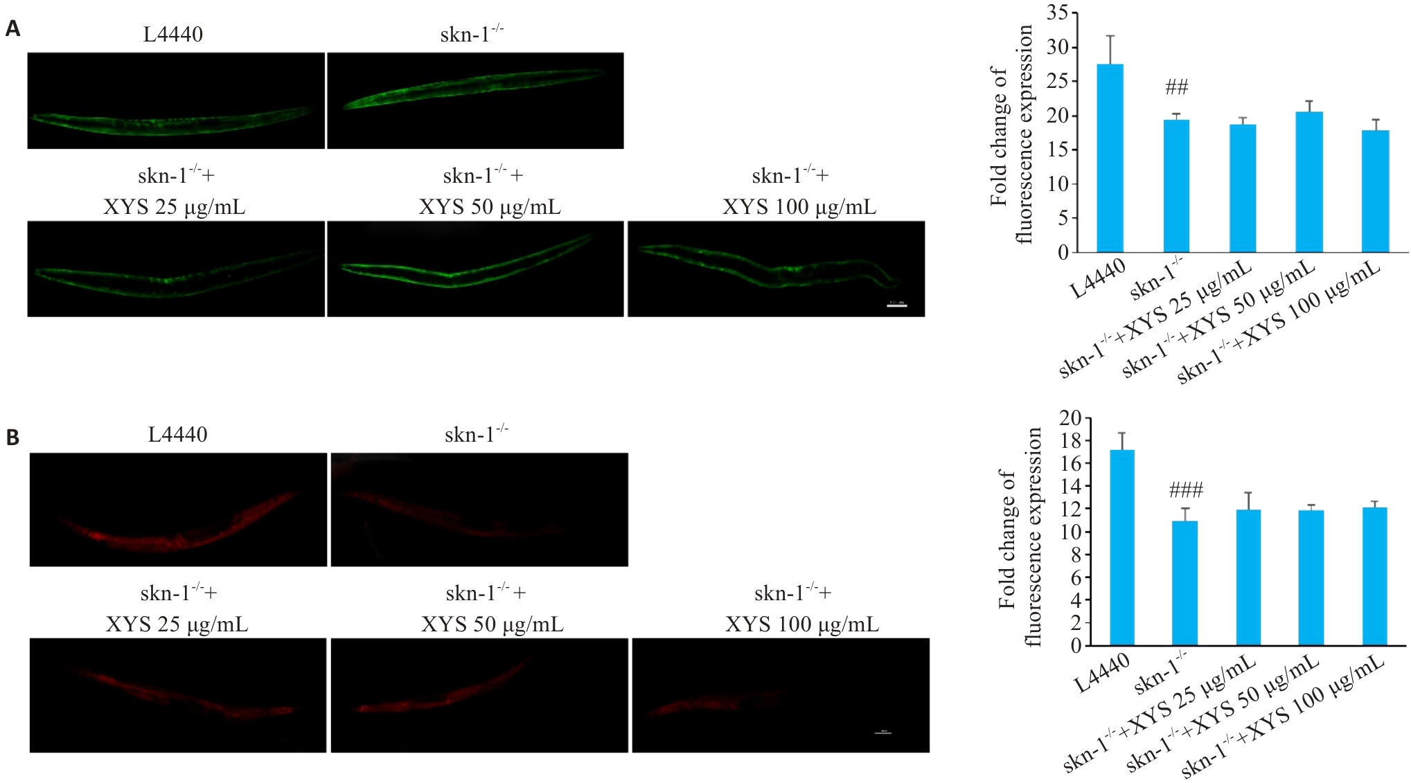
Fig.9 XYS regulates α-synuclein accumulation and lipid deposition via skn-1. A: Representative fluorescence images of skn-1 RNAi worms and quantification of fluorescence intensity. B: Representative Nile Red staining images of skn-1 RNAi worms and quantification of lipid accumulation based on Nile Red staining. Scale bar=100 μm. Data represent Mean±SD from at least 3 independent biological replicates (n=30). ##P<0.01, ###P<0.001 vs control group.
| [1] | Tanner CM, Ostrem JL. Parkinson's disease[J]. N Engl J Med, 2024, 391(5): 442-52. doi:10.1056/nejmra2401857 |
| [2] | Chen Y, Luo XX, Yin YK, et al. The interplay of iron, oxidative stress, and α-synuclein in Parkinson's disease progression[J]. Mol Med, 2025, 31(1): 154. doi:10.1186/s10020-025-01208-3 |
| [3] | Blackwell TK, Steinbaugh MJ, Hourihan JM, et al. SKN-1/Nrf, stress responses, and aging in Caenorhabditis elegans [J]. Free Radic Biol Med, 2015, 88: 290-301. doi:10.1016/j.freeradbiomed.2015.06.008 |
| [4] | Chen JB, Lei CF, Li XJ, et al. Research progress on classical traditional Chinese medicine formula Xiaoyaosan in the treatment of depression[J]. Front Pharmacol, 2022, 13: 925514. doi:10.3389/fphar.2022.925514 |
| [5] | Liu XY, Yan YZ, Jiang AJ, et al. Xiaoyao San against depression via Glia-mediated neuroinflammation using network pharmacology and experimental validation[J]. Sci Rep, 2025, 15: 39708. doi:10.1038/s41598-025-23296-1 |
| [6] | Wei YY, Lin WF, Zhang TH, et al. Effectiveness of traditional Chinese medicineas as an adjunct therapy for refractory schizophrenia: a systematic review and meta analysis[J]. Sci Rep, 2018, 8: 6230. doi:10.1038/s41598-018-24547-0 |
| [7] | Chen JX, Xu JK, Huang P, et al. The potential applications of traditional Chinese medicine in Parkinson's disease: a new opportunity[J]. Biomed Pharmacother, 2022, 149: 112866. doi:10.1016/j.biopha.2022.112866 |
| [8] | Zhang Y, Xu XM. Chinese herbal medicine in the treatment of depression in Parkinson's disease: from molecules to systems[J]. Front Pharmacol, 2022, 13: 879459. doi:10.3389/fphar.2022.879459 |
| [9] | Xu Y, Chen WY, Chen ZR, et al. Mechanism of action of Xiaoyao San in treatment of ischemic stroke is related to anti-apoptosis and activation of PI3K/Akt pathway[J]. Drug Des Devel Ther, 2021, 15: 753-67. doi:10.2147/dddt.s280217 |
| [10] | Magalingam KB, Radhakrishnan A, Haleagrahara N. Protective effects of quercetin glycosides, rutin, and isoquercetrin against 6-hydroxydopamine (6-OHDA)‑induced neurotoxicity in rat pheo-chromocytoma (PC-12) cells[J]. Int J Immunopathol Pharmacol, 2016, 29(1): 30-9. doi:10.1177/0394632015613039 |
| [11] | Singh S, Kumar P. Piperine in combination with quercetin halt 6-OHDA induced neurodegeneration in experimental rats: Biochemical and neurochemical evidences[J]. Neurosci Res, 2018, 133: 38-47. doi:10.1016/j.neures.2017.10.006 |
| [12] | Hu K, Zhu SS, Wu FY, et al. Aureusidin ameliorates 6-OHDA-induced neurotoxicity via activating Nrf2/HO-1 signaling pathway and preventing mitochondria-dependent apoptosis pathway in SH-SY5Y cells and Caenorhabditis elegans [J]. Chem Biol Interact, 2024, 387: 110824. doi:10.1016/j.cbi.2023.110824 |
| [13] | Tamuli R, Mellick GD, Schirra HJ, et al. Mode of action of toxin 6-hydroxydopamine in SH-SY5Y using NMR metabolomics[J]. Molecules, 2025, 30(16): 3352. doi:10.3390/molecules30163352 |
| [14] | Kimble J, Nüsslein-Volhard C. The great small organisms of developmental genetics: Caenorhabditis elegans and Drosophila melanogaster [J]. Dev Biol, 2022, 485: 93-122. doi:10.1016/j.ydbio.2022.02.013 |
| [15] | Ma JY, Li KL, Shi SL, et al. The application of UHPLC-HRMS for quality control of traditional Chinese medicine[J]. Front Pharmacol, 2022, 13: 922488. doi:10.3389/fphar.2022.922488 |
| [16] | 陈周雯, 任雨敏, 唐 鲜, 等. 西南手参醇提物通过抗氧化途径延缓秀丽隐杆线虫衰老的机制[J]. 食品科学, 2026, 47(6): 214-25. doi:10.7506/spkx1002-6630-20250910-079 |
| [17] | Offenburger SL, Ho XY, Tachie-Menson T, et al. 6-OHDA-induced dopaminergic neurodegeneration in Caenorhabditis elegans is promoted by the engulfment pathway and inhibited by the transthyretin-related protein TTR-33[J]. PLoS Genet, 2018, 14(1): e1007125. doi:10.1371/journal.pgen.1007125 |
| [18] | Tsai RT, Tsai CW, Liu SP, et al. Maackiain ameliorates 6-hydroxydopamine and SNCA pathologies by modulating the PINK1/parkin pathway in models of Parkinson's disease in Caenorhabditis elegans and the SH-SY5Y cell line[J]. Int J Mol Sci, 2020, 21(12): 4455. doi:10.3390/ijms21124455 |
| [19] | Palikaras K, SenGupta T, Nilsen H, et al. Assessment of dopaminergic neuron degeneration in a C. elegans model of Parkinson's disease[J]. STAR Protoc, 2022, 3(2): 101264. doi:10.1016/j.xpro.2022.101264 |
| [20] | Balkrishna A, Pathak N, Singh R, et al. Neurogrit gold attenuates 6-OHDA-induced dopaminergic neurodegeneration in Parkinson's model of Caenorhabditis elegans by reducing α‑synuclein accumulation and pink/pdr-1 driven mitochondrial dysfunction[J]. CNS Neurosci Ther, 2025, 31(5): e70401. doi:10.1111/cns.70401 |
| [21] | Sawin ER, Ranganathan R, Horvitz HR. C. elegans locomotory rate is modulated by the environment through a dopaminergic pathway and by experience through a serotonergic pathway[J]. Neuron, 2000, 26(3): 619-31. doi:10.1016/s0896-6273(00)81199-x |
| [22] | Picca A, Calvani R, Coelho-Junior HJ, et al. Mitochondrial dysfunction, oxidative stress, and neuroinflammation: intertwined roads to neurodegeneration[J]. Antioxidants, 2020, 9(8): 647. doi:10.3390/antiox9080647 |
| [23] | Fu RH, Tsai CW, Liu SP, et al. Neuroprotective capability of narcissoside in 6-OHDA-exposed Parkinson's disease models through enhancing the MiR200a/nrf-2/GSH axis and mediating MAPK/Akt associated signaling pathway[J]. Antioxidants, 2022, 11(11): 2089. doi:10.3390/antiox11112089 |
| [24] | Kwon TG, Kim YJ, Hong JY, et al. A review of antidepressant and anxiolytic effects of Soyo-San (Xiaoyao-San) and modified Soyo-San in animal models[J]. Phytomedicine, 2024, 135: 155387. doi:10.1016/j.phymed.2024.155387 |
| [25] | Bastien BL, Haury WR, Smisko WR, et al. Nlr-1/CNTNAP regulates dopamine circuit structure and foraging behaviors in C. elegans[J]. Commun Biol, 2024, 7: 1248. doi:10.1038/s42003-024-06936-6 |
| [26] | Lee I, Knickerbocker AC, Depew CR, et al. Effect of altered production and storage of dopamine on development and behavior in C. elegans[J]. Front Toxicol, 2024, 6: 1374866. doi:10.3389/ftox.2024.1374866 |
| [27] | Jeong H, Park JY, Lee JH, et al. Deficiency in RCAT-1 function causes dopamine metabolism related behavioral disorders in Caenorhabditis elegans [J]. Int J Mol Sci, 2022, 23(4): 2393. doi:10.3390/ijms23042393 |
| [28] | Maulik M, Mitra S, Bult-Ito A, et al. Behavioral phenotyping and pathological indicators of Parkinson's disease in C. elegans models[J]. Front Genet, 2017, 8: 77. doi:10.3389/fgene.2017.00077 |
| [29] | Abidar S, Nhiri M, Hritcu L. Phytochemicals against the 6-hydroxydopamine-induced Parkinson's disease in rodents: a systematic review[J]. Next Res, 2025, 2(3): 100473. doi:10.1016/j.nexres.2025.100473 |
| [30] | Kim KH, Lee SY, Shin J, et al. Dose-dependent neuroprotective effect of standardized bee venom phospholipase a(2) against MPTP-induced Parkinson's disease in mice[J]. Front Aging Neurosci, 2019, 11: 80. doi:10.3389/fnagi.2019.00080 |
| [31] | Liu XJ, Zheng ZJ, Xue C, et al. LRRK2 mediates α‑synuclein-induced neuroinflammation and ferroptosis through the p62-Keap1-Nrf2 pathway in Parkinson's disease[J]. Inflammation, 2025, 48(5): 3666-91. doi:10.1007/s10753-025-02291-8 |
| [32] | Chakraborty S, Bornhorst J, Nguyen TT, et al. Oxidative stress mechanisms underlying Parkinson's disease-associated neuro-degeneration in C. elegans[J]. Int J Mol Sci, 2013, 14(11): 23103-28. doi:10.3390/ijms141123103 |
| [33] | Phan HD, Nguyen TTM, Lee S, et al. The metabolic contribution of SKN-1/Nrf2 to the lifespan of Caenorhabditis elegans [J]. Metabolomics, 2023, 19(6): 58. doi:10.1007/s11306-023-02022-w |
| [34] | Pang SS, Lynn DA, Lo JY, et al. SKN-1 and Nrf2 couples proline catabolism with lipid metabolism during nutrient deprivation[J]. Nat Commun, 2014, 5: 5048. doi:10.1038/ncomms6048 |
| [35] | Zhao CY, Tu J, Wang CC, et al. Lysophosphatidylcholine binds α‑synuclein and prevents its pathological aggregation[J]. Natl Sci Rev, 2024, 11(6): nwae182. doi:10.1093/nsr/nwae182 |
| [36] | Lastres-Becker I, Ulusoy A, Innamorato NG, et al. α‑Synuclein expression and Nrf2 deficiency cooperate to aggravate protein aggregation, neuronal death and inflammation in early-stage Parkinson's disease[J]. Hum Mol Genet, 2012, 21(14): 3173-92. doi:10.1093/hmg/dds143 |
| [37] | Idi W, Sheta R, Oueslati A. Interplay between lipid droplets and alpha-synuclein: implication in Parkinson's disease pathogenesis[J]. Front Mol Neurosci, 2025, 18: 1681039. doi:10.3389/fnmol.2025.1681039 |
| [38] | Coles NP, Elsheikh S, Quesnel A, et al. Alpha-synuclein aggregation induces prominent cellular lipid changes as revealed by Raman spectroscopy and machine learning analysis[J]. Brain Commun, 2025, 7(2): fcaf133. doi:10.1093/braincomms/fcaf133 |
| [1] | GUI Jianjun, SUN Xiaodong, WEN Shu, LIU Xin, QIN Bingqing, SANG Ming. Resveratrol protects dopaminergic neurons in a mouse model of Parkinson's disease by regulating the gut-brain axis via inhibiting the TLR4 signaling pathway [J]. Journal of Southern Medical University, 2024, 44(2): 270-279. |
| [2] | MU Feifei, CHEN Xi, DU Xixun, JIAO Qian, BI Mingxia, JIANG Hong. Regulatory mechanism of interferon regulatory factor 1 by α-synuclein in Parkinson's disease models [J]. Journal of Southern Medical University, 2021, 41(11): 1641-1648. |
| [3] | . Voxel-based analysis of cerebral blood flow changes in Parkinson disease using arterial spin labeling technique [J]. Journal of Southern Medical University, 2018, 38(01): 117-. |
| [4] | LI Rui, PENG Ning, DU Fang, LI Xu-ping, LE Wei-dong Department of Neurology, People’s Hospital of Shaanxi Province, Xi’an 710068, China; Health Science Center, Shanghai Institute for Biological Science, Chinese Academy of Science, Shanghai 200025, China. Epigallocatechin gallate protects dopaminergic neurons against 1-methyl-4-phenyl-1,2,3,6-tetrahydropyridine-induced neurotoxicity by inhibiting microglial cell activation [J]. Journal of Southern Medical University, 2006, 26(04): 376-380. |
| [5] | JIANG Xue-mei, HUANG Yong, ZHUO Ying, GAO Yan-ping Department of Acupuncture and Moxibustion, Department of Orthopedics, College of Traditional Chinese Medicine, Southern Medical University, Guangzhou 510515, China. Therapeutic effect of scalp electroacupuncture on Parkinson disease [J]. Journal of Southern Medical University, 2006, 26(01): 114-116. |
| Viewed | ||||||
|
Full text |
|
|||||
|
Abstract |
|
|||||
