
Journal of Southern Medical University ›› 2026, Vol. 46 ›› Issue (10): 2558-2574.doi: 10.12122/j.issn.1673-4254.2026.10.23
Lun LI1( ), Yuqin ZHANG3(), Qingli ZHAN1, Wei ZHAO2(), Junbing FAN1()
Received:2026-05-25
Online:2026-10-20
Published:2026-10-26
Contact:
Wei ZHAO, Junbing FAN
E-mail:864852592@qq.com;791369631@qq.com;pummpa@163.com;fjb2012@smu.edu.cn
Lun LI, Yuqin ZHANG, Qingli ZHAN, Wei ZHAO, Junbing FAN. Transarterial embolization microspheres for liver cancer: current applications and advances[J]. Journal of Southern Medical University, 2026, 46(10): 2558-2574.
Add to citation manager EndNote|Ris|BibTeX
URL: https://www.j-smu.com/EN/10.12122/j.issn.1673-4254.2026.10.23
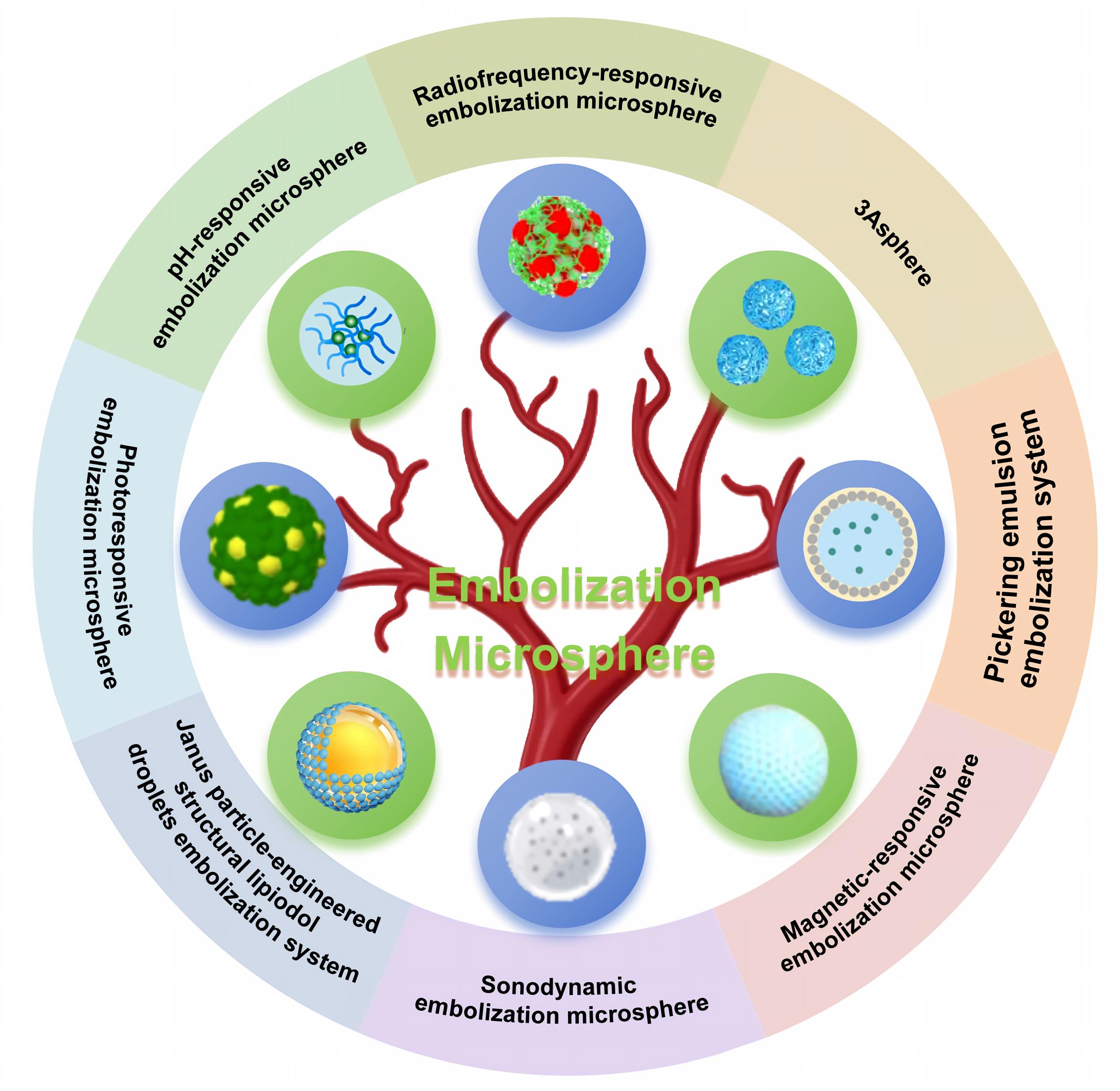
Fig.1 Classification of transarterial embolization microspheres for liver cancer.
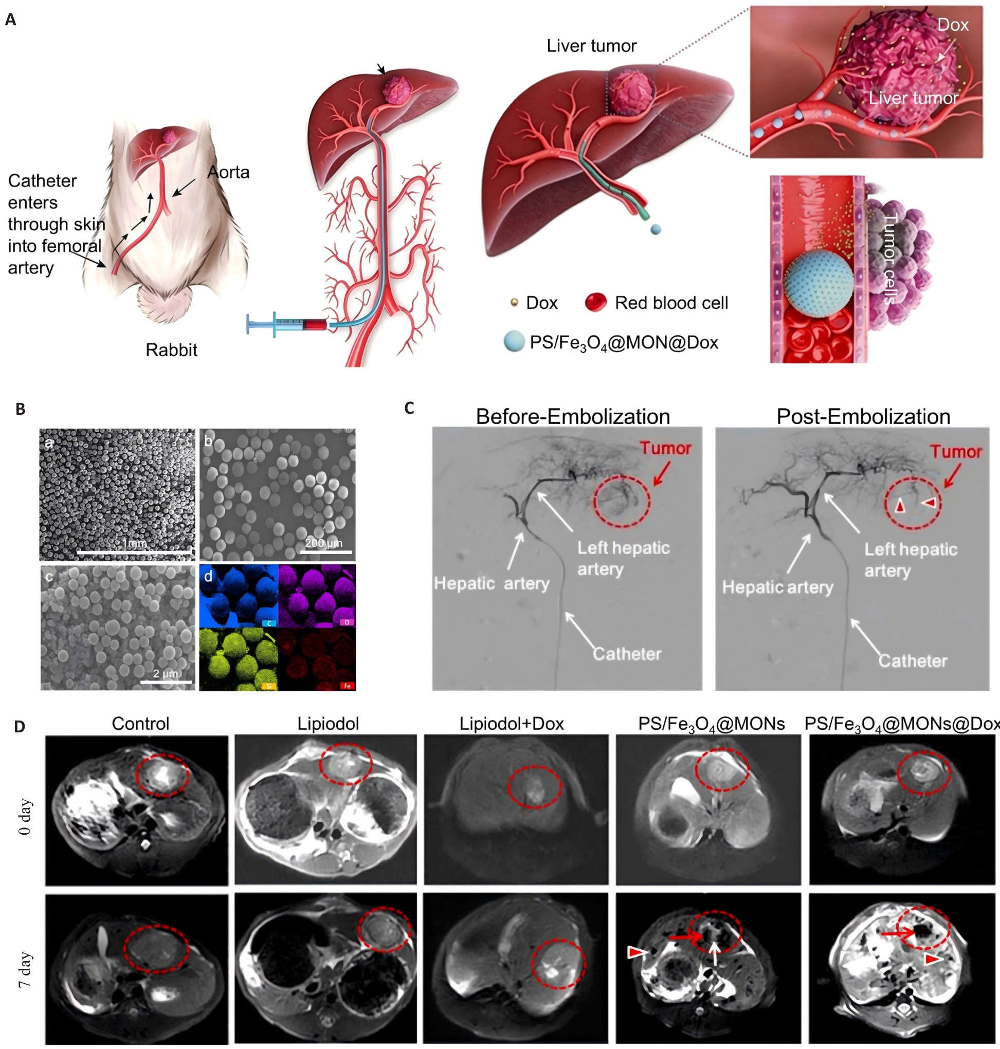
Fig.2 Magnetic-responsive embolization microspheres. A: Schematic diagram of embolization of PS/Fe₃O₄@MONs microspheres. The microspheres are fabricated by loading polystyrene/Fe₃O₄ particles onto mesoporous silica microspheres (MONs). B: Scanning electron microscope (SEM) image and elemental mapping of PS/Fe₃O₄@MONs microspheres. Blue, purple, yellow, and red indicate carbon, oxygen, silicon, and iron elements, respectively. C: DSA images before and after embolization with PS/Fe₃O₄@MONs microspheres. The red dotted lines indicate the visualized tumor blood vessel. D: MRI images showing tumor changes on day 0 and day 7 after treatment in the control group, Lipiodol group, Lipiodol+Dox group, PS/Fe₃O₄@MONs group and PS/Fe₃O₄@MONs@Dox group. The red dotted lines indicate the tumor.
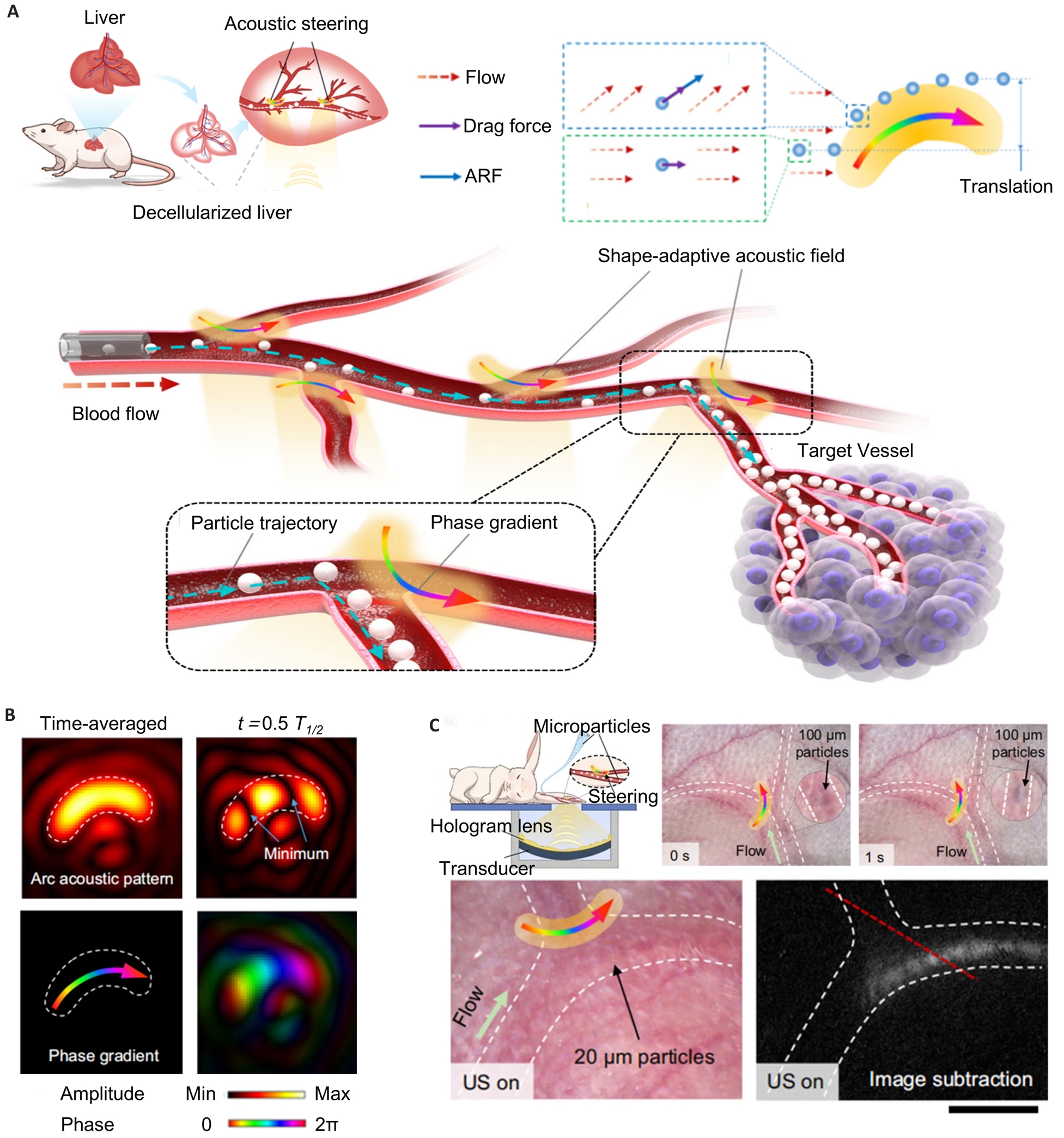
Fig.3 Sonodynamic embolization microspheres. A: Schematic diagram of the sonodynamic embolization process of sonodynamic embolization microspheres. B: Transient air pressure diagrams at different time points inside simulated blood vessels (T1/2 represents half of the acoustic cycle). C: Embolization process of sonodynamic embolization microspheres in the rabbit ear artery under ultrasonic guidance: movement trajectories of 100 μm and 20 μm microspheres during acoustic steering. Image subtraction reveals microsphere size distribution. Red dotted lines indicate the positions for grayscale analysis (The black arrows indicate microspheres, and the multicolored arrows denote acoustic pressure phase gradient). Scale bar=100 μm.
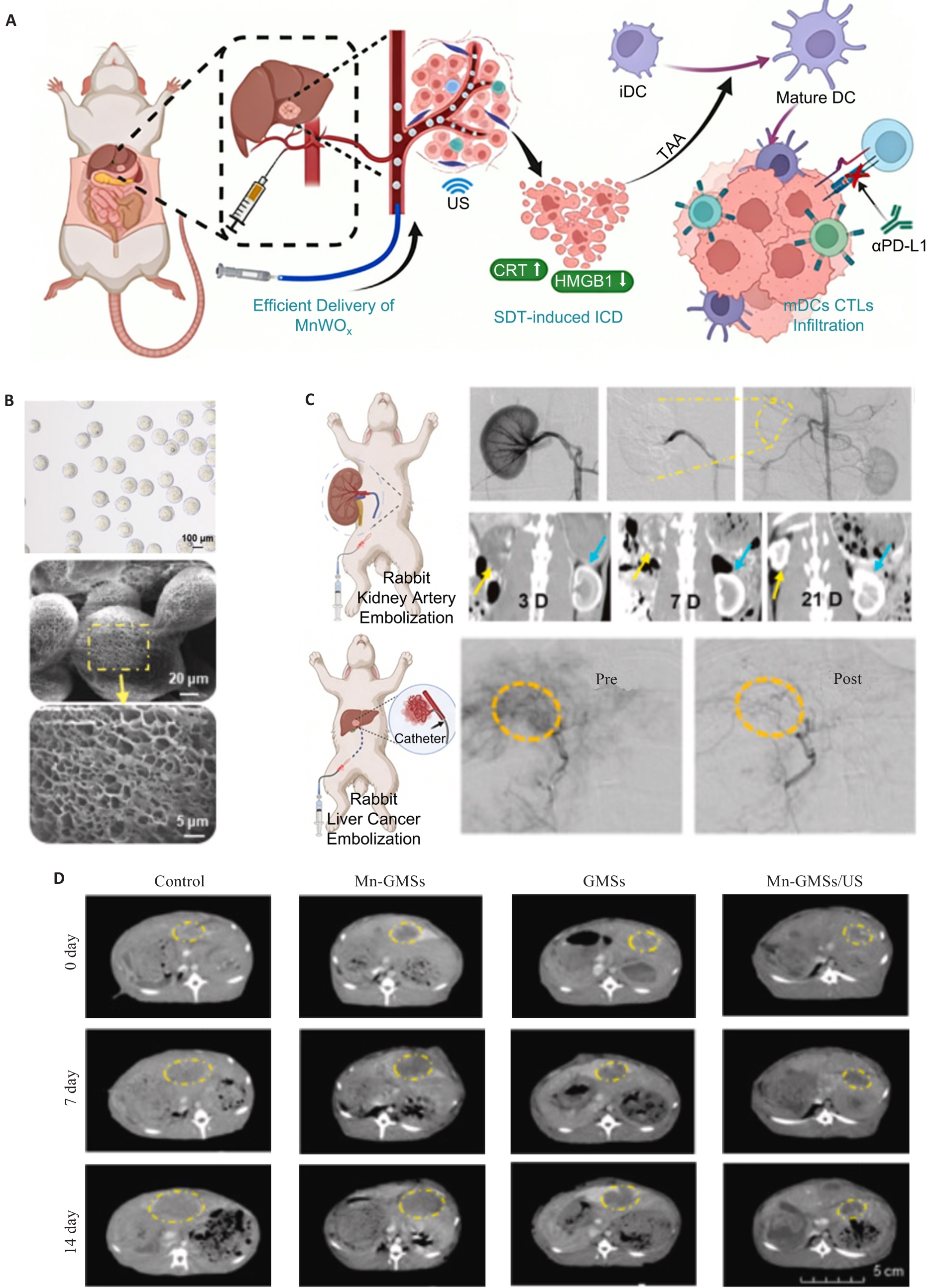
Fig.4 Sonodynamic immunological-embolization microspheres. A: Schematic illustration of combined sonodynamic immunotherapy and embolization using gelatin microspheres (Mn-GMSs) loaded with hypoxia-responsive manganese tungstate (MnWOₓ) sonosensitizer nanoparticles. B: Optical microscopy (OM) and scanning electron microscopy images of Mn-GMSs microspheres. C: DSA images of rabbit renal artery and hepatocellular carcinoma blood vessels before and after embolization with Mn-GMSs (The yellow dotted lines indicate the embolized region). CT images of kidneys acquired on days 3, 7, and 21 post-renal artery embolization (blue arrow: right kidney, yellow arrow: left kidney). D: CT images evaluating hepatocellular carcinoma tumor changes on days 0, 7, and 14 after treatment in the control group, GMSs group, Mn-GMSs group, and Mn-GMSs/US group (The yellow dotted lines indicate the tumor).
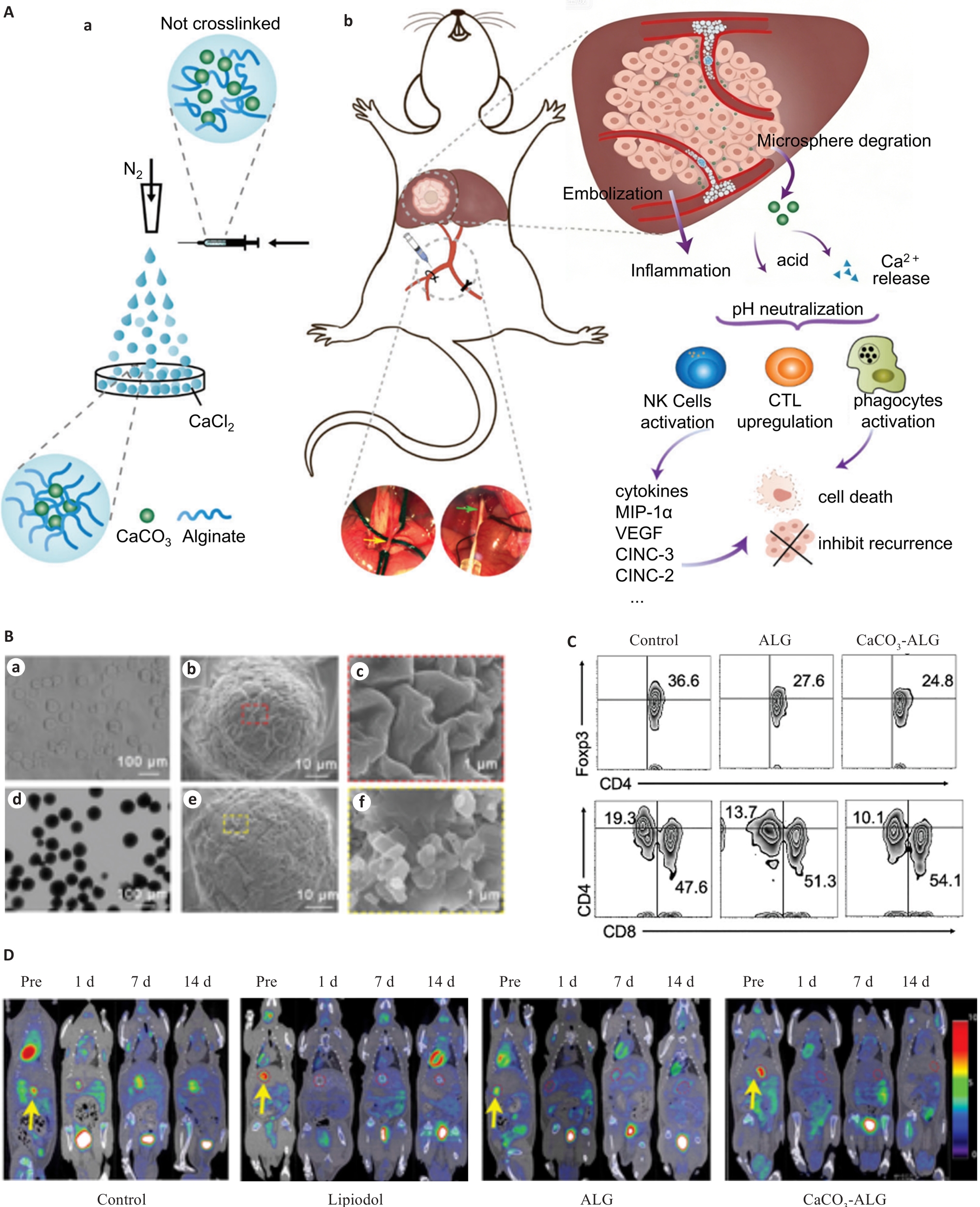
Fig.5 pH-responsive embolization microspheres. A: Schematic diagram and mechanism of pH-responsive calcium carbonate-alginate (CaCO₃-ALG) embolization microspheres for hepatocellular carcinoma embolization in SD rats. The puncture site is upstream of the ligation point at the distal gastroduodenal artery (yellow arrow). Microspheres are injected via the catheter placed in the hepatic artery (green arrow). B: Characterization of CaCO₃-ALG microspheres. a-c) Optical and scanning electron microscopic images of ALG and CaCO₃-ALG microspheres; d-f) Optical and scanning electron microscopic images of CaCO₃-ALG microspheres. C: Changes in the counts of regulatory T cells (Treg) and CD4⁺ T cells in the tumor microenvironment after embolization with ALG and CaCO₃-ALG microspheres in SD rat liver tumors. D: PET imaging evaluation of tumor changes in SD rat liver cancer after treatment with lipiodol, ALG microspheres, and CaCO₃-ALG microspheres (The yellow arrows indicate the tumor)
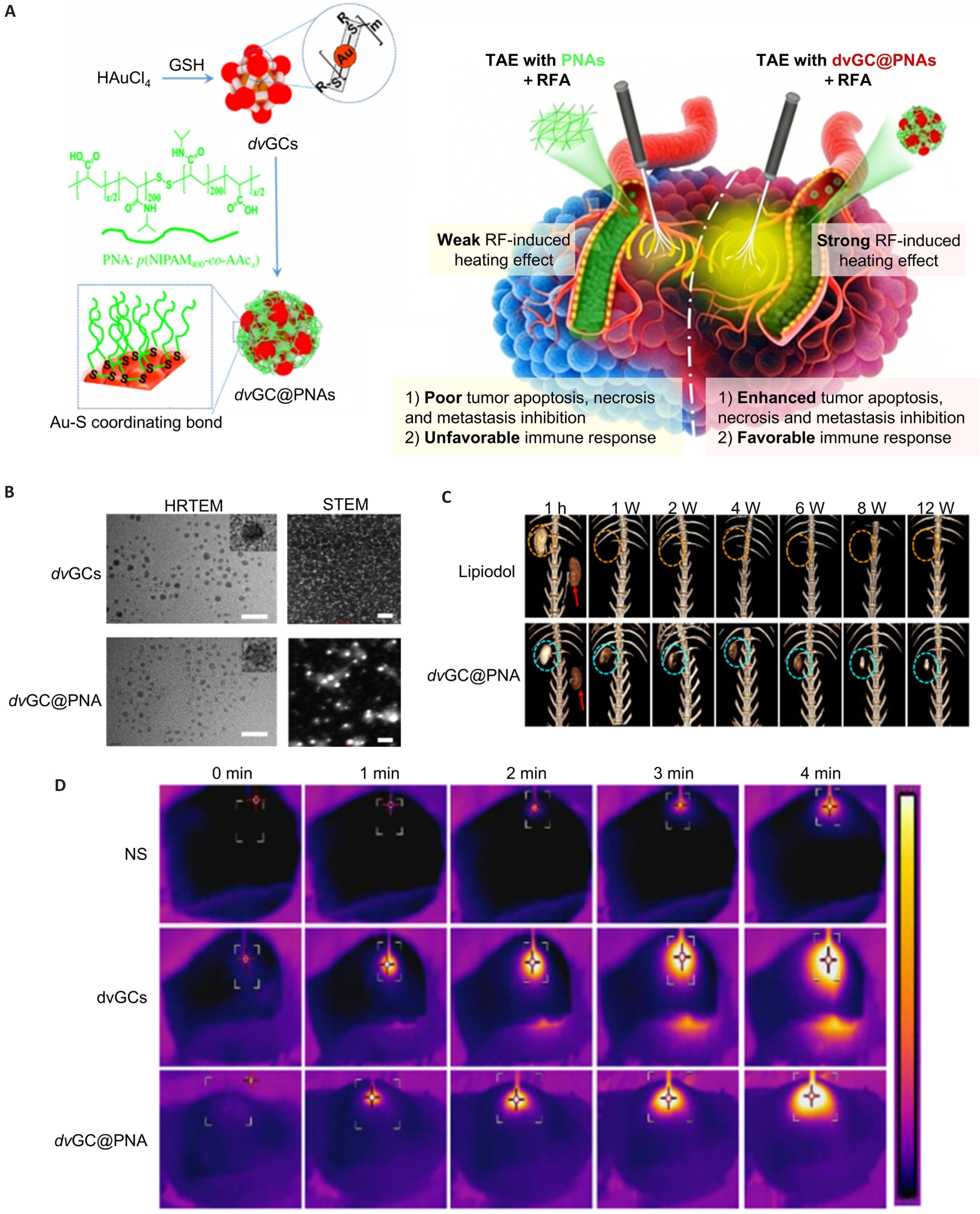
Fig.6 Radiofrequency-responsive embolization microspheres. A: Schematic illustration of the preparation of divalent gold colloidal nanoparticle microspheres (dvGC@PNAs) and their synergistic anti-tumor therapy combining radiofrequency ablation (RFA) and transarterial embolization (TAE). B: High-resolution transmission electron microscope images of dvGC particles and dvGC@PNAs embolization microspheres. C: Three-dimensional CT reconstructed images of rabbit renal arteries at different time points after embolization in the Lipiodol group and dvGC@PNAs group. D: Ex vivo thermal imaging comparison of porcine liver tissues in the NS group, dvGCs group, and dvGC@PNAs group at 0, 1, 2, 3, and 4 minutes under radiofrequency current stimulation (0.8 mmol Au/mL, 100 W, 4.0 min).
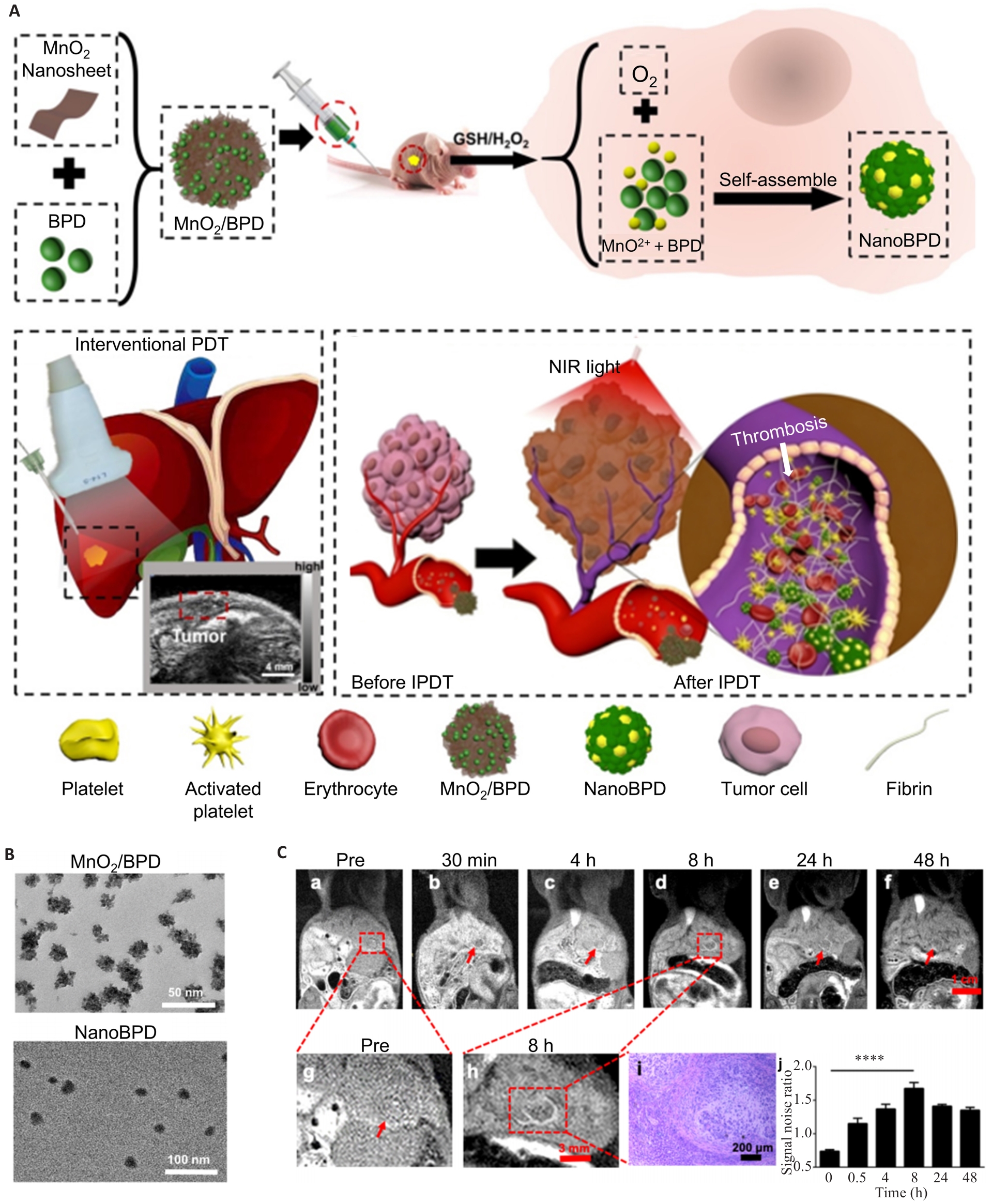
Fig.7 Photoresponsive embolization microspheres. A: Schematic preparation of MnO₂/verteporfin (BPD)-based embolization microspheres (NanoBPD) and their application in liver cancer embolization (The white arrows indicate thrombosis). B: Transmission electron microscopy (TEM) images of MnO₂/BPD and NanoBPD embolization microspheres. C: Tumor changes in mice at different time points after NanoBPD embolization. a-i) MRI observations of tumor evolution at different times post-embolization (The red frames and the arrows indicate the tumor; magnified details and corresponding pathological images are presented). j) Magnetic resonance signal-to-noise ratio (SNR) of tumor and normal liver tissues at 0, 0.5, 4, 8, 24, 48h after NanoBPD embolization (P<0.0001).
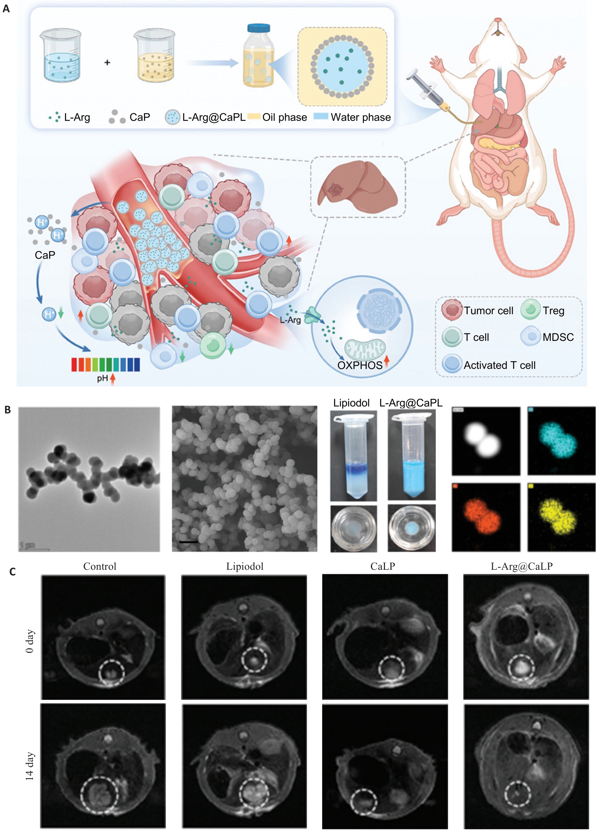
Fig.8 Pickering emulsion embolization system. A: Preparation principle of calcium phosphate nanoparticle-based Pickering emulsion microspheres loaded with L-arginine (L-Arg@CaPL) and schematic diagram of liver cancer embolization in SD rats. B: TEM and SEM images of calcium phosphate nanoparticles, photograph of L-Arg@CaPL emulsion, and elemental distribution of Ca (blue), P (red), and O (yellow) in microspheres. Scale bar: 1 μm. C: MRI-based evaluation of tumor changes in SD rat liver cancer on day 0 and day 14 after embolization with Lipiodol, CaLP, and L-Arg@CaPL (The white dotted lines indicate the tumor).
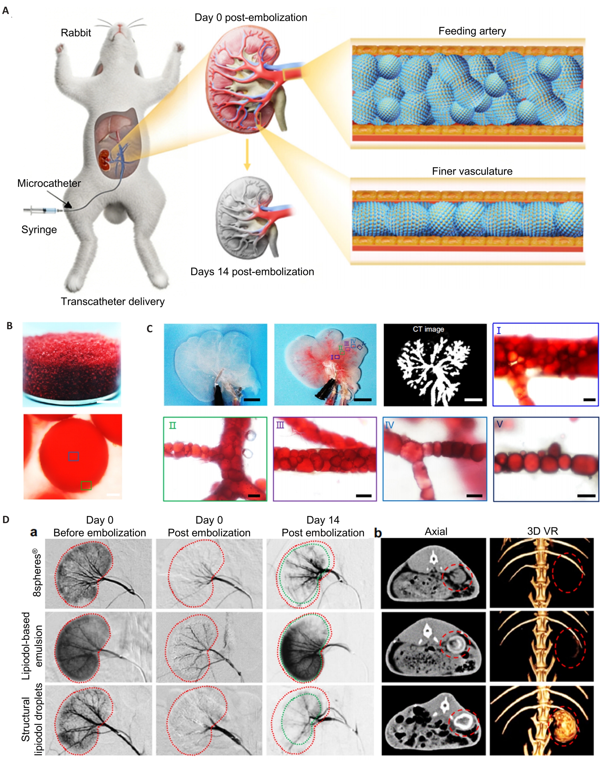
Fig.9 Janus particle-engineered structural lipiodol droplets embolization system. A: Schematic diagram of embolization with Janus particle-based lipiodol droplet microspheres. B: Photographs (upper) and optical microscopic images (lower) of Janus particle-based lipiodol droplet microspheres. C: Distribution of Janus particle-based lipiodol droplet microspheres in decellularized liver blood vessels of SD rats. D: Evaluation of renal artery embolization efficacy in rabbits. a) In vivo DSA images of rabbit renal arteries before embolization and on day 14 after treatment with 8spheres®, lipiodol, and Janus particle-based lipiodol droplet microspheres (The red dotted lines indicate renal outline before embolization, and the green dotted lines indicate renal outline after embolization). b) DSA images showing the embolization effects of the three groups on day 14 post-operation. The red dotted lines indicate the pre-embolization renal outline.
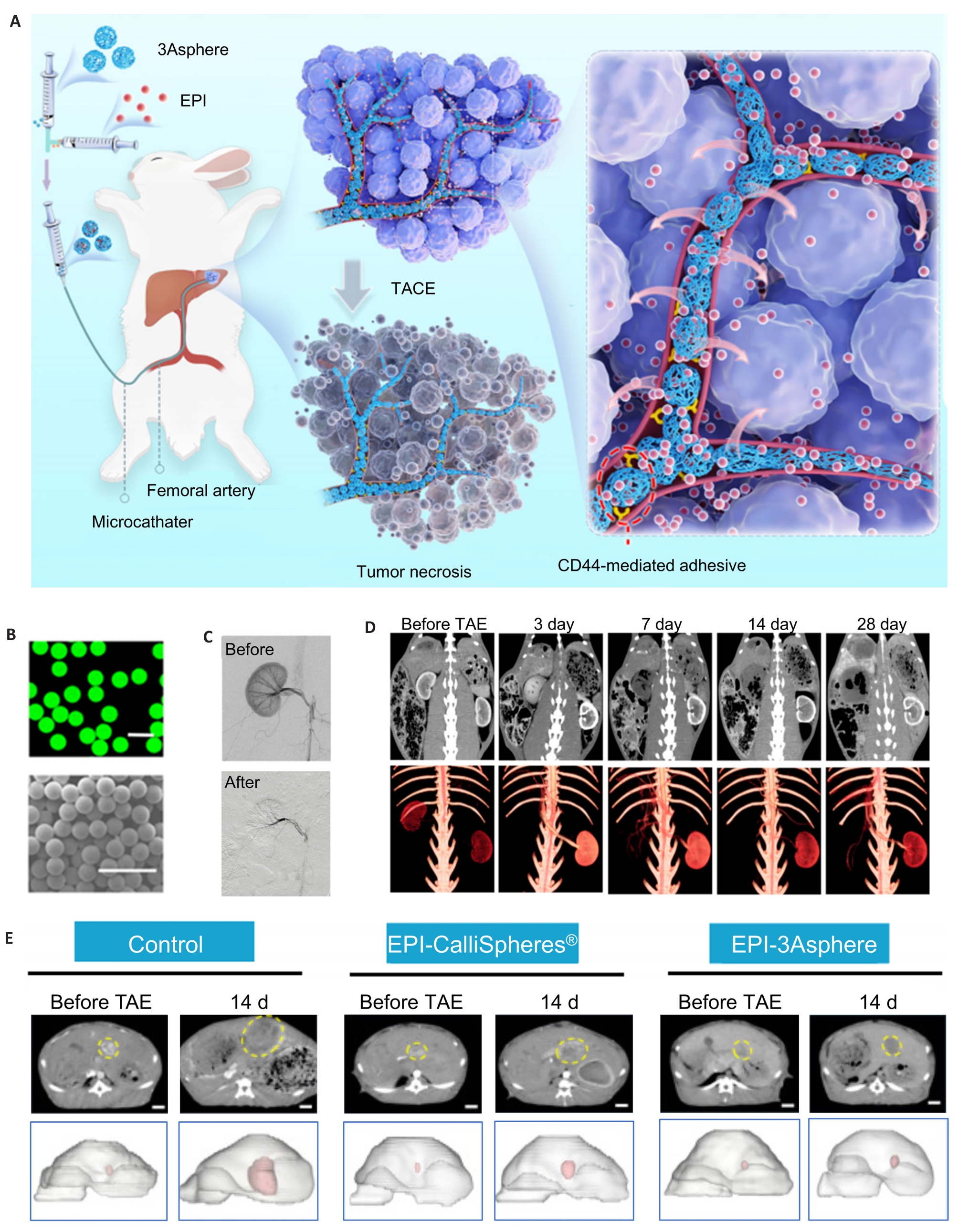
Fig.10 Tumor vessel-adaptive, adhesive, and absorbable (3Asphere) embolization microspheres. A: Schematic diagram of rabbit liver cancer embolization with hyaluronic acid derivative-based 3Asphere microspheres. B: Fluorescence and scanning electron microscopic images of 3Asphere microspheres. Scale bar: 100 μm. C: Renal artery embolization effect of 3Asphere microspheres in rabbits observed via DSA imaging. D: Imaging evaluation (upper) and 3D reconstruction (lower) of rabbit renal arteries before embolization and on days 3, 7, 14, and 28 after 3Asphere embolization under CT scanning. E: CT imaging assessment of tumor changes in rabbit liver cancer before embolization and on day 14 post treatment with EPI-CalliSpheres® and EPI 3Asphere microspheres (The yellow dotted lines indicate the tumor).
| [1] | Pesapane F, Nezami N, Patella F, et al. New concepts in embolotherapy of HCC[J]. Med Oncol, 2017, 34(4): 58. doi:10.1007/s12032-017-0917-2 |
| [2] | Shao GL, Zou YH, Lucatelli P, et al. Chinese expert consensus on technical recommendations for the standard operation of drug-eluting beads for transvascular embolization[J]. Ann Transl Med, 2021, 9(8): 714. doi:10.21037/atm-21-1678 |
| [3] | Rao AK, Sobel J, Chatham-Stephens K, et al. Clinical guidelines for diagnosis and treatment of botulism, 2021[J]. MMWR Recomm Rep, 2021, 70(2): 1-30. doi:10.15585/mmwr.rr7002a1 |
| [4] | Yuan HJ, Liu FY, Li X, et al. Application of nanomedicines in interventional targeted therapy for hepatocellular carcinoma[J]. Chin J Hepatob Surg, 2018, 24(6): 427-30. |
| [5] | Sheth RA, Sabir S, Krishnamurthy S, et al. Endovascular embolization by transcatheter delivery of particles: past, present, and future[J]. J Funct Biomater, 2017, 8(2): 12. doi:10.3390/jfb8020012 |
| [6] | Llovet JM, Real MI, Montaña X, et al. Arterial embolisation or chemoembolisation versus symptomatic treatment in patients with unresectable hepatocellular carcinoma: a randomised controlled trial[J]. Lancet, 2002, 359(9319): 1734-9. doi:10.1016/s0140-6736(02)08649-x |
| [7] | Zeng J, He DF. Research progress on interventional embolic materials in liver cancer treatment[J]. Chin J Interven Radiol, 2023, 11(1): 62-7. |
| [8] | Cloft HJ, Kallmes DF. Aneurysm packing with HydroCoil Embolic System versus platinum coils: initial clinical experience[J]. Am J Neuroradiol, 2004, 25(1): 60-2. |
| [9] | Peng C, Diao YH, Cai SF, et al. Endovascular coiling versus microsurgical clipping for ruptured intracranial aneurysms: a meta-analysis and systematic review[J]. Chin Neurosurg J, 2022, 8(1): 17. doi:10.1186/s41016-022-00283-3 |
| [10] | Chiesa AG, Hart WR. Uterine artery embolization of leiomyomas with trisacryl gelatin microspheres (TGM): pathologic features and comparison with polyvinyl alcohol emboli[J]. Int J Gynecol Pathol, 2004, 23(4): 386-92. doi:10.1097/01.pgp.0000139663.13539.e1 |
| [11] | Scullen T, Mathkour M, Nerva JD, et al. Commentary: middle meningeal artery embolization for chronic subdural hematoma: a multi-center experience of 154 consecutive embolizations[J]. Neurosurgery, 2021, 88(2): E130-2. doi:10.1093/neuros/nyaa432 |
| [12] | Molyneux AJ, Kerr RSC, Yu LY. International subarachnoid aneurysm trial (ISAT) of neurosurgical clipping versus endovascular coiling in 2143 patients with ruptured intracranial aneurysms: a randomised comparison of effects on survival, dependency, seizures, rebleeding, subgroups, and aneurysm occlusion[J]. ACC Curr J Rev, 2005, 14(12): 52. doi:10.1016/j.accreview.2005.11.083 |
| [13] | Xiao YD, Ma C, Zhang ZS, et al. Safety and efficacy assessment of transarterial chemoembolization using drug-eluting beads in patients with hepatocellular carcinoma and arterioportal shunt: a single-center experience[J]. Cancer Manag Res, 2019, 11: 1551-7. doi:10.2147/cmar.s193948 |
| [14] | Gao YN, Li ZH, Hong Y, et al. Decellularized liver as a translucent ex vivo model for vascular embolization evaluation[J]. Biomaterials, 2020, 240: 119855. doi:10.1016/j.biomaterials.2020.119855 |
| [15] | Facciorusso A. Drug-eluting beads transarterial chemoembolization for hepatocellular carcinoma: Current state of the art[J]. World J Gastroenterol, 2018, 24(2): 161-9. doi:10.3748/wjg.v24.i2.161 |
| [16] | Chang PY, Huang CC, Hung CH, et al. Multidisciplinary Taiwan consensus recommendations for the use of DEBDOX-TACE in hepatocellular carcinoma treatment[J]. Liver Cancer, 2018, 7(4): 312-22. doi:10.1159/000487608 |
| [17] | Zhu ZM, Chen YY, Yang YJ, et al. Research progress on the application of X-ray visualizing microspheres in TACE for hepatocellular carcinoma[J]. Chin J Hepatob Surg, 2025, 31(11): 863-7. |
| [18] | Dong H, Yang DL, Hu YL, et al. Recent advances in smart nanoplatforms for tumor non-interventional embolization therapy[J]. J Nanobiotechnol, 2022, 20(1): 337. doi:10.1186/s12951-022-01548-w |
| [19] | Liu L, Liang XX, Xu XX, et al. Magnetic mesoporous embolic microspheres in transcatheter arterial chemoembolization for liver cancer[J]. SSRN Journal, 2020,130: 374-84. doi:10.1016/j.actbio.2021.05.031 |
| [20] | Virdis F, Reccia I, Di Saverio S, et al. Clinical outcomes of primary arterial embolization in severe hepatic trauma: a systematic review[J]. Diagn Interv Imag, 2019, 100(2): 65-75. doi:10.1016/j.diii.2018.10.004 |
| [21] | Ramaswamy RS, Akinwande O, Tiwari T. Renal embolization: current recommendations and rationale for clinical practice[J]. Curr Urol Rep, 2018, 19(3): 5. doi:10.1007/s11934-018-0756-5 |
| [22] | Huang XQ. Severe biliary complications after hepatic artery embolization[J]. World J Gastroenterol, 2002, 8(1): 119. doi:10.3748/wjg.v8.i1.119 |
| [23] | Luo YC, Yin Q, Chen KK, et al. Superselective embolic particle guidance in vessel networks via shape-adaptive acoustic manipulation[J]. Nat Commun, 2025, 16: 254. doi:10.1038/s41467-024-55478-2 |
| [24] | Athanassiadis AG, Ma ZC, Moreno-Gomez N, et al. Ultrasound-responsive systems as components for smart materials[J]. Chem Rev, 2022, 122(5): 5165-208. doi:10.1021/acs.chemrev.1c00622 |
| [25] | Xu JC, Pei ZF, Wang YJ, et al. Bioactive microspheres to enhance sonodynamic-embolization-metalloimmune therapy for orthotopic liver cancer[J]. Biomaterials, 2025, 317: 123063. doi:10.1016/j.biomaterials.2024.123063 |
| [26] | Tian SJ, Li HY, Niu YX, et al. Photopolymerized pH-responsive GelMA microspheres with doxorubicin modification for embolization therapy[J]. J Drug Deliv Sci Technol, 2025, 114: 107446. doi:10.1016/j.jddst.2025.107446 |
| [27] | Zhang AM, Xiao ZS, Liu QF, et al. CaCO3-encapuslated microspheres for enhanced transhepatic arterial embolization treatment of hepatocellular carcinoma[J]. Adv Healthc Mater, 2021, 10(19): 2100748. doi:10.1002/adhm.202100748 |
| [28] | Zhu YJ, Yang ZJ, Dong ZL, et al. CaCO3-assisted preparation of pH-responsive immune-modulating nanoparticles for augmented chemo-immunotherapy[J]. Nano Micro Lett, 2020, 13(1): 29. doi:10.1007/s40820-020-00549-4 |
| [29] | Sato Y, Hasegawa T, Chatani S, et al. Percutaneous radiofrequency ablation for liver tumors: technical tips[J]. Interv Radiol, 2020, 5(2): 50-7. doi:10.22575/interventionalradiology.2020-0008 |
| [30] | Mertyna P, Hines-Peralta A, Liu ZJ, et al. Radiofrequency ablation: variability in heat sensitivity in tumors and tissues[J]. J Vasc Interv Radiol, 2007, 18(5): 647-54. doi:10.1016/j.jvir.2007.02.033 |
| [31] | Zhao M, Wang JP, Pan CC, et al. CT-guided radiofrequency ablation after with transarterial chemoembolization in treating unresectable hepatocellular carcinoma with long overall survival improvement[J]. Eur J Radiol, 2012, 81(10): 2717-25. doi:10.1016/j.ejrad.2011.10.023 |
| [32] | Llovet JM, Vilana R, Brú C, et al. Increased risk of tumor seeding after percutaneous radiofrequency ablation for single hepatocellular carcinoma[J]. Hepatology, 2001, 33(5): 1124-9. doi:10.1053/jhep.2001.24233 |
| [33] | Li L, Guo XP, Peng XL, et al. Radiofrequency-responsive dual-valent gold nanoclusters for enhancing synergistic therapy of tumor ablation and artery embolization[J]. Nano Today, 2020, 35: 100934. doi:10.1016/j.nantod.2020.100934 |
| [34] | Luo ZT, Yuan X, Yu Y, et al. From aggregation-induced emission of Au(I)-thiolate complexes to ultrabright Au(0)@Au(I)-thiolate core-shell nanoclusters[J]. J Am Chem Soc, 2012, 134(40): 16662-70. doi:10.1021/ja306199p |
| [35] | Ji AQ, Yang PJ, Ma B, et al. Photothermal-responsive curcumin-loaded copper-based nanocomposites for targeted drug release and combined immunotherapy[J]. J Colloid Interface Sci, 2025, 698: 138042. doi:10.1016/j.jcis.2025.138042 |
| [36] | Gore S, Are V, Chanchlani B, et al. Multifunctional nanoplatforms for combined photothermal and photodynamic therapy: Tumor-responsive strategies for enhanced precision[J]. Int J Pharm, 2025, 684: 126131. doi:10.1016/j.ijpharm.2025.126131 |
| [37] | Wang YQ, Shang WT, Zhong HS, et al. Tumor vessel targeted self-assemble nanoparticles for amplification and prediction of the embolization effect in hepatocellular carcinoma[J]. ACS Nano, 2020, 14(11): 14907-18. doi:10.1021/acsnano.0c04341 |
| [38] | Ethirajan M, Chen YH, Joshi P, et al. The role of porphyrin chemistry in tumor imaging and photodynamic therapy[J]. Chem Soc Rev, 2011, 40(1): 340-62. doi:10.1039/b915149b |
| [39] | Scott LJ, Goa KL. Verteporfin[J]. Drugs Aging, 2000, 16(2): 139-46. doi:10.2165/00002512-200016020-00005 |
| [40] | Vander Heiden MG, Cantley LC, Thompson CB. Understanding the Warburg effect: the metabolic requirements of cell proliferation[J]. Science, 2009, 324(5930): 1029-33. doi:10.1126/science.1160809 |
| [41] | Tran A, Moon JT, Shaikh J, et al. Acetazolamide enhanced drug-eluting beads: manipulating the hepatocellular carcinoma microenvironment[J]. Minim Invasive Ther Allied Technol, 2022, 31(6): 973-7. doi:10.1080/13645706.2022.2040535 |
| [42] | Lv YF, Deng ZQ, et al. Intratumoral Pi deprivation benefits chemoembolization therapy via increased accumulation of intracellular doxorubicin[J]. Drug Deliv, 2022, 29(1): 1743-53. doi:10.1080/10717544.2022.2081384 |
| [43] | Chen MJ, Guo XJ, Shen L, et al. Monodisperse CaCO3-loaded gelatin microspheres for reversing lactic acid-induced chemotherapy resistance during TACE treatment[J]. Int J Biol Macromol, 2023, 231: 123160. doi:10.1016/j.ijbiomac.2023.123160 |
| [44] | Razi M, Gu JP, He X, et al. Conventional versus drug-eluting bead transarterial chemoembolization: a better option for treatment of unresectable hepatocellular carcinoma[J]. J Interv Med, 2021, 4(1): 11-4. doi:10.1016/j.jimed.2020.10.006 |
| [45] | Deschamps F, Farouil G, Gonzalez W, et al. Stabilization improves theranostic properties of lipiodol®‑based emulsion during liver trans-arterial chemo-embolization in a VX2 rabbit model[J]. CardioVascular Interv Radiol, 2017, 40(6): 907-13. doi:10.1007/s00270-017-1616-2 |
| [46] | Deschamps F, Isoardo T, Denis S, et al. Biodegradable Pickering emulsions of Lipiodol for liver trans-arterial chemo-embolization[J]. Acta Biomater, 2019, 87: 177-86. doi:10.1016/j.actbio.2019.01.054 |
| [47] | Li H, Qian K, Zhang HS, et al. Pickering gel emulsion of lipiodol stabilized by hairy nanogels for intra-artery embolization antitumor therapy[J]. Chem Eng J, 2021, 418: 129534. doi:10.1016/j.cej.2021.129534 |
| [48] | Wang D, Zhang L, Yang WH, et al. Arginine-loaded nano-calcium-phosphate-stabilized lipiodol Pickering emulsions potentiates transarterial embolization-immunotherapy[J]. Adv Sci, 2025, 12(6): 2410484. doi:10.1002/advs.202410484 |
| [49] | Tao SJ, Lin BQ, Zhou HW, et al. Janus particle-engineered structural lipiodol droplets for arterial embolization[J]. Nat Commun, 2023, 14: 5575. doi:10.1038/s41467-023-41322-6 |
| [50] | Haney B, Chen D, Cai LH, et al. Millimeter-size Pickering emulsions stabilized with Janus microparticles[J]. Langmuir, 2019, 35(13): 4693-701. doi:10.1021/acs.langmuir.9b00058 |
| [51] | Zhang X, Fu QR, Duan HW, et al. Janus nanoparticles: from fabrication to (bio)applications[J]. ACS Nano, 2021, 15(4): 6147-91. doi:10.1021/acsnano.1c01146 |
| [52] | Zhu YJ, Yang ZJ, Pan ZJ, et al. Metallo-alginate hydrogel can potentiate microwave tumor ablation for synergistic cancer treatment[J]. Sci Adv, 2022, 8(31): eabo5285. doi:10.1126/sciadv.abo5285 |
| [53] | Xian J, University HN, et al. Elemene hydrogel modulates the tumor immune microenvironment for enhanced treatment of postoperative cancer recurrence and metastases[J]. J Am Chem Soc, 2024, 146(51): 35252-63. doi:10.1021/jacs.4c12531 |
| [54] | Nam J, Kim A, Kim K, et al. Engineered polysaccharides for controlling innate and adaptive immune responses[J]. Nat Rev Bioeng, 2024, 2(9): 733-51. doi:10.1038/s44222-024-00193-2 |
| [55] | Guo JK, Huang JT, Huang ZL, et al. Tumor vessel-adaptable adhesive and absorbable microspheres for sustainable transarterial chemoembolization therapy[J]. Nat Commun, 2025, 16: 6239. doi:10.1038/s41467-025-61621-4 |
| [56] | Zheng DW, Deng WW, Song WF, et al. Biomaterial-mediated modulation of oral microbiota synergizes with PD-1 blockade in mice with oral squamous cell carcinoma[J]. Nat Biomed Eng, 2022, 6(1): 32-43. doi:10.1038/s41551-021-00807-9 |
| [57] | Su Y, Zhang BL, Sun RW, et al. PLGA-based biodegradable microspheres in drug delivery: recent advances in research and application[J]. Drug Deliv, 2021, 28(1): 1397-418. doi:10.1080/10717544.2021.1938756 |
| [58] | Jeon SI, Kim MS, Kim HJ, et al. Biodegradable poly(lactide-co-glycolide) microspheres encapsulating hydrophobic contrast agents for transarterial chemoembolization[J]. J Biomater Sci Polym Ed, 2022, 33(4): 409-25. doi:10.1080/09205063.2021.1990472 |
| [59] | Owen J, Negussie AH, Burks SR, et al. Microbubbles bound to drug-eluting beads enable ultrasound imaging and enhanced delivery of therapeutics[J]. Sci Rep, 2024, 14: 20929. doi:10.1038/s41598-024-71831-3 |
| [60] | Moschovaki-Zeiger O, Arkoudis NA, Giannakis A, et al. Biodegradable microspheres for transarterial chemoembolization in malignant liver disease[J]. Medicina, 2024, 60(4): 678. doi:10.3390/medicina60040678 |
| No related articles found! |
| Viewed | ||||||
|
Full text |
|
|||||
|
Abstract |
|
|||||
