
Journal of Southern Medical University ›› 2026, Vol. 46 ›› Issue (10): 2482-2493.doi: 10.12122/j.issn.1673-4254.2026.10.17
Guannan ZHANG1( ), Xiang LI2, Yuan MI2, Danyang TIAN2, Jinqi MENG3()
Received:2026-03-06
Online:2026-10-20
Published:2026-10-26
Contact:
Jinqi MENG
E-mail:13483111223@163.com;MengJinqi@hebmu.edu.cn
Supported by:Guannan ZHANG, Xiang LI, Yuan MI, Danyang TIAN, Jinqi MENG. Aerobic exercise improves metabolic disorders and multi-organ damage in diabetic rats by activating the Sirt1/Creb1/Ppargc1a axis[J]. Journal of Southern Medical University, 2026, 46(10): 2482-2493.
Add to citation manager EndNote|Ris|BibTeX
URL: https://www.j-smu.com/EN/10.12122/j.issn.1673-4254.2026.10.17
| Primer | Sequence |
|---|---|
| Rat-Btubulin-F1 | TAGCAGCGTTGCTGGGTATC |
| Rat-Btubulin-R1 | GACTGGAATGCCTCGCTCTT |
| Rat-Sirt1-F1 | TCAGAACCACCAAAGCGGAA |
| Rat-Sirt1-R1 | GCAAGGCGAGCATAAATACCA |
| Rat-Creb1-F1 | TCCTCCCCAGCACTTCCTAC |
| Rat-Creb1-R1 | CCAATCCAGTCCATTTTCCA |
| Rat-Ppargc1a-F2 | AACAGAAACAGCAGCAGAGACAA |
| Rat-Ppargc1a-R2 | GCAGTTCCAGAGAGTTCCACAC |
Tab.1 Primer sequences for qPCR of the target genes
| Primer | Sequence |
|---|---|
| Rat-Btubulin-F1 | TAGCAGCGTTGCTGGGTATC |
| Rat-Btubulin-R1 | GACTGGAATGCCTCGCTCTT |
| Rat-Sirt1-F1 | TCAGAACCACCAAAGCGGAA |
| Rat-Sirt1-R1 | GCAAGGCGAGCATAAATACCA |
| Rat-Creb1-F1 | TCCTCCCCAGCACTTCCTAC |
| Rat-Creb1-R1 | CCAATCCAGTCCATTTTCCA |
| Rat-Ppargc1a-F2 | AACAGAAACAGCAGCAGAGACAA |
| Rat-Ppargc1a-R2 | GCAGTTCCAGAGAGTTCCACAC |
| Variables | Blank control group | Diabetes control group | Diabetes aerobic group | Diabetes exhaustion group |
|---|---|---|---|---|
| Weight (g) | 328.90±10.73 | 379.50±11.88** | 350.10±12.68## | 296.60±14.16## |
| Liver wet weight (g) | 14.22±1.52 | 26.49±2.64** | 19.26±2.49## | 29.60±3.12# |
| Liver index (%) | 4.33±0.51 | 6.99±0.75** | 5.50±0.64## | 9.97±0.78## |
Tab.2 Changes in body weight, liver wet weight, and liver index of the rats in each group (Mean±SD, n=10)
| Variables | Blank control group | Diabetes control group | Diabetes aerobic group | Diabetes exhaustion group |
|---|---|---|---|---|
| Weight (g) | 328.90±10.73 | 379.50±11.88** | 350.10±12.68## | 296.60±14.16## |
| Liver wet weight (g) | 14.22±1.52 | 26.49±2.64** | 19.26±2.49## | 29.60±3.12# |
| Liver index (%) | 4.33±0.51 | 6.99±0.75** | 5.50±0.64## | 9.97±0.78## |
| Variables | Blank control group | Diabetes control group | Diabetes aerobic group | Diabetes exhaustion group |
|---|---|---|---|---|
| FBG (mmol/L) | 6.24±0.72 | 13.09±1.06** | 8.11±0.85## | 10.29±0.72## |
| Lactic acid (mmol/L) | 2.55±0.39 | 3.97±0.50** | 3.05±0.46## | 6.46±0.65## |
| Pyruvic acid (mmol/L) | 0.42±0.05 | 0.82±0.11** | 0.61±0.10## | 1.11±0.17## |
| Creatinine (mg/dL) | 0.61±0.08 | 1.55±0.16** | 0.82±0.13## | 2.35±0.27## |
| Urea nitrogen (mmol/L) | 3.70±0.28 | 6.19±0.98** | 4.73±0.65## | 7.46±0.97# |
| Creatine kinase (U/L) | 115.70±24.00 | 305.30±30.42** | 191.30±24.41## | 508.90±58.60## |
Tab. 3 Changes in fasting blood glucose, blood lactate, pyruvate, creatinine, blood urea nitrogen, and creatine kinase levels in each group of the rats (Mean±SD, n=10)
| Variables | Blank control group | Diabetes control group | Diabetes aerobic group | Diabetes exhaustion group |
|---|---|---|---|---|
| FBG (mmol/L) | 6.24±0.72 | 13.09±1.06** | 8.11±0.85## | 10.29±0.72## |
| Lactic acid (mmol/L) | 2.55±0.39 | 3.97±0.50** | 3.05±0.46## | 6.46±0.65## |
| Pyruvic acid (mmol/L) | 0.42±0.05 | 0.82±0.11** | 0.61±0.10## | 1.11±0.17## |
| Creatinine (mg/dL) | 0.61±0.08 | 1.55±0.16** | 0.82±0.13## | 2.35±0.27## |
| Urea nitrogen (mmol/L) | 3.70±0.28 | 6.19±0.98** | 4.73±0.65## | 7.46±0.97# |
| Creatine kinase (U/L) | 115.70±24.00 | 305.30±30.42** | 191.30±24.41## | 508.90±58.60## |
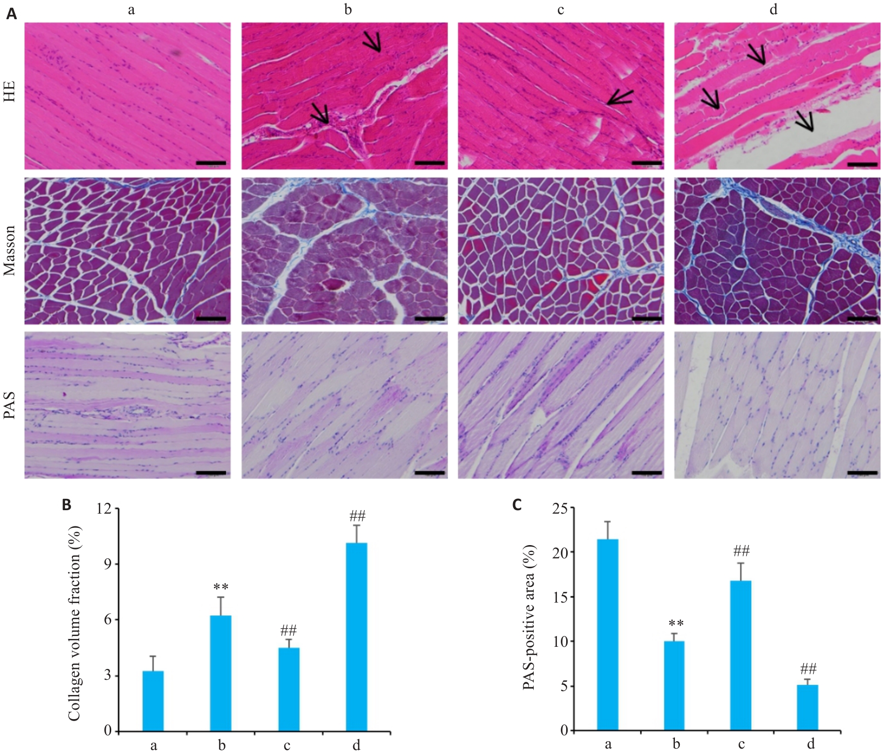
Fig.1 Histopathological changes in skeletal muscles of the rats in each group. A: HE staining, Masson staining, and PAS staining of mouse skeletal muscle tissues (Scale bar=100 μm). B: Collagen volume fraction of the rats in each group. C: Percentage of PAS-positive area in each group of rats. Data are presented as Mean±SD (n=10). a: Blank control group; b: Diabetes control group; c: Diabetes aerobic group; d: Diabetes exhaustive group. Arrows indicate damaged muscle fibers, inflammatory cell infiltration, and vacuolar degeneration. **P<0.01 vs blank control group; ##P<0.01 vs the diabetes control group.
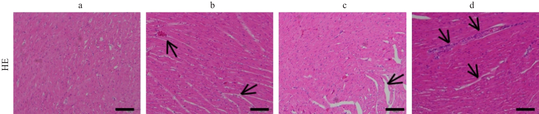
Fig.2 HE staining of myocardial tissue from rats in each group (Scale bar=100 μm). a: Blank control group; b: Diabetes control group; c: Diabetes aerobic group; d: Diabetes exhaustive group. Arrows indicate disordered and ruptured myocardial fibers, widened intercellular spaces, and necrotic areas.
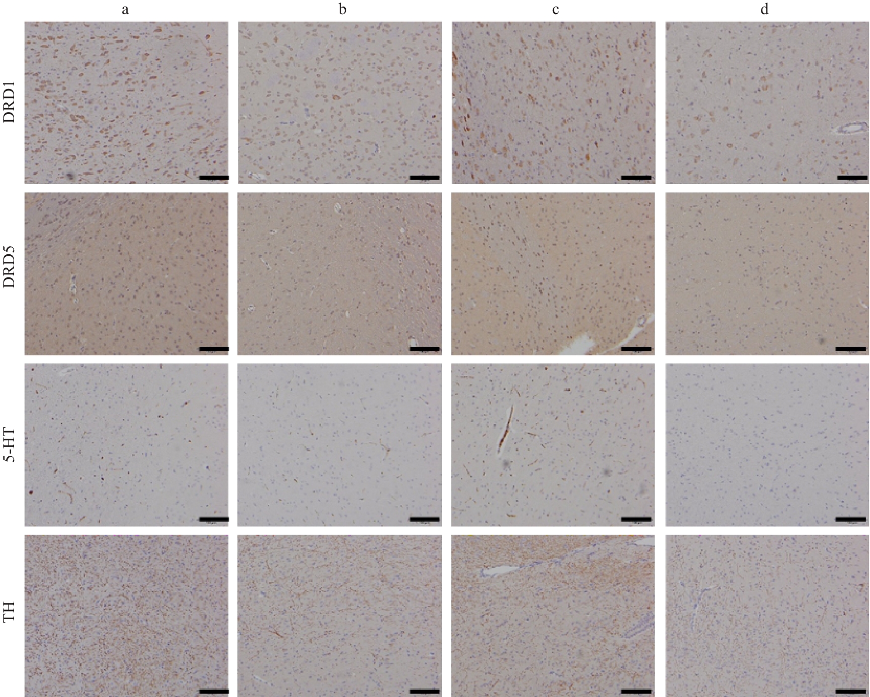
Fig.3 Expression distribution of DRD1, DRD5, 5-HT, and TH in the prefrontal cortex of rats in each group (Scale bar=100 μm. a: Blank control group; b: Diabetes control group; c: Diabetes aerobic group; d: Diabetes exhaustive group.
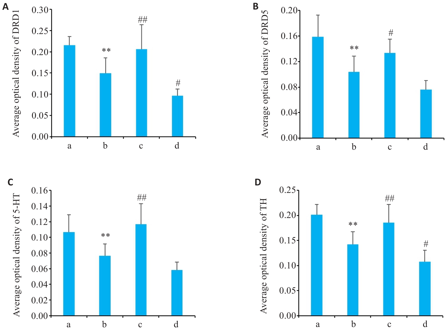
Fig.4 Average optical density of DRD1 (A), DRD5 (B), 5-HT (C) and TH (D) in the prefrontal cortex of the rats in each group (Mean±SD, n=10). **P<0.01 vs the blank control group; #P<0.05, ##P<0.01 vs the diabetes control group. a. Blank control group; b. Diabetes control group; c. Diabetes aerobic group; d. Diabetes exhaustive group.
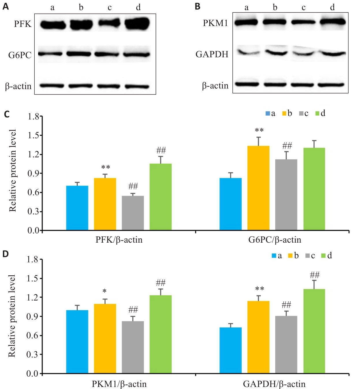
Fig.5 Protein expressions of G6PC, PFK, GAPDH and PKM1 in liver tissues of the rats in each group. A: Protein expression levels of G6PC and PFK detected by Western blotting. B: Protein expression levels of GAPDH and PKM1 detected by Western blotting. C: Expression levels of G6PC and PFK in the liver tissues of different groups. D: Expression levels of GAPDH and PKM1 in liver tissues of different groups. Data are presented as Mean±SD (n=10). *P<0.05, **P<0.01 vs the blank control group; ##P<0.01 vs the diabetes control group. a: Blank control group; b: Diabetes control group; c: Diabetes aerobic group; d: Diabetes exhaustive group.
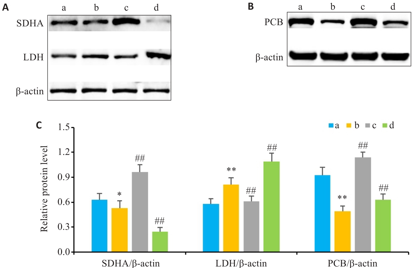
Fig.6 Protein expression of LDH, SDHA and PCB in liver tissues of rats in each group. A: Detection of protein expression levels of LDH and SDHA by Western blotting. B: Detection of protein expression levels of PCB by Western blotting. C: Expression levels of SDHA, LDH and PCB in liver tissues of different groups (Mean±SD, n=10).*P<0.05, **P<0.01 vs blank control group; ##P<0.01 vs the diabetes control group. a: Blank control group; b: Diabetes control group; c: Diabetes aerobic group; d: Diabetes exhaustive group.
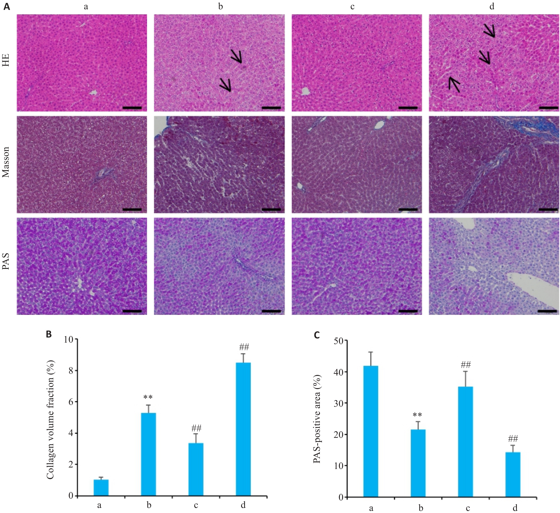
Fig.7 HE, Masson, and PAS staining (A) of liver tissues, collagen volume fraction (B) and percentage of PAS-positive area in each group (Scale bar=100 μm). Data are presented as Mean±SD (n=10). a: Blank control group; b: Diabetes control group; c: Diabetes aerobic group; d: Diabetes exhaustive group. Arrows indicate lipid vacuoles of varying sizes and inflammatory cell infiltration in hepatocytes. **P<0.01 vs blank control group; ##P<0.01 vs the diabetes control group.
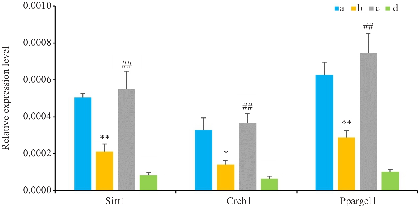
Fig.8 Changes in mRNA expression of Sirt1, Creb1, and Ppargc1a in the livers of rats in each group (Mean±SD, n=10).*P<0.05, **P<0.01 vs the blank control group; ##P<0.01 vs the diabetes control group. a: Blank control group; b: Diabetes control group; c: Diabetes aerobic group; d: Diabetes exhaustive group.
| [1] | Risk Factor CollaborationNCD. Worldwide trends in diabetes prevalence and treatment from 1990 to 2022: a pooled analysis of 1108 population-representative studies with 141 million participants[J]. Lancet, 2024, 404(10467): 2077-93. |
| [2] | Cao R, Tian HM, Zhang Y, et al. Signaling pathways and intervention for therapy of type 2 diabetes mellitus[J]. MedComm, 2023, 4(3): e283. doi:10.1002/mco2.283 |
| [3] | Zhou Z, Sun B, Yu DS, et al. Gut microbiota: an important player in type 2 diabetes mellitus[J]. Front Cell Infect Microbiol, 2022, 12: 834485. doi:10.3389/fcimb.2022.834485 |
| [4] | Dutta BJ, Singh S, Seksaria S, et al. Inside the diabetic brain: Insulin resistance and molecular mechanism associated with cognitive impairment and its possible therapeutic strategies[J]. Pharmacol Res, 2022, 182: 106358. doi:10.1016/j.phrs.2022.106358 |
| [5] | Liu RJ, Shi XF, Zhang X. The effects of resistance training on glycemic control, cardiometabolic health, and body composition in middle-aged and older women with type 2 diabetes and overweight or obesity: a systematic review and meta-analysis[J]. Maturitas, 2025, 201: 108691. doi:10.1016/j.maturitas.2025.108691 |
| [6] | Mohd S, Kumar LL, Harish V, et al. Diabetes mellitus: Complications, emerging therapeutic targets, and evolving treatment approaches[J]. Obes Med, 2025, 58: 100652. doi:10.1016/j.obmed.2025.100652 |
| [7] | Xing HM, Lu JH, Yoong SQ, et al. Effect of aerobic and resistant exercise intervention on inflammaging of type 2 diabetes mellitus in middle-aged and older adults: a systematic review and meta-analysis[J]. J Am Med Dir Assoc, 2022, 23(5): 823-30.e13. doi:10.1016/j.jamda.2022.01.055 |
| [8] | Zhao RX, Li JY, Du FB, et al. Effects of long-term moderate-intensity exercise on autonomic nervous system dysfunction induced by cardiac neurovascular interface deterioration[J]. Rev Cardiovasc Med, 2025, 26(10): 40714. doi:10.31083/rcm40714 |
| [9] | Gao HY, Zhao YL, Zhao LL, et al. The role of oxidative stress in multiple exercise-regulated bone homeostasis[J]. Aging Dis, 2023, 14(5): 1555-82. doi:10.14336/ad.2023.0223 |
| [10] | Amar D, Gay NR, Jimenez-Morales D, et al. The mitochondrial multi-omic response to exercise training across rat tissues[J]. Cell Metab, 2024, 36(6): 1411-29.e10. |
| [11] | Araki E, Goto A, Kondo T, et al. Japanese clinical practice guideline for diabetes 2019[J]. Diabetol Int, 2020, 11(3): 165-223. doi:10.1007/s13340-020-00439-5 |
| [12] | Sheikh R, Shakerian S, Fatemi Tabatabaei SR, et al. Moderate and high-intensity interval training protect against diabetes-induced modulation of hepatic CD86 and CD206 expression associated with the amelioration of insulin resistance and inflammation in rats[J]. Immunobiology, 2023, 228(6): 152745. doi:10.1016/j.imbio.2023.152745 |
| [13] | Yap KH, Yee GS, Candasamy M, et al. Catalpol ameliorates insulin sensitivity and mitochondrial respiration in skeletal muscle of type-2 diabetic mice through insulin signaling pathway and AMPK/SIRT1/PGC-1α/PPAR-γ activation[J]. Biomolecules, 2020, 10(10): 1360. doi:10.3390/biom10101360 |
| [14] | Ma XT, Gao HY, Wang Z, et al. Beneficial effects of different types of exercise on diabetic cardiomyopathy[J]. Biomolecules, 2025, 15(9): 1223. doi:10.3390/biom15091223 |
| [15] | Wang GD. Aerobic exercise ameliorates myocardial ischemia/reperfusion injury and thrombosis of diabetic rats via activation of AMPK/Sirt1/PGC-1α pathway[J]. Gen Physiol Biophys, 2022, 41(4): 319-28. doi:10.4149/gpb_2022010 |
| [16] | Zheng LF, Qin RT, Rao ZJ, et al. High-intensity interval training induces renal injury and fibrosis in type 2 diabetic mice[J]. Life Sci, 2023, 324: 121740. doi:10.1016/j.lfs.2023.121740 |
| [17] | Sahin AM, Sonmez OF, et al. Effects of acute exhaustive exercise on oxidant and antioxidant system parameters in rats with streptozotocin induced diabetes mellitus[J]. TurkJSportsMed, 2020, 55(2): 131-7. doi:10.5152/tjsm.2020.169 |
| [18] | Tan LS, Lau HH, Abdelalim EM, et al. The role of glucose-6-phosphatase activity in glucose homeostasis and its potential for diabetes therapy[J]. Trends Mol Med, 2025, 31(2): 152-64. doi:10.1016/j.molmed.2024.09.005 |
| [19] | Luu L, Dai FF, Prentice KJ, et al. The loss of Sirt1 in mouse pancreatic beta cells impairs insulin secretion by disrupting glucose sensing[J]. Diabetologia, 2013, 56(9): 2010-20. doi:10.1007/s00125-013-2946-5 |
| [20] | Yu Y, Zhao Y, Teng F, et al. Berberine improves cognitive deficiency and muscular dysfunction via activation of the AMPK/SIRT1/PGC-1a pathway in skeletal muscle from naturally aging rats[J]. J Nutr Health Aging, 2018, 22(6): 710-7. doi:10.1007/s12603-018-1015-7 |
| [21] | Chen C, Zhou M, Ge YC, et al. SIRT1 and aging related signaling pathways[J]. Mech Ageing Dev, 2020, 187: 111215. doi:10.1016/j.mad.2020.111215 |
| [22] | Shen YH, Mao ZJ, Chen HM, et al. Exercise-specific post-translational modification signatures: unveiling precise regulatory mechanisms of molecular exercise language and cellular adaptation[J]. Front Sports Act Living, 2026, 8: 1765170. doi:10.3389/fspor.2026.1765170 |
| [23] | Rodrigues B, Figueroa DM, Mostarda CT, et al. Maximal exercise test is a useful method for physical capacity and oxygen consumption determination in streptozotocin-diabetic rats[J]. Cardiovasc Diabetol, 2007, 6: 38. doi:10.1186/1475-2840-6-38 |
| [24] | Qin F, Dong YN, Wang ST, et al. Maximum oxygen consumption and quantification of exercise intensity in untrained male Wistar rats[J]. Sci Rep, 2020, 10: 11520. doi:10.1038/s41598-020-68455-8 |
| [25] | Arifin WN, Zahiruddin WM. Sample size calculation in animal studies using resource equation approach[J]. Malays J Med Sci, 2017, 24(5): 101-5. doi:10.21315/mjms2017.24.5.11 |
| [26] | Charan J, Kantharia ND. How to calculate sample size in animal studies?[J]. J Pharmacol Pharmacother, 2013, 4(4): 303-6. doi:10.4103/0976-500x.119726 |
| [27] | Gao HY, Ma XT, Wang Z, et al. The effects of different types of exercise on pulmonary inflammation and fibrosis in mice with type 2 diabetes mellitus[J]. Cells, 2025, 14(13): 1026. doi:10.3390/cells14131026 |
| [28] | Tomiga Y, Tanaka K, Kusuyama J, et al. Exercise training ameliorates carbon tetrachloride-induced liver fibrosis and anxiety-like behaviors[J]. Am J Physiol Gastrointest Liver Physiol, 2024, 327(6): G850-60. doi:10.1152/ajpgi.00161.2024 |
| [29] | Festing MF. On determining sample size in experiments involving laboratory animals[J]. Lab Anim, 2018, 52(4): 341-50. doi:10.1177/0023677217738268 |
| [30] | Yu Y, Xu LS, Wu Y, et al. The antihypertensive effect of MK on spontaneously hypertensive rats through the AMPK/Akt/ENOS/NO and ERK1/2/Cx43 signaling pathways[J]. Hypertens Res, 2021, 44(7): 781-90. doi:10.1038/s41440-021-00638-w |
| [31] | Bortolon JR, Murata GM, Borges L, et al. Recovery of diabetic rats after physical exhaustion: kinetic alterations in muscle inflammation and muscle-signaling proteins to atrophy and hypertrophy[J]. Front Physiol, 2020, 11: 573416. doi:10.3389/fphys.2020.573416 |
| [32] | Oka SI, Byun J, Huang CY, et al. Nampt potentiates antioxidant defense in diabetic cardiomyopathy[J]. Circ Res, 2021, 129(1): 114-30. doi:10.1161/circresaha.120.317943 |
| [33] | Wang SY, Zhu SY, Wu J, et al. Exercise enhances cardiac function by improving mitochondrial dysfunction and maintaining energy homoeostasis in the development of diabetic cardiomyopathy[J]. J Mol Med, 2020, 98(2): 245-61. doi:10.1007/s00109-019-01861-2 |
| [34] | De Sousa RAL, Improta-Caria AC, de Freitas Souza BS. Exercise–linked irisin: consequences on mental and cardiovascular health in type 2 diabetes[J]. Int J Mol Sci, 2021, 22(4): 2199. doi:10.3390/ijms22042199 |
| [35] | Al-Sayyar A, Hammad MM, Williams MR, et al. Neurotransmitters in type 2 diabetes and the control of systemic and central energy balance[J]. Metabolites, 2023, 13(3): 384. doi:10.3390/metabo13030384 |
| [36] | Hajiluian G, Abbasalizad Farhangi M, Nameni G, et al. Oxidative stress-induced cognitive impairment in obesity can be reversed by vitamin D administration in rats[J]. Nutr Neurosci, 2018, 21(10): 744-52. doi:10.1080/1028415x.2017.1348436 |
| [37] | He HJ, Zhong Y, Wang HL, et al. Smad3 mediates diabetic dyslipidemia and fatty liver in db/db mice by targeting PPARδ[J]. Int J Mol Sci, 2023, 24(14): 11396. doi:10.3390/ijms241411396 |
| [38] | Adam SY, Eltahir HA, Mohammed Ismaeel FA, et al. Gum Arabic regulates glucose metabolic enzymes through modification of hepatic phosphoenolpyruvate carboxykinase expression in diabetic rats[J]. Bioact Carbohydr Diet Fibre, 2025, 34: 100490. doi:10.1016/j.bcdf.2025.100490 |
| [39] | He LN, Li Y, Zeng N, et al. Regulation of basal expression of hepatic PEPCK and G6Pase by AKT2[J]. Biochem J, 2020, 477(5): 1021-31. doi:10.1042/bcj20190570 |
| [40] | Pietrowska-Borek M, Dobrogojski J, Sobieszczuk-Nowicka E, et al. New insight into plant signaling: extracellular ATP and uncommon nucleotides[J]. Cells, 2020, 9(2): 345. doi:10.3390/cells9020345 |
| [1] | Xiaolong WANG, Jian YANG, Shuang LIANG, Zheyi DONG. Buffering effect of intensive lipid lowering on mortality risk in type 2 diabetic patients with chronic kidney disease and challenging glycemic management: a cohort study based on dual metabolic trajectories [J]. Journal of Southern Medical University, 2026, 46(7): 1634-1647. |
| [2] | Guodong ZHANG, Siang WEI, Hong WANG, Yanli XIE, Hui HE, Rong LI. Aerobic exercise produces cardioprotective effects in mice by regulating the oxidative stress-inflammation-Hippo/YAP signaling axis [J]. Journal of Southern Medical University, 2026, 46(4): 753-760. |
| [3] | Xiaosong ZOU, Xing ZHANG, Ping LI, Ruixue TIAN, Xiaomiao LU. Curcumin improves osteoporosis in type 2 diabetic mice by inhibiting RANBP3L expression [J]. Journal of Southern Medical University, 2026, 46(2): 325-334. |
| [4] | Nan LI, Liang ZHANG, Qiaofeng GUO, Yue ZHOU, Changjiang LIU. Aerobic exercise regulates macrophage polarization and improves insulin resistance in mice: the mediating role of miR-221-3p [J]. Journal of Southern Medical University, 2026, 46(1): 74-82. |
| [5] | Zhaoyong LI, Fenghua ZHOU, Xiaomin SUN, Huashan ZHAO, Yao JIN, Peikun HE, Yuhua JIA. Jiangzhi Quban Recipe improves type 2 diabetes mellitus complicated with hyperlipidemia by multi-target regulation of the inflammation-metabolism network: network pharmacology analysis and clinical validation [J]. Journal of Southern Medical University, 2026, 46(1): 83-93. |
| [6] | Ruimin HAN, Manke ZHAO, Junfang YUAN, Zhenhong SHI, Zhen WANG, Defeng WANG. Live combined Bacillus subtilis and Enterococcus faecium improves glucose and lipid metabolism in type 2 diabetic mice with circadian rhythm disruption via the SCFAs/GPR43/GLP-1 pathway [J]. Journal of Southern Medical University, 2025, 45(7): 1490-1497. |
| [7] | Huaiwen XU, Li WENG, Hong XUE. CXCL12 is a potential therapeutic target for type 2 diabetes mellitus complicated by chronic obstructive pulmonary disease [J]. Journal of Southern Medical University, 2025, 45(1): 100-109. |
| [8] | Yan WANG, Yuqing RUAN, Can CUI, Xiu WANG. Jiaotaiwan improves brain glucose metabolism in a mouse model of Alzheimer's disease by activating the PI3K/AKT signaling pathway [J]. Journal of Southern Medical University, 2024, 44(5): 894-903. |
| [9] | Jingjing YANG, Lixia YIN, Ting DUAN, Minzhu NIU, Zhendong HE, Xinrui CHEN, Xiaofeng ZHANG, Jing LI, Zhijun GENG, Lugen ZUO. High expression of ATP5A1 in gastric carcinoma is correlated with a poor prognosis and enhanced glucose metabolism in tumor cells [J]. Journal of Southern Medical University, 2024, 44(5): 974-980. |
| [10] | FANG Fusheng, WANG Ning, LIU Xingyu, WANGWei, SUN Jing, LI Hong, SUN Banruo, GU Zhaoyan, FU Xiaomin, YAN Shuangtong. Value of C-peptide-based insulin resistance index for evaluating correlation between insulin resistance and serum uric acid level in individuals undergoing health examination [J]. Journal of Southern Medical University, 2023, 43(9): 1509-1514. |
| [11] | LIU Ze, HE Wenxia, FANG Ruizhe, XIAO Gang, YAN Fang, QIU Qiguang, FANG Huilong, WANG Junjie. Lifetide biofeedback intervention in type 2 diabetic patients: a pilot observation [J]. Journal of Southern Medical University, 2023, 43(9): 1622-1628. |
| [12] | ZHOU Bei, LI Jing, FANG Chenyuan, HUANG Yanan, SANG Guirui, TAO Shaoping, HE Chunling. Comparison of therapeutic effect of metformin hydrochloride/vildagliptin and liraglutide on type 2 diabetes mellitus in obese patients [J]. Journal of Southern Medical University, 2023, 43(3): 436-442. |
| [13] | LIU Siyu, LIU Qing, PENG Qunlong, ZHANG Yuanfang, WANG Junjie. Dihydromyricetin improves cardiac insufficiency by inhibiting HMGB1 in diabetic rats [J]. Journal of Southern Medical University, 2022, 42(5): 641-648. |
| [14] | . Correlation of baseline serum 25-hydroxyvitamin D level with the risk of type 2 diabetes mellitus: a prospective cohort study [J]. Journal of Southern Medical University, 2021, 41(6): 811-819. |
| [15] | . Research progress in the role and mechanism of polysaccharides in regulating glucose and lipid metabolism [J]. Journal of Southern Medical University, 2021, 41(3): 471-474. |
| Viewed | ||||||
|
Full text |
|
|||||
|
Abstract |
|
|||||
