
Journal of Southern Medical University ›› 2026, Vol. 46 ›› Issue (10): 2405-2414.doi: 10.12122/j.issn.1673-4254.2026.10.10
Mengru HE1( ), Chuan JIN1, Yixuan YU3, Jingxian WANG1, Xu TONG1, Muyan CHEN3, Hongwei SONG2, Ying WANG2, Li ZHOU2, Jun WANG1, Hongju WANG1, Miaonan LI1()
Received:2026-03-20
Online:2026-10-20
Published:2026-10-26
Contact:
Miaonan LI
E-mail:3021997475@qq.com;13855265385@163.com
Mengru HE, Chuan JIN, Yixuan YU, Jingxian WANG, Xu TONG, Muyan CHEN, Hongwei SONG, Ying WANG, Li ZHOU, Jun WANG, Hongju WANG, Miaonan LI. Myocardium global longitudinal strain and peripheral blood neutrophil-to-lymphocyte ratio for predicting MACE risks following percutaneous coronary intervention for first-time acute myocardial infarction: a prospective cohort study[J]. Journal of Southern Medical University, 2026, 46(10): 2405-2414.
Add to citation manager EndNote|Ris|BibTeX
URL: https://www.j-smu.com/EN/10.12122/j.issn.1673-4254.2026.10.10
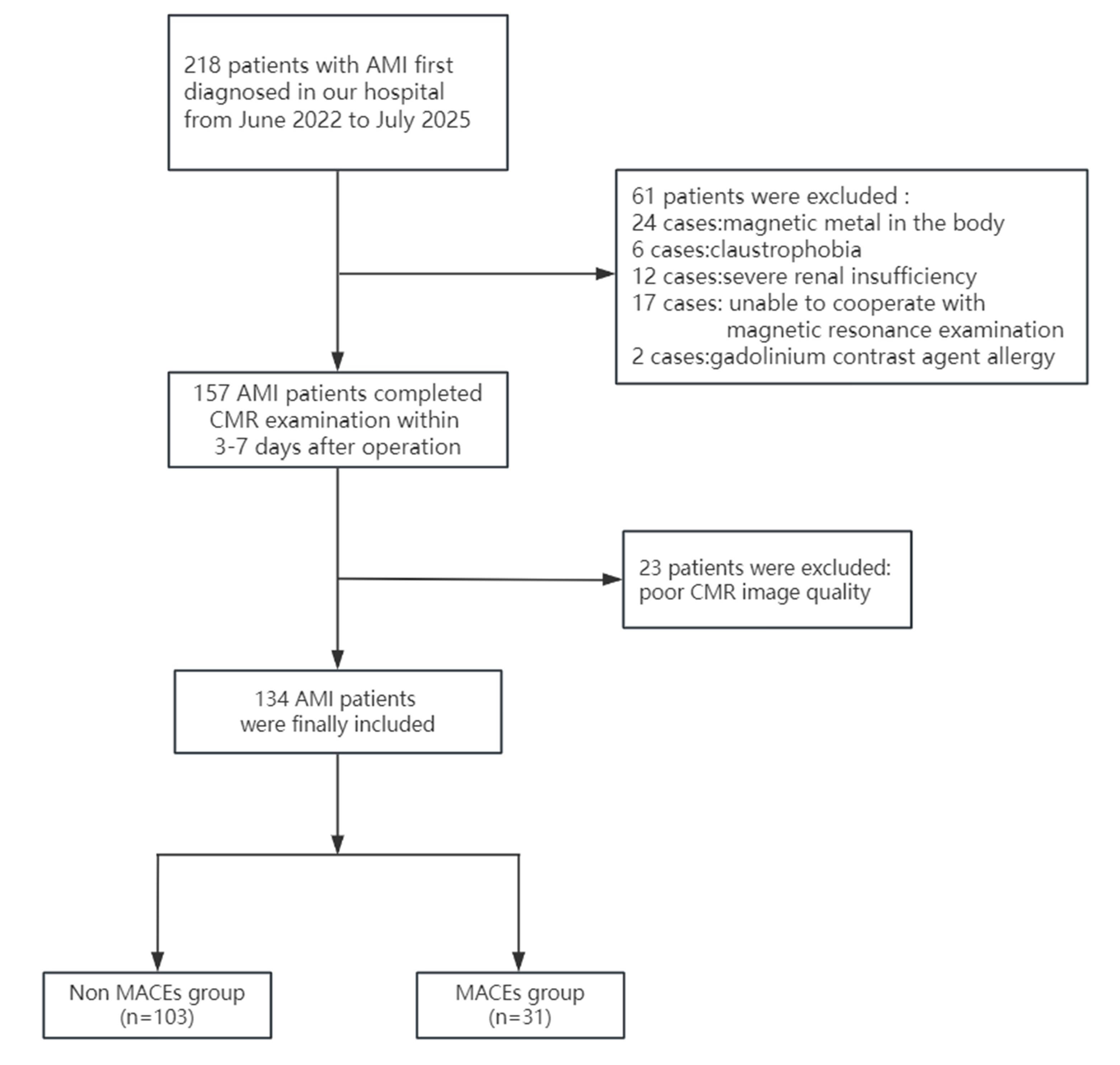
Fig.1 Flow chart of patient enrollment and grouping.
| Characteristics | Non-MACEs group (n=103) | MACEs group (n=31) | P |
|---|---|---|---|
| Age (year) | 59±11 | 64±12 | 0.041 |
| Male [n (%)] | 79 (76.7%) | 20 (64.5%) | 0.176 |
| BMI (kg/m2) | 25.0±3.7 | 25.0±3.2 | 0.921 |
| Hypertension [n (%)] | 61 (59.2%) | 15 (48.4%) | 0.286 |
| Diabetes [n (%)] | 25 (24.3%) | 9 (29.0%) | 0.593 |
| AF [n (%)] | 4 (3.9%) | 4 (12.9%) | 0.083 |
| Previous stroke [n (%)] | 8 (7.8%) | 4 (12.9%) | 0.472 |
| Previous smoking [n (%)] | 59 (57.3%) | 17 (54.8%) | 0.810 |
| WBC (109/L) | 9.69 (8.04, 11.47) | 11.47 (10.29, 13.49) | <0.001 |
| Neutrophil (109/L) | 7.37 (5.71, 9.22) | 9.56 (8.55, 11.66) | <0.001 |
| Lymphocyte (109/L) | 0.48 (0.36, 0.67) | 0.48 (0.27, 0.61) | 0.364 |
| Monocyte (109/L) | 1.43 (1.13, 2.01) | 1.02 (0.88, 1.39) | <0.001 |
| Platelet (109/L) | 205 (168, 240) | 214 (171, 254) | 0.388 |
| NLR | 5.3 (3.2, 7.0) | 9.2 (7.4, 12.4) | <0.001 |
| PLR | 142 (93, 182) | 173 (142, 254) | <0.001 |
| FBG (mmol/L) | 6.5 (5.4, 8.2) | 6.3 (5.1, 8.8) | 0.959 |
| UA (µmol/L) | 315 (259, 381) | 334 (260, 394) | 0.513 |
| SCr (µmol/L) | 62 (51, 74) | 66 (51, 82) | 0.593 |
| TC (mmol/L) | 4.35 (3.73, 5.25) | 4.80 (4.13, 5.53) | 0.110 |
| TG (mmol/L) | 1.52 (1.02, 2.09) | 1.32 (0.89, 2.08) | 0.754 |
| LDL-C (mmol/L) | 2.77 (2.07, 3.38) | 2.93 (2.40, 3.66) | 0.297 |
| HDL-C (mmol/L) | 1.05 (0.89, 1.22) | 1.13 (0.98, 1.39) | 0.129 |
| CK-MB(U/L) | 40 (20, 100) | 38 (20, 141) | 0.829 |
| cTn I (μg/L) | 11 (3, 22) | 8 (1, 47) | 0.827 |
| NT-proBNP(pg/mL) | 258 (61, 697) | 1,030 (123, 2,330) | 0.001 |
Tab.1 Baseline characteristics of the two groups of AMI patients
| Characteristics | Non-MACEs group (n=103) | MACEs group (n=31) | P |
|---|---|---|---|
| Age (year) | 59±11 | 64±12 | 0.041 |
| Male [n (%)] | 79 (76.7%) | 20 (64.5%) | 0.176 |
| BMI (kg/m2) | 25.0±3.7 | 25.0±3.2 | 0.921 |
| Hypertension [n (%)] | 61 (59.2%) | 15 (48.4%) | 0.286 |
| Diabetes [n (%)] | 25 (24.3%) | 9 (29.0%) | 0.593 |
| AF [n (%)] | 4 (3.9%) | 4 (12.9%) | 0.083 |
| Previous stroke [n (%)] | 8 (7.8%) | 4 (12.9%) | 0.472 |
| Previous smoking [n (%)] | 59 (57.3%) | 17 (54.8%) | 0.810 |
| WBC (109/L) | 9.69 (8.04, 11.47) | 11.47 (10.29, 13.49) | <0.001 |
| Neutrophil (109/L) | 7.37 (5.71, 9.22) | 9.56 (8.55, 11.66) | <0.001 |
| Lymphocyte (109/L) | 0.48 (0.36, 0.67) | 0.48 (0.27, 0.61) | 0.364 |
| Monocyte (109/L) | 1.43 (1.13, 2.01) | 1.02 (0.88, 1.39) | <0.001 |
| Platelet (109/L) | 205 (168, 240) | 214 (171, 254) | 0.388 |
| NLR | 5.3 (3.2, 7.0) | 9.2 (7.4, 12.4) | <0.001 |
| PLR | 142 (93, 182) | 173 (142, 254) | <0.001 |
| FBG (mmol/L) | 6.5 (5.4, 8.2) | 6.3 (5.1, 8.8) | 0.959 |
| UA (µmol/L) | 315 (259, 381) | 334 (260, 394) | 0.513 |
| SCr (µmol/L) | 62 (51, 74) | 66 (51, 82) | 0.593 |
| TC (mmol/L) | 4.35 (3.73, 5.25) | 4.80 (4.13, 5.53) | 0.110 |
| TG (mmol/L) | 1.52 (1.02, 2.09) | 1.32 (0.89, 2.08) | 0.754 |
| LDL-C (mmol/L) | 2.77 (2.07, 3.38) | 2.93 (2.40, 3.66) | 0.297 |
| HDL-C (mmol/L) | 1.05 (0.89, 1.22) | 1.13 (0.98, 1.39) | 0.129 |
| CK-MB(U/L) | 40 (20, 100) | 38 (20, 141) | 0.829 |
| cTn I (μg/L) | 11 (3, 22) | 8 (1, 47) | 0.827 |
| NT-proBNP(pg/mL) | 258 (61, 697) | 1,030 (123, 2,330) | 0.001 |
| Characteristics | Non-MACEs group (n=103) | MACEs group (n=31) | P |
|---|---|---|---|
| Gensini score | 72 (46, 102) | 88 (75, 166) | 0.004 |
| Number of diseased vessels [n (%)] | 0.205 | ||
| One vessel disease | 26 (25.2%) | 4 (12.9%) | 0.148 |
| Two vessel disease | 25 (24.3%) | 6 (19.4%) | 0.569 |
| Three vessel disease | 52 (50.5%) | 21 (67.7%) | 0.091 |
| Anterior myocardial infarction | 47 (45.6%) | 27 (87.1%) | <0.001 |
| Culprit vessel [n (%)] | <0.001 | ||
| LAD | 44 (42.7%) | 24 (77.4%) | <0.001 |
| LCX | 15 (14.6%) | 4 (12.9%) | 0.815 |
| RCA | 44 (42.7%) | 3 (9.7%) | <0.001 |
| Chest pain TIMI classification [n (%)] | 0.011 | ||
| Low risk | 60 (58.3%) | 9 (29.0%) | 0.004 |
| Intermediate risk | 31 (30.1%) | 14 (45.2%) | 0.119 |
| High risk | 12 (11.7%) | 8 (25.8%) | 0.099 |
Tab.2 Comparison of coronary artery lesions between the two groups of AMI patients
| Characteristics | Non-MACEs group (n=103) | MACEs group (n=31) | P |
|---|---|---|---|
| Gensini score | 72 (46, 102) | 88 (75, 166) | 0.004 |
| Number of diseased vessels [n (%)] | 0.205 | ||
| One vessel disease | 26 (25.2%) | 4 (12.9%) | 0.148 |
| Two vessel disease | 25 (24.3%) | 6 (19.4%) | 0.569 |
| Three vessel disease | 52 (50.5%) | 21 (67.7%) | 0.091 |
| Anterior myocardial infarction | 47 (45.6%) | 27 (87.1%) | <0.001 |
| Culprit vessel [n (%)] | <0.001 | ||
| LAD | 44 (42.7%) | 24 (77.4%) | <0.001 |
| LCX | 15 (14.6%) | 4 (12.9%) | 0.815 |
| RCA | 44 (42.7%) | 3 (9.7%) | <0.001 |
| Chest pain TIMI classification [n (%)] | 0.011 | ||
| Low risk | 60 (58.3%) | 9 (29.0%) | 0.004 |
| Intermediate risk | 31 (30.1%) | 14 (45.2%) | 0.119 |
| High risk | 12 (11.7%) | 8 (25.8%) | 0.099 |
| Characteristics | Non-MACEs group (n=103) | MACEs group (n=31) | P |
|---|---|---|---|
| Myocardial mass (g) | 101 (85, 117) | 100 (87, 120) | 0.667 |
| LGE (%) | 16 (12, 25) | 22 (17, 33) | 0.011 |
| LVEDV/BSA (mL/m2 ) | 72 (61, 84) | 72 (61, 95) | 0.478 |
| LVESV/BSA (mL/m2 ) | 37 (28, 45) | 41 (35, 60) | 0.017 |
| LVEF (%) | 49 ± 10 | 40±11 | <0.001 |
| Reverse motion [n (%)] | 75 (72.8%) | 28 (90.3%) | 0.043 |
| MVO [n (%)] | 57 (55.3%) | 23 (74.2%) | 0.061 |
| MVO volume (mL) | 2.87 (1.03, 4.70) | 2.08 (0.92, 5.47) | 0.698 |
| Ventricular aneurysm [n (%)] | 16 (15.5%) | 16 (51.6%) | <0.001 |
| Myocardial strain in infarct segment [n (%)] | |||
| Radial strain | 16 (13, 22) | 12 (11, 17) | 0.003 |
| Circumferential strain | 11.5 (9.4, 14.1) | 10.1 (9.0, 11.4) | 0.018 |
| Longitudinal strain | 11.2 (9.0, 14.3) | 7.7 (6.7, 9.5) | <0.001 |
| Global myocardial strain | |||
| Radial strain | 26 (21, 32) | 24 (19, 26) | 0.018 |
| Circumferential strain | 15.11±3.10 | 13.57±2.77 | 0.011 |
| Longitudinal strain | 11.6 (9.3, 14.4) | 8.3 (6.8, 9.4) | <0.001 |
Tab.3 Comparison of cardiac magnetic resonance (CMR) parameters between the two groups of AMI patients
| Characteristics | Non-MACEs group (n=103) | MACEs group (n=31) | P |
|---|---|---|---|
| Myocardial mass (g) | 101 (85, 117) | 100 (87, 120) | 0.667 |
| LGE (%) | 16 (12, 25) | 22 (17, 33) | 0.011 |
| LVEDV/BSA (mL/m2 ) | 72 (61, 84) | 72 (61, 95) | 0.478 |
| LVESV/BSA (mL/m2 ) | 37 (28, 45) | 41 (35, 60) | 0.017 |
| LVEF (%) | 49 ± 10 | 40±11 | <0.001 |
| Reverse motion [n (%)] | 75 (72.8%) | 28 (90.3%) | 0.043 |
| MVO [n (%)] | 57 (55.3%) | 23 (74.2%) | 0.061 |
| MVO volume (mL) | 2.87 (1.03, 4.70) | 2.08 (0.92, 5.47) | 0.698 |
| Ventricular aneurysm [n (%)] | 16 (15.5%) | 16 (51.6%) | <0.001 |
| Myocardial strain in infarct segment [n (%)] | |||
| Radial strain | 16 (13, 22) | 12 (11, 17) | 0.003 |
| Circumferential strain | 11.5 (9.4, 14.1) | 10.1 (9.0, 11.4) | 0.018 |
| Longitudinal strain | 11.2 (9.0, 14.3) | 7.7 (6.7, 9.5) | <0.001 |
| Global myocardial strain | |||
| Radial strain | 26 (21, 32) | 24 (19, 26) | 0.018 |
| Circumferential strain | 15.11±3.10 | 13.57±2.77 | 0.011 |
| Longitudinal strain | 11.6 (9.3, 14.4) | 8.3 (6.8, 9.4) | <0.001 |
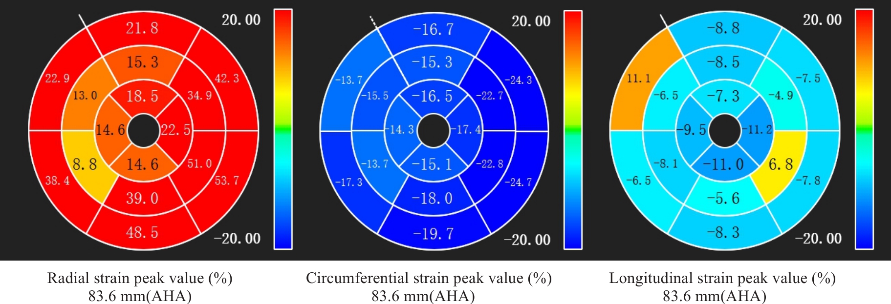
Fig.2 A 71-year-old female patient with anterior myocardial infarction showing significantly reduced global longitudinal strain with paradoxical motion. She experienced a major adverse cardiovascular events (MACEs) 17 months after percutaneous coronary intervention (PCI).
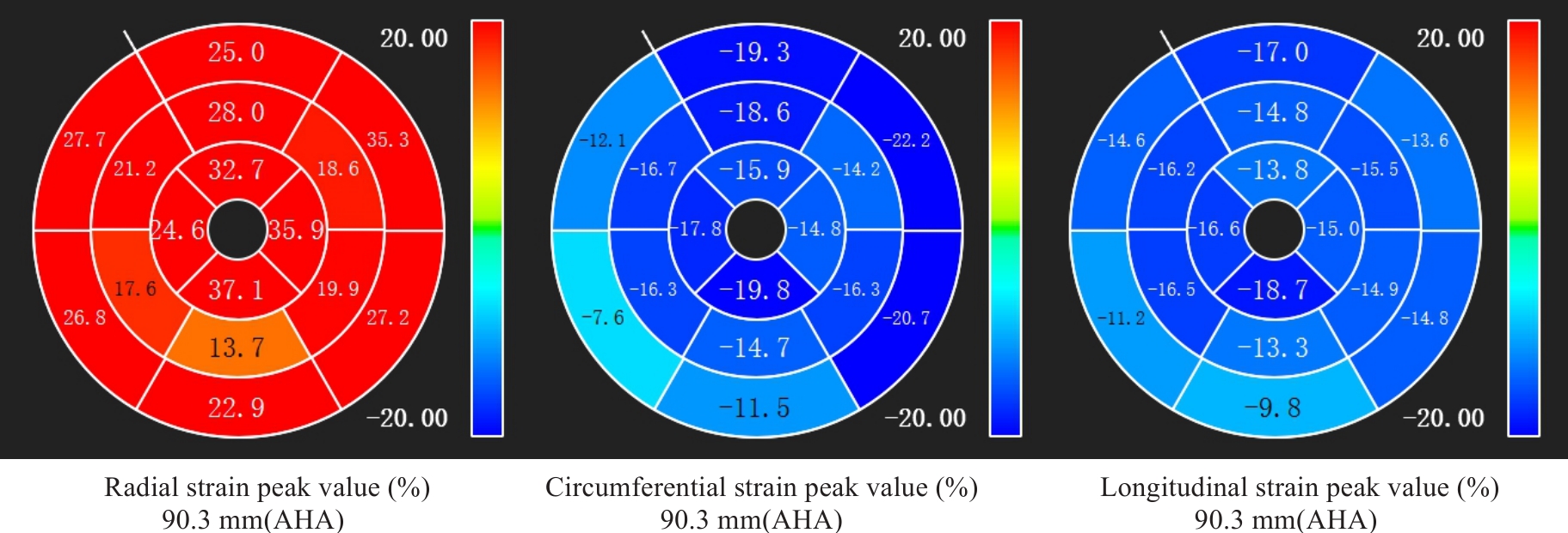
Fig.3 A 70-year-old male patient with inferior wall myocardial infarction showing no significant decrease in global longitudinal strain. He remained free of MACEs 22 months after PCI.
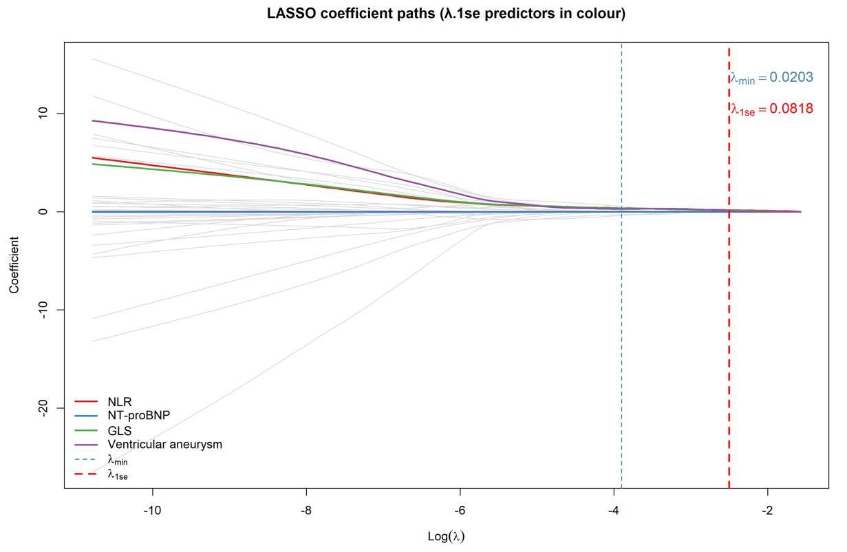
Fig.4 Based on the variable screening results of LASSO regression (λ.1se criterion), 4 indicators were selected: NLR, GLS, NT proBNP, and ventricular aneurysm (n=134).
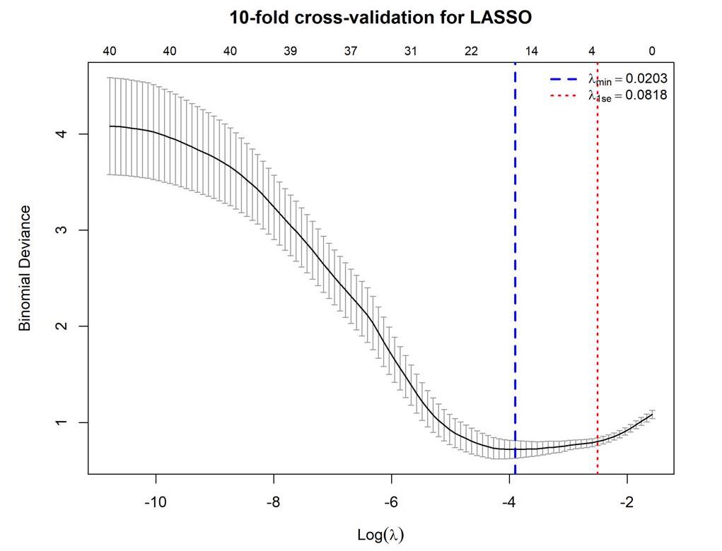
Fig.5 10-fold cross validation curve of LASSO regression (n=134).
| Index | β | SE | Wald | P | OR | 95% CI |
|---|---|---|---|---|---|---|
| NLR | 0.371 | 0.096 | 15.037 | <0.001 | 1.449 | 1.201-1.747 |
| GLS | -0.475 | 0.132 | 12.946 | <0.001 | 0.622 | 0.480-0.805 |
| NT-proBNP | 0.020 | 0.015 | 1.732 | 0.188 | 1.020* | 0.990-1.051 |
| Ventricular aneurysm | 0.563 | 1.282 | 0.870 | 0.351 | 1.755 | 0.538-5.726 |
Tab.4 Multivariate logistic regression analysis of factors affecting post-PCI MACEs
| Index | β | SE | Wald | P | OR | 95% CI |
|---|---|---|---|---|---|---|
| NLR | 0.371 | 0.096 | 15.037 | <0.001 | 1.449 | 1.201-1.747 |
| GLS | -0.475 | 0.132 | 12.946 | <0.001 | 0.622 | 0.480-0.805 |
| NT-proBNP | 0.020 | 0.015 | 1.732 | 0.188 | 1.020* | 0.990-1.051 |
| Ventricular aneurysm | 0.563 | 1.282 | 0.870 | 0.351 | 1.755 | 0.538-5.726 |
| Index | β | SE | Wald | P | OR | 95% CI |
|---|---|---|---|---|---|---|
| NLR | 0.388 | 0.092 | 17.946 | <0.001 | 1.475 | 1.232-1.765 |
| GLS | -0.531 | 0.129 | 16.804 | <0.001 | 0.588 | 0.456-0.758 |
| Constant | 1.160 | 1.224 | 0.898 | - | 3.190 | - |
Tab.5 Binary logistic regression analysis of GLS and NLR for predicting MACEs
| Index | β | SE | Wald | P | OR | 95% CI |
|---|---|---|---|---|---|---|
| NLR | 0.388 | 0.092 | 17.946 | <0.001 | 1.475 | 1.232-1.765 |
| GLS | -0.531 | 0.129 | 16.804 | <0.001 | 0.588 | 0.456-0.758 |
| Constant | 1.160 | 1.224 | 0.898 | - | 3.190 | - |
| Parameter | Cutoff value | Sensitivity(%) | Specificity(%) | AUC | 95% CI | Youden index |
|---|---|---|---|---|---|---|
| NLR | 7.223 | 0.774 | 0.777 | 0.820 | 0.735-0.905 | 0.551 |
| GLS | -9.65 | 0.839 | 0.718 | 0.825 | 0.740-0.910 | 0.557 |
| Joint prediction | 0.303 | 0.839 | 0.883 | 0.911 | 0.858-0.965 | 0.722 |
Tab.6 Predictive model for the risk of MACEs
| Parameter | Cutoff value | Sensitivity(%) | Specificity(%) | AUC | 95% CI | Youden index |
|---|---|---|---|---|---|---|
| NLR | 7.223 | 0.774 | 0.777 | 0.820 | 0.735-0.905 | 0.551 |
| GLS | -9.65 | 0.839 | 0.718 | 0.825 | 0.740-0.910 | 0.557 |
| Joint prediction | 0.303 | 0.839 | 0.883 | 0.911 | 0.858-0.965 | 0.722 |
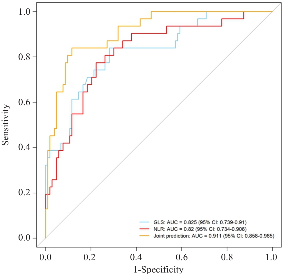
Fig.6 ROC curve of MACEs predicted by NLR, GLS and their combination (n=134).
| [1] | 国家心血管病中心. 中国心血管健康与疾病报告2024[M]. 北京: 中国协和医科大学出版社, 2025. |
| [2] | 中华医学会心血管病学分会, 中华心血管病杂志编辑委员会. 急性ST段抬高型心肌梗死诊断和治疗指南 [J]. 中华心血管病杂志, 2010, (8): 675-690. |
| [3] | 张丽伟, 赵 赫, 曾辰华. 血浆C型凝集素样受体2水平与急性ST段抬高型心肌梗死患者经皮冠状动脉介入术后发生主要不良心血管事件的关系[J]. 中华实用诊断与治疗杂志, 2020, 34(12): 1239-43. |
| [4] | Yang J, Yin P, Li MX, et al. Long-term cardiovascular and noncardiovascular mortality after acute coronary syndrome a nationwide competing-risks analysis of 1.56 million patients[J]. JACC, 2026, 87(12): 1511-22. doi:10.1016/j.jacc.2025.12.064 |
| [5] | Yang XY, Wen L, Sun M, et al. Prediction of cardiac deterioration in acute heart failure patients: Evaluation of the efficacy of single laboratory indicator models versus comprehensive models[J]. Medicine, 2024, 103(44): e40266. doi:10.1097/md.0000000000040266 |
| [6] | von Knobelsdorff-Brenkenhoff F, Schulz-Menger J. Cardiovascular magnetic resonance in the guidelines of the European Society of Cardiology: a comprehensive summary and update[J]. J Cardiovasc Magn Reson, 2023, 25(1): 42. doi:10.1186/s12968-023-00950-z |
| [7] | Zhang Y, Ko SM. The imaging spectrum of myocardial infarction and its associated complications: a contemporary pictorial review of computed tomography and magnetic resonance imaging[J]. Diagn Interv Radiol, 2026. DOI:10.4274/dir.2026.263576 . |
| [8] | Pan JL, Ng SM, Neubauer S, et al. Phenotyping heart failure by cardiac magnetic resonance imaging of cardiac macro- and microscopic structure: state of the art review[J]. Eur Heart J Cardiovasc Imaging, 2023, 24(10): 1302-17. doi:10.1093/ehjci/jead124 |
| [9] | Thomas KE, Fotaki A, Botnar RM, et al. Imaging methods: magnetic resonance imaging[J]. Circ Cardiovasc Imaging, 2023, 16(1): e014068. doi:10.1161/circimaging.122.014068 |
| [10] | Ren Y, Li S, Chen YC, et al. Prognostic value of left ventricular strain determined using CMR-feature tracking in patients with alcoholic cardiomyopathy[J]. Int J Cardiol, 2025, 435: 133391. doi:10.1016/j.ijcard.2025.133391 |
| [11] | Frișan AC, Lazăr MA, Șoșdean R, et al. Myocardial work analysis in ST-elevation myocardial infarction: insights into left ventricular ejection fraction: a pilot study[J]. Life, 2025, 15(3): 338. doi:10.3390/life15030338 |
| [12] | Alhawamdeh N, Bani Hani DA, Alshraideh JA, et al. Novel inflammatory markers predict cardiovascular events after percutaneous coronary intervention in patients with acute coronary syndrome[J]. Heart Lung, 2026, 75: 111-6. doi:10.1016/j.hrtlng.2025.09.014 |
| [13] | 张 慧, 王 黎, 上官海娟, 等. 基于心脏磁共振评估的冠状动脉微血管阻塞对急性ST段抬高型心肌梗死患者急诊经皮冠状动脉介入治疗术后预后的临床研究[J]. 中国介入心脏病学杂志, 2025, 33(6): 327-33. |
| [14] | Hu YN, Liu YS, Wang YP, et al. Dual-pathway model of IL-6 and HSP22/HSP27 dynamics predicts post-PCI MACE in STEMI patients[J]. BMC Cardiovasc Disord, 2025, 25(1): 863. doi:10.1186/s12872-025-05304-4 |
| [15] | Thygesen K, Alpert JS, Jaffe AS, et al. Fourth universal definition of myocardial infarction (2018)[J]. Circulation, 2018, 138(20): e618-51. doi:10.1161/cir.0000000000000632 |
| [16] | Byrne RA, Rossello X, Coughlan JJ, et al. 2023 ESC guidelines for the management of acute coronary syndromes[J]. Eur Heart J, 2023, 44(38): 3720-826. |
| [17] | Norman S, Koshy A, Biswas S, et al. A systematic comparison of ACC/AHA, ESC and JCS guidelines on intravascular imaging: level of evidence and clinical use scenarios[J]. Heart Lung Circ, 2024, 33: S549-50. doi:10.1016/j.hlc.2024.06.927 |
| [18] | Tudela Martínez JI, Alcaraz Pérez P, Martínez Encarnación L, et al. Cardiac magnetic resonance in adults: an updated review of the diagnostic approach to major heart diseases[J]. J Clin Med, 2025, 14(20): 7323. doi:10.3390/jcm14207323 |
| [19] | Thrane PG, Olesen KKW, Thim T, et al. Temporal trends in cardiovascular events after primary percutaneous coronary intervention for ST-segment elevation myocardial infarction[J]. JACC Adv, 2025, 4(3): 101614. doi:10.1016/j.jacadv.2025.101614 |
| [20] | 杨彦杰, 王 鸽, 严 研, 等. 低风险急性ST段抬高型心肌梗死患者早期出院的可行性分析[J]. 中国心血管病研究, 2023, 21(8): 680-3. doi:10.3969/j.issn.1672-5301.2023.08.002 |
| [21] | 杜晓彬, 王方方, 杨耀峰. 血清S100A4、ANGPTK4、CX3CL1水平联合检测对急性心肌梗死患者PCI术后MACE发生的预测价值[J]. 黑龙江医药科学, 2024, 47(4): 41-4. |
| [22] | Peles I, Barrett O, Cafri C, et al. Predictors of adverse outcome in high-risk percutaneous coronary interventions patients[J]. Can J Cardiol, 2023, 39(10): 1369-79. doi:10.1016/j.cjca.2023.04.021 |
| [23] | Chen YF, Jiang M, Xia C, et al. A novel deep learning system for STEMI prognostic prediction from multi-sequence cardiac magnetic resonance[J]. Sci Bull, 2025, 70(24): 4241-52. doi:10.1016/j.scib.2025.11.027 |
| [24] | Wang CP, Zhou L, Liang Y, et al. Interactions of ST-elevation myocardial infarction, age, and sex and the risk of major adverse cardiovascular events among Chinese adults: a secondary analysis of a single-centre prospective cohort[J]. BMJ Open, 2022, 12(7): e058494. doi:10.1136/bmjopen-2021-058494 |
| [25] | 李洋洋, 梁力晖, 陈柏林, 等. 急性冠脉综合征患者经皮冠状动脉介入术后院内MACE发作风险预测模型构建[J]. 心脏杂志, 2022, 34(5): 531-6. |
| [26] | 郭智超, 范春雨. 全身免疫炎症指数对急性心肌梗死病人PCI术后院内不良预后的预测价值[J]. 中西医结合心脑血管病杂志, 2024, 22(24): 4530-3. |
| [27] | Fang CY, Chen ZF, Zhang J, et al. The predictive value of neutrophil-lymphocyte ratio combined with the Global Registry of Acute Coronary Events score for inhospital adverse cardiovascular events in patients with acute ST-elevation myocardial infarction[J]. J Res Med Sci, 2024, 29: 41. doi:10.4103/jrms.jrms_485_22 |
| [28] | Matter MA, Paneni F, Libby P, et al. Inflammation in acute myocardial infarction: the good, the bad and the ugly[J]. Eur Heart J, 2024, 45(2): 89-103. doi:10.1093/eurheartj/ehad486 |
| [29] | Mornar Jelavić M, Babić Z, Babić D, et al. Ten-year outcomes after acute stemi treated with primary pci - the role of acute biomarkers and other parameters in predicting clinical severity and prognosis[J]. Acta Clin Croat, 2024, 63(3/4): 733-44. |
| [30] | Zhang C, Zeng S, Ji WJ, et al. Synergistic role of circulating CD14++CD16+ monocytes and fibrinogen in predicting the cardiovascular events after myocardial infarction[J]. Clin Cardiol, 2023, 46(5): 521-8. doi:10.1002/clc.24005 |
| [31] | Yang F, Yu M, Yuan YF, et al. A new comprehensive parameter combining residual C-reactive protein and neutrophil risk predicting adverse events in Chinese patients after percutaneous coronary intervention: a multi-center prospective cohort study[J]. Chin Med J, 2026, 139(1): 139-41. doi:10.1097/cm9.0000000000003885 |
| [32] | Helali J, Ramesh K, Brown J, et al. Late gadolinium enhancement on cardiac MRI: a systematic review and meta-analysis of prognosis across cardiomyopathies[J]. Int J Cardiol, 2025, 419: 132711. doi:10.1016/j.ijcard.2024.132711 |
| [33] | Troger F, Pamminger M, Poskaite P, et al. Clinical impact of persistent microvascular obstruction in CMR after reperfused STEMI[J]. Circ Cardiovasc Imag, 2025, 18(6): e017645. doi:10.1161/circimaging.124.017645 |
| [34] | Li M, Wang J, Ding S, et al. Cardiac magnetic resonance imaging-derived pathophysiology and prognosis of diabetes mellitus with acute myocardial infarction after revascularization: a prospective cohort study. Ann Med. 2024, 56(1): 2399751. doi:10.1080/07853890.2024.2399751 |
| [35] | Guglielmo M, Fusini L, Baessato F, et al. PROGnostic RolE of strain measurements in stress cardiac MRI in predicting major adverse cardiac events[J]. Int J Cardiol, 2024, 412: 132337. doi:10.1016/j.ijcard.2024.132337 |
| [36] | Jering KS, Claggett BL, Braunwald E, et al. NT-proBNP in the early convalescent phase after high-risk myocardial infarction is associated with adverse cardiovascular outcomes: the PARADISE-MI trial[J]. J Card Fail, 2026, 32(3): 586-95. doi:10.1016/j.cardfail.2025.03.018 |
| [37] | Charkiewicz-Szeremeta K, Sawicka-Śmiarowska E, Dubatówka M, et al. Predictors of long-term prognosis focused on kidney function in patients with chronic coronary syndrome[J]. Diseases, 2026, 14(2): 78. doi:10.3390/diseases14020078 |
| [38] | Feng YL, Zhuo YF, Cheng HJ. Predictive value of circulating cardiomyocyte-specific cell-free DNA levels for heart failure risk after acute ST-segment elevation myocardial infarction[J]. Postepy Kardiol Interwencyjnej, 2025, 21(4): 565-76. doi:10.5114/aic.2025.156807 |
| [39] | Zornitzki L, Barlev Y, Gefen S, et al. Left ventricular global longitudinal strain in patients with coronary microvascular dysfunction and preserved ejection fraction: a single-center study[J]. Pol Heart J, 2026, 84(2): 208-16. doi:10.33963/v.phj.110652 |
| [40] | Shereef AS, Abdelmajeed MG, Alshair MH, et al. Coronary slow flow and its correlation with reduced left ventricle global longitudinal strain: a case–control study[J]. Echo Res Pract, 2024, 11(1): 2. doi:10.1186/s44156-023-00037-6 |
| [41] | Ersbøll M, Valeur N, Mogensen UM, et al. Global left ventricular longitudinal strain is closely associated with increased neurohormonal activation after acute myocardial infarction in patients with both reduced and preserved ejection fraction: a two-dimensional speckle tracking study[J]. Eur J Heart Fail, 2012, 14(10): 1121-9. doi:10.1093/eurjhf/hfs107 |
| [42] | Kolenda Zloić S, Martins da Fonseca J, Ayogu CI, et al. Prognostic impact of left ventricular strain by feature tracking in acute myocardial infarction treated with PCI: a meta-analysis[J]. Clin Imaging, 2025, 124: 110514. doi:10.1016/j.clinimag.2025.110514 |
| [43] | Li XB, Yu C, Liu XW, et al. A prediction model based on systemic immune-inflammatory index combined with other predictors for major adverse cardiovascular events in acute myocardial infarction patients[J]. J Inflamm Res, 2024, 17: 1211-25. doi:10.2147/jir.s443153 |
| [44] | Aghezzaf S, Coisne A, Bauters C, et al. Feasibility and prognostic significance of ventricular-arterial coupling after myocardial infarction: the RIGID-MI cohort[J]. Eur Heart J Cardiovasc Imaging, 2024, 25(5): 668-77. doi:10.1093/ehjci/jead342 |
| [45] | Jigoranu RA, Mitu O, Oancea AF, et al. Diagnostic and severity assessment of coronary artery disease using ApoB/ApoA-I ratio: insights from a statin-treated eastern European cohort[J]. Medicina, 2026, 62(2): 297. doi:10.3390/medicina62020297 |
| [46] | Zhang Y, Luo R, Sun XC, et al. The relationship between Gensini score and rates of 30-day mortality in acute coronary syndrome patients in China[J]. BMC Cardiovasc Disord, 2025, 26(1): 3. doi:10.1186/s12872-025-05303-5 |
| [47] | 李萌玫, 孙洪巧, 万雪梅, 等. 急性心肌梗死急诊PCI术后罪犯血管部位对心力衰竭及心肌标志物的影响[J]. 临床心血管病杂志, 2025, 41(10): 774-80. |
| [48] | Vidal-Calés P, Bujak K, Rinaldi R, et al. Long-term prognostic impact of the left anterior descending coronary artery as the STEMI-related culprit vessel: subanalysis of the EXAMINATION-EXTEND trial[J]. REC Interv Cardiol, 2025, 7(2): 99-108. |
| Viewed | ||||||
|
Full text |
|
|||||
|
Abstract |
|
|||||
