
Journal of Southern Medical University ›› 2026, Vol. 46 ›› Issue (10): 2338-2356.doi: 10.12122/j.issn.1673-4254.2026.10.05
Huixin TAN1( ), Junguo JIN2, Manqi YANG3, Jingli WANG3, Fenghua ZHOU4(), Ping ZENG1(), Xuegang SUN4()
Received:2026-04-11
Online:2026-10-20
Published:2026-10-26
Contact:
Fenghua ZHOU, Ping ZENG, Xuegang SUN
E-mail:tanhuixinx@163.com;Wendyzhou515@126.com;zengping@gdph.org.cn;sun_smu@126.com
Supported by:Huixin TAN, Junguo JIN, Manqi YANG, Jingli WANG, Fenghua ZHOU, Ping ZENG, Xuegang SUN. PGAM5 deficiency attenuates atherosclerosis, enhances oxidized LDL uptake by liver sinusoidal endothelial cells and reshapes circulating lipidome in mice[J]. Journal of Southern Medical University, 2026, 46(10): 2338-2356.
Add to citation manager EndNote|Ris|BibTeX
URL: https://www.j-smu.com/EN/10.12122/j.issn.1673-4254.2026.10.05
| Primers | Sequence | Length (bp) |
|---|---|---|
| β-actin | Upstream5'-CACCCAGCACAATGAAGATC AAGAT-3' Downstream5'CCAGTTTTTAAATCCTGAGT CAAGC-3' | 317 |
| PGAM5 | Upstream5'-CTCAATAATGGCAGCATCACCC-3' Downstream5'-ATCTTGTCGGGAGGCATGAA-3' | 98 |
Tab.1 Primer sequences
| Primers | Sequence | Length (bp) |
|---|---|---|
| β-actin | Upstream5'-CACCCAGCACAATGAAGATC AAGAT-3' Downstream5'CCAGTTTTTAAATCCTGAGT CAAGC-3' | 317 |
| PGAM5 | Upstream5'-CTCAATAATGGCAGCATCACCC-3' Downstream5'-ATCTTGTCGGGAGGCATGAA-3' | 98 |
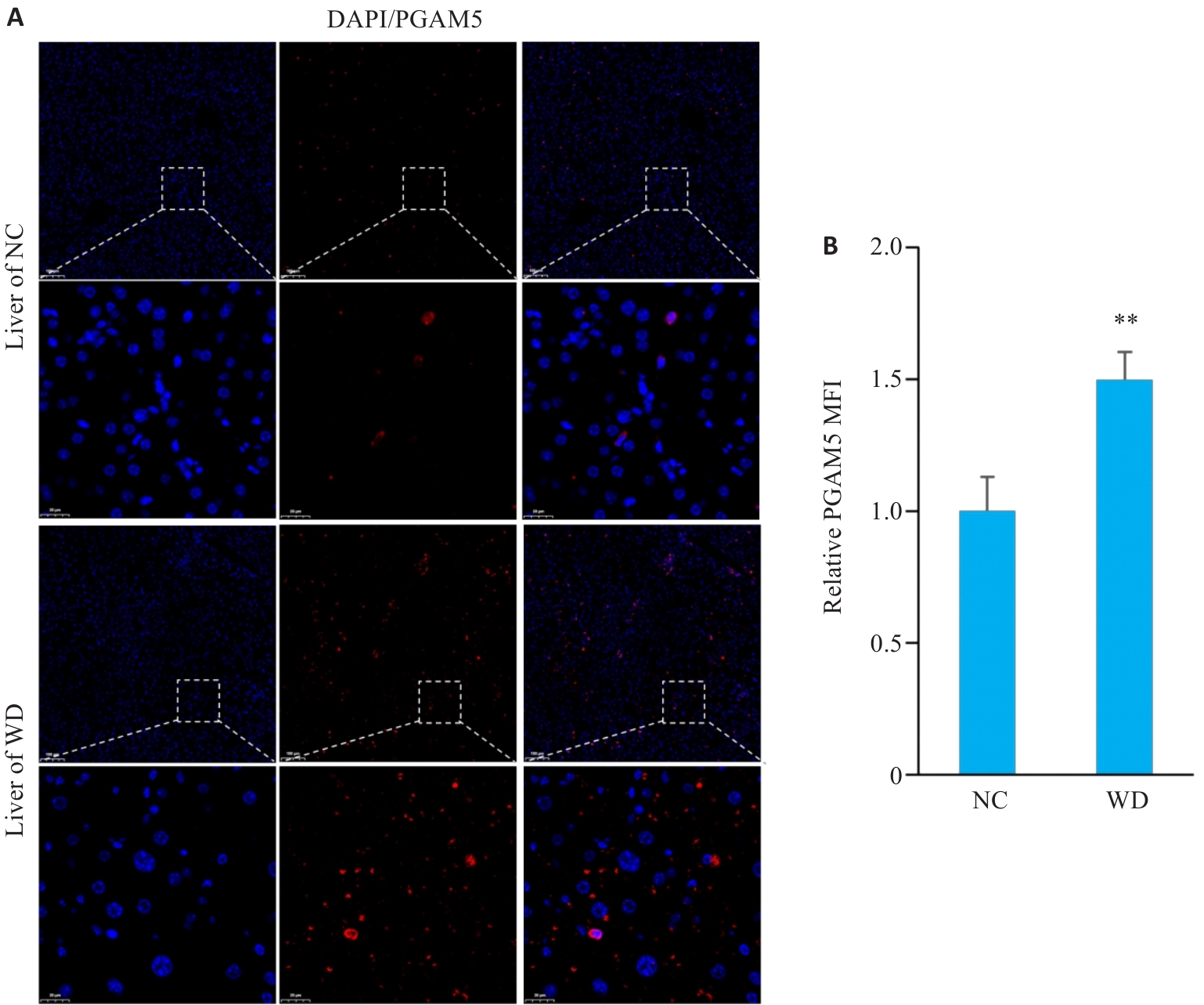
Fig.1 Increased hepatic PGAM5 expression in atherosclerotic mice. A: Immunofluorescence staining with DAPI (blue) and PGAM5 (red) of mouse liver tissues (Original magnification: ×100; inset magnification: ×600). B: Quantitative analysis of relative mean fluorescence intensity (MFI) of PGAM5 in mouse liver tissues (n=4). **P<0.01 vs NC group.
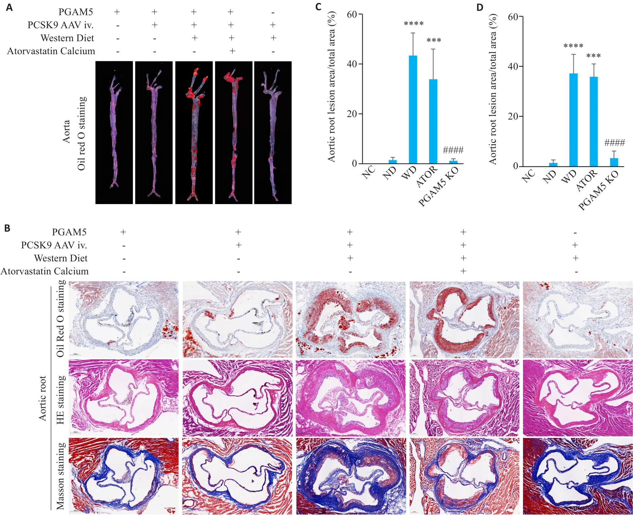
Fig.2 PGAM5 deficiency attenuates atherosclerosis in mice. A: Oil Red O staining of mouse whole aorta. B: Oil Red O, HE, and Masson staining of mouse aortic root sections (×60). C: Quantification of aortic root lipid deposition with the lesion area presented as percentage of total vessel area (NC, n=3; ND, n=4; WD, n=6; ATOR, n=5; PGAM5 KO, n=3). D: Quantification of aortic root lesion area in HE staining (NC, n=5; ND, n=4; WD, n=6; ATOR, n=5; PGAM5 KO, n=5). ***P<0.001, ****P<0.0001 vs NC group; ####P<0.0001 vs WD group.
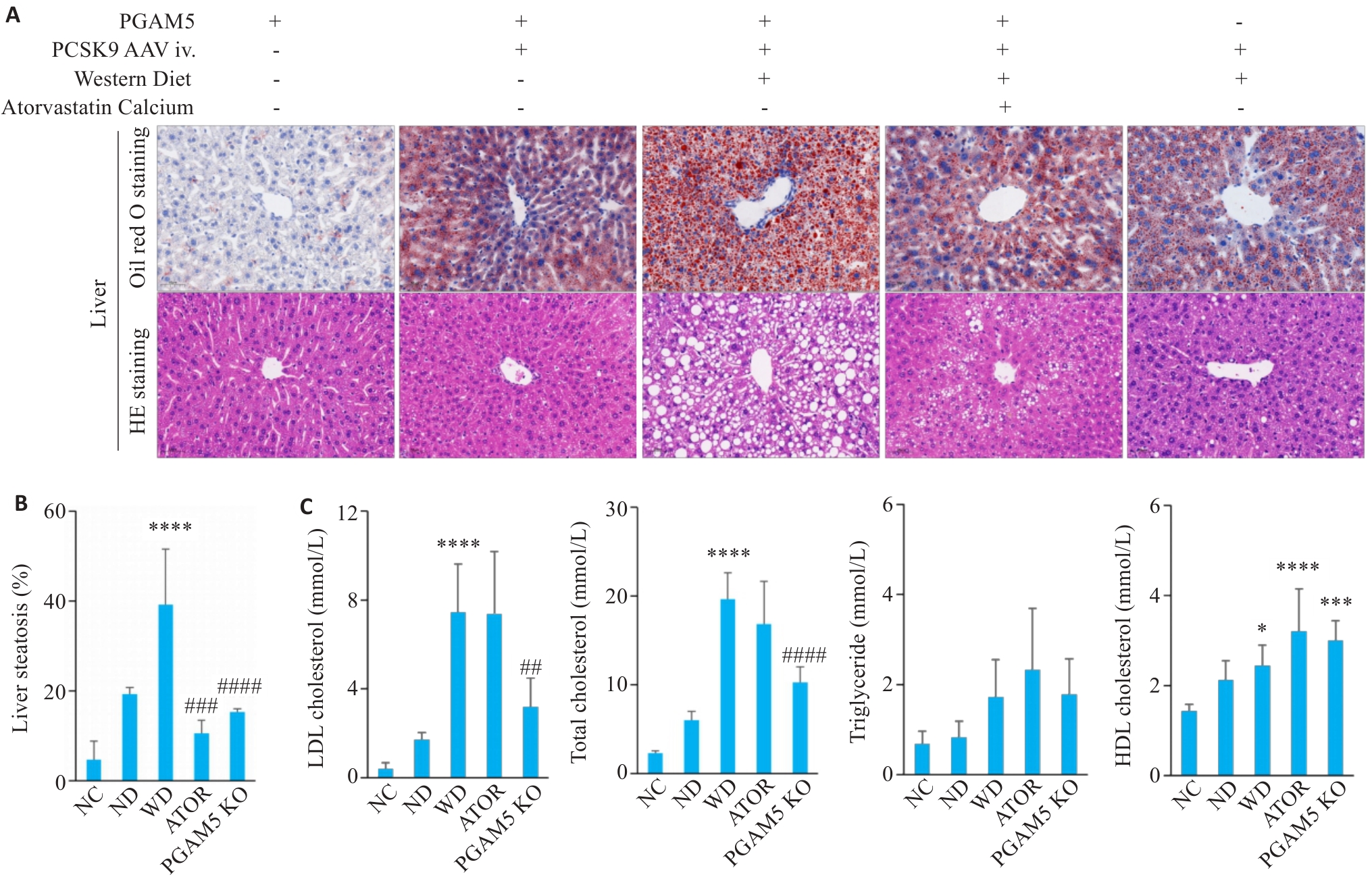
Fig.3 PGAM5 deficiency alleviates hepatic lipid deposition and plasma lipid abnormalities in atherosclerotic mice. A: Oil Red O staining and HE staining of mouse liver tissues (×20). B: Quantitative analysis of hepatic lipid accumulation area (expressed as the percentage of lipid-positive area relative to total tissue area) (NC, n=7; ND, n=3; WD, n=7; ATOR, n=3; PGAM5 KO, n=5). C: Plasma levels of LDL-C (n=6), TC (n=6), TG (n=6), and HDL-C (NC, n=7; ND, n=6; WD, n=6; ATOR, n=6; PGAM5 KO, n=6) in mice of each group. *P<0.05, ***P<0.001, ****P<0.0001 vs NC group; ##P<0.01, ###P<0.001, ####P<0.0001 vs WD group.
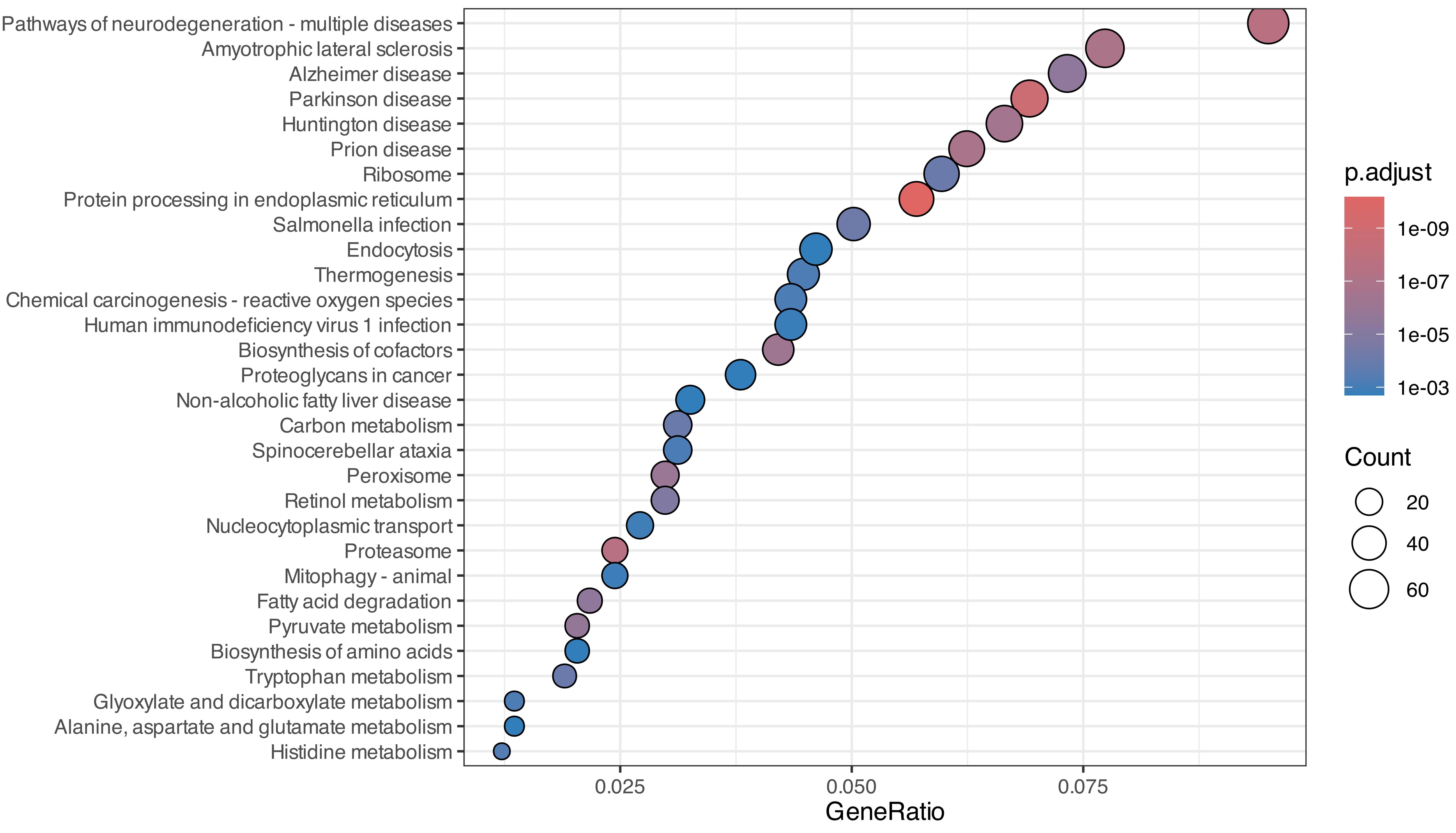
Fig.4 Enrichment analysis of differential liver proteins between the two groups using KEGG pathways.
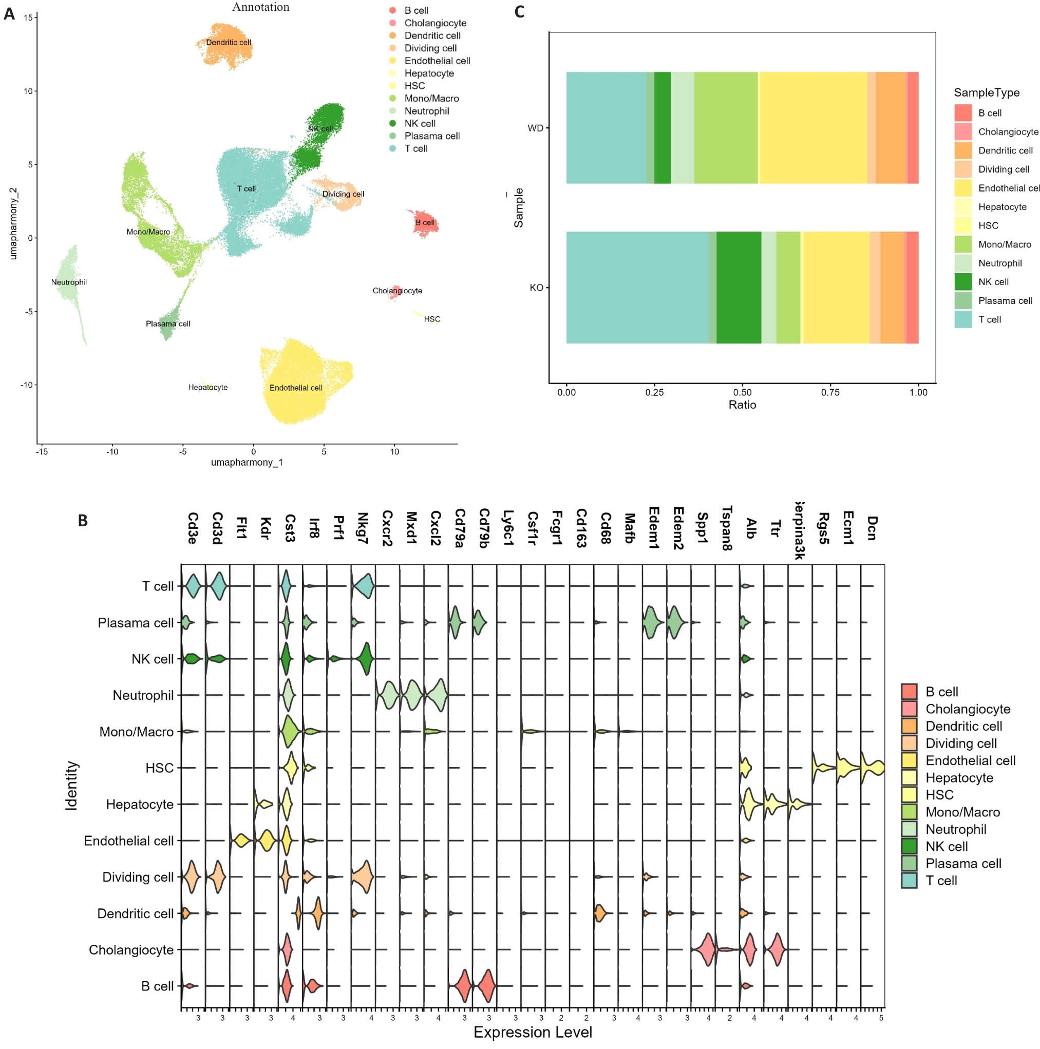
Fig.5 Single-cell RNA sequencing analysis of mouse liver tissues. A: UMAP visualization of major hepatic cell clusters in WD and PGAM5 KO groups. B: Marker gene expression used for cell-type annotation. C: Proportional distribution of major cell populations between groups (n=4).
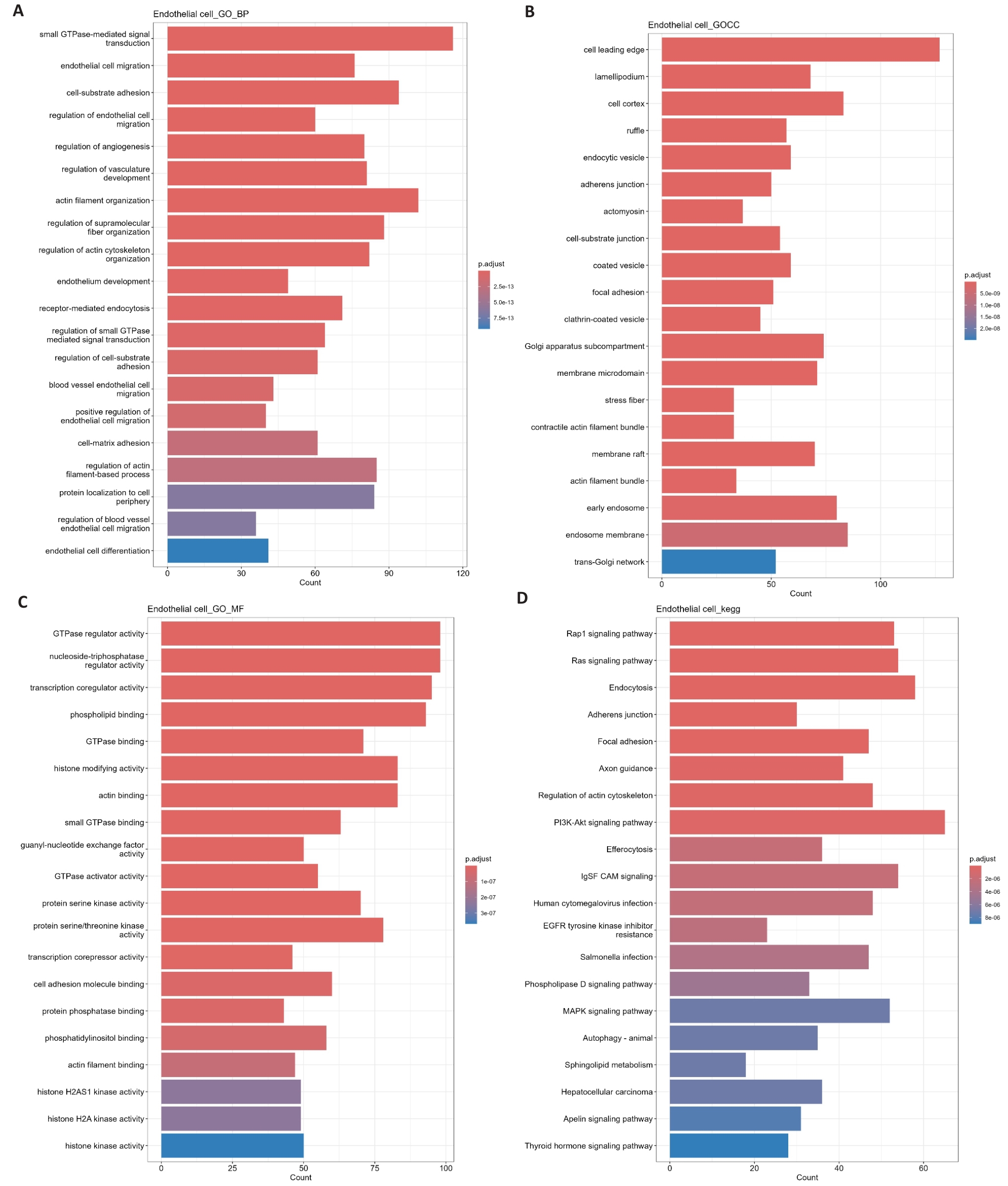
Fig.6 GO and KEGG enrichment analysis of differential genes between endothelial cells and other hepatic cell populations. A-C: Gene Ontology (GO) enrichment analysis of differentially expressed genes in ECs, including biological process (A), cellular component (B), and molecular function (C). D: Kyoto Encyclopedia of Genes and Genomes (KEGG) pathway enrichment analysis of differentially expressed genes in ECs.
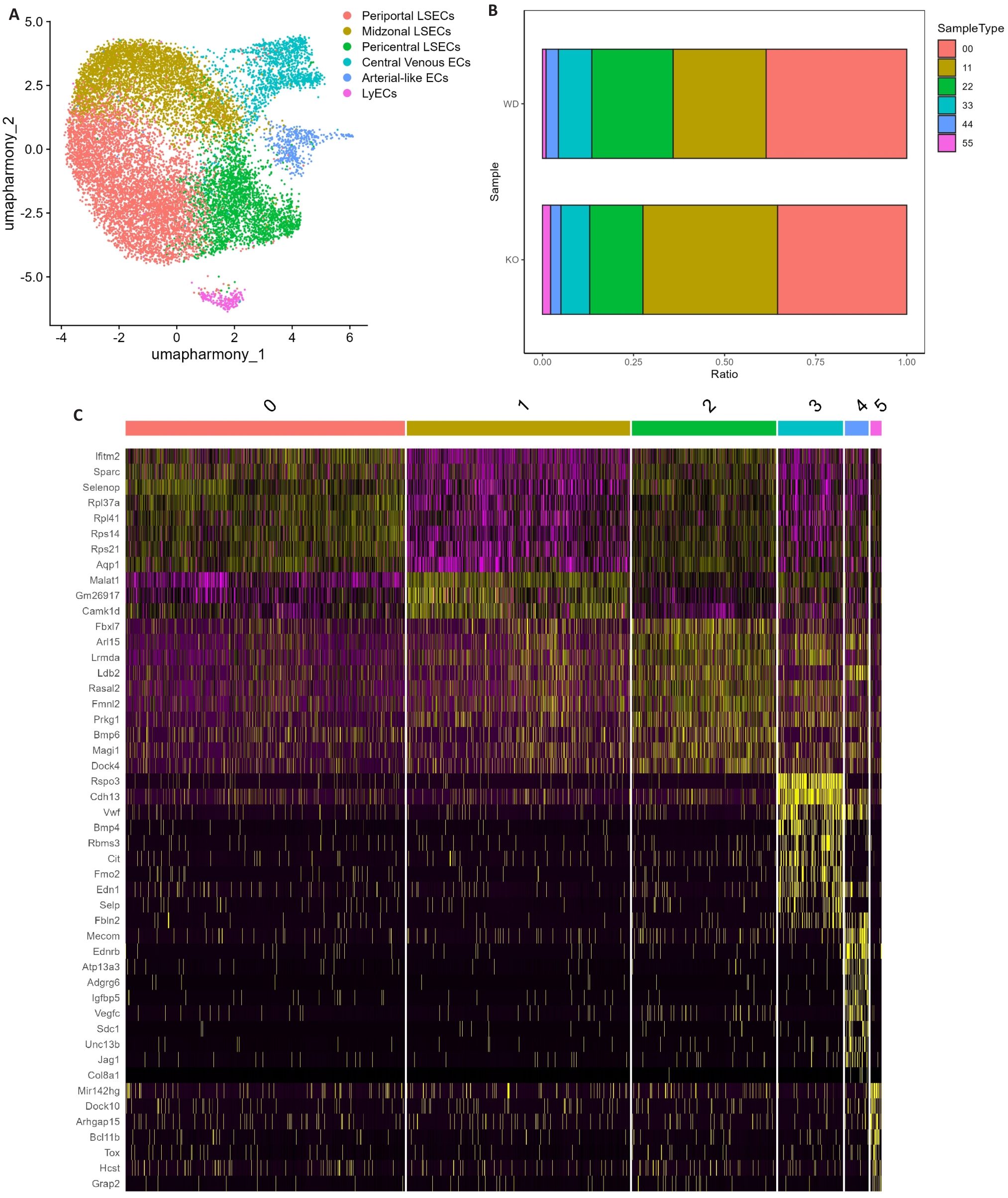
Fig.7 Transcriptional characteristics of hepatic endothelial subpopulations. A: UMAP dimensionality reduction plot of liver endothelial cells subpopulations. B: Proportional distribution of different endothelial cells subpopulations across samples. C: Heatmap of differentially expressed genes among endothelial cells subpopulations.
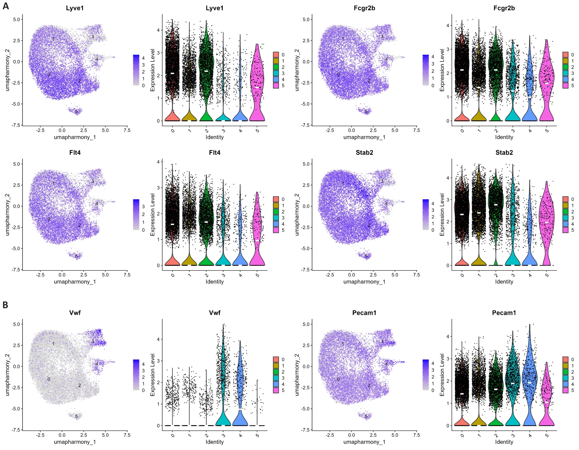
Fig.8 Expression of marker genes in hepatic endothelial subpopulations. A: UMAP visualization and violin plots of Lyve1, Fcgr2b, Flt4, and Stab2 expression across clusters. B: UMAP visualization and violin plots of Vwf and Pecam1 expression across clusters.
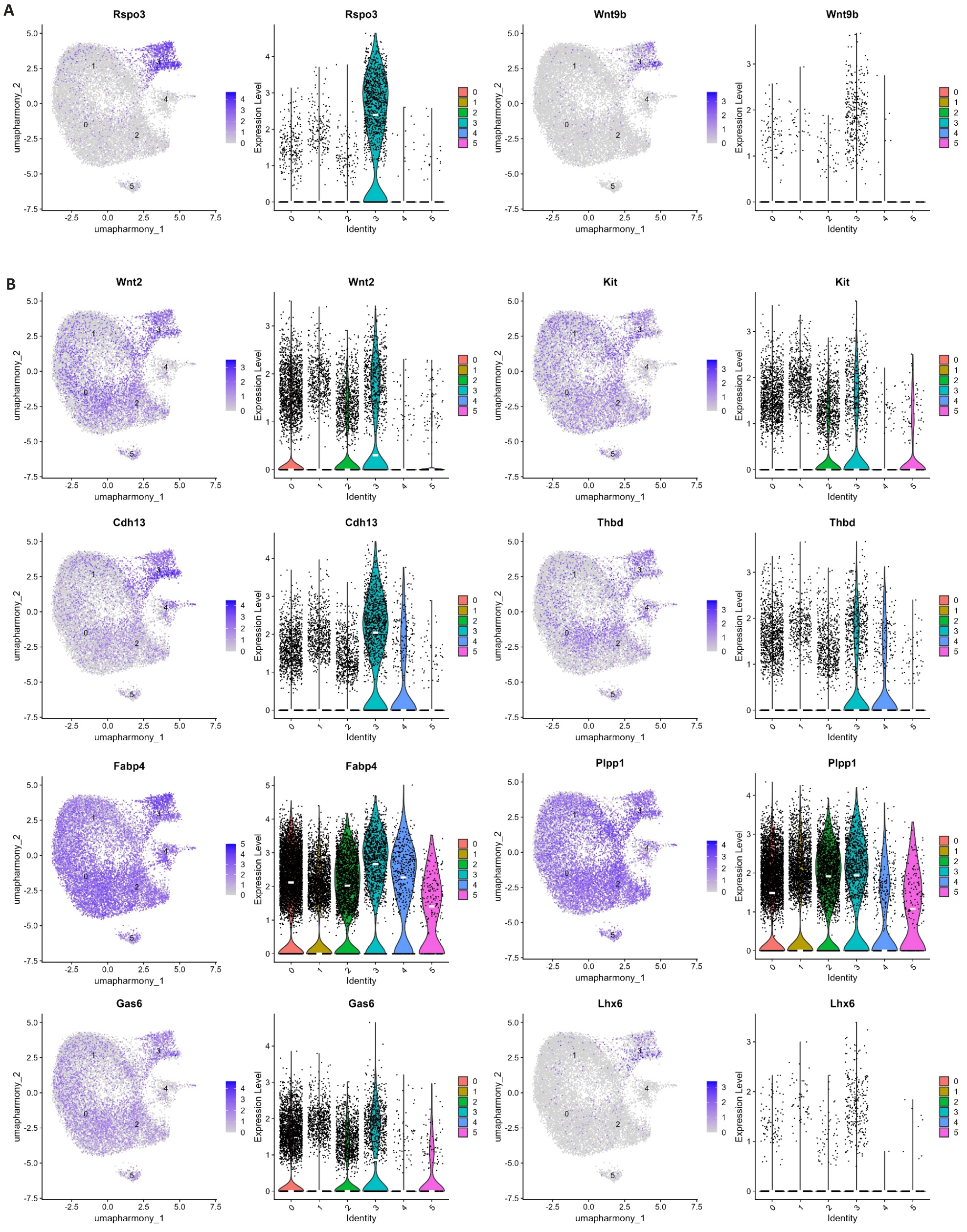
Fig.9 Expression of central vein endothelial cells marker genes. A: UMAP visualization and violin plots of Rspo3 and Wnt9b expression across clusters. B: UMAP visualization and violin plots of Wnt2, Kit, Cdh13, Thbd, Fabp4, Plpp1, Gas6, and Lhx6 expression across clusters.
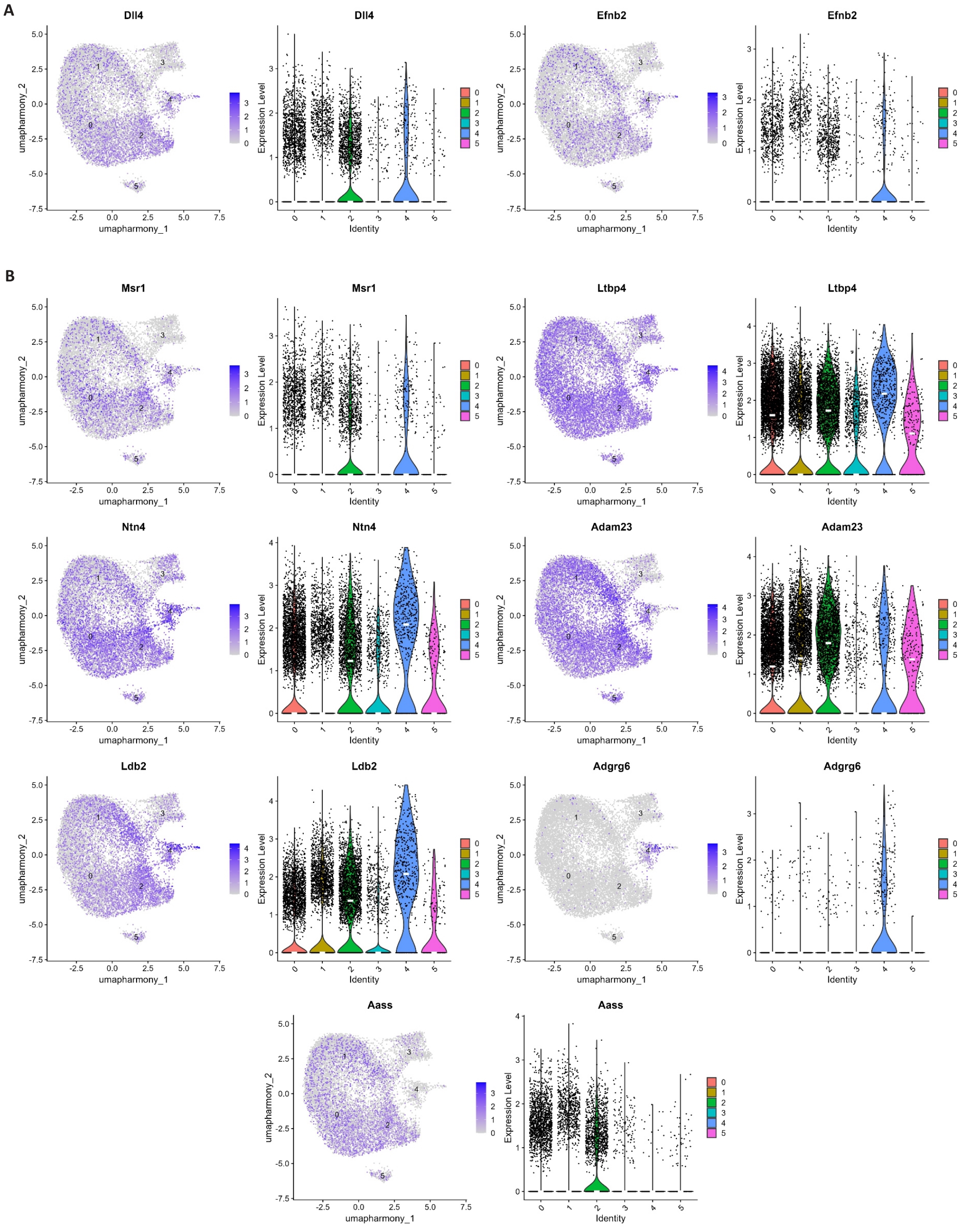
Fig.10 Expression profile of marker genes in arterial-like endothelial cells. A: UMAP visualization and violin plots of Dll4 and Efnb2 expression across clusters. B: UMAP visualization and violin plots of Msr1, Ltbp4, Ntn4, Adam23, Ldb2, Adgrg6, and Aass expression across clusters.
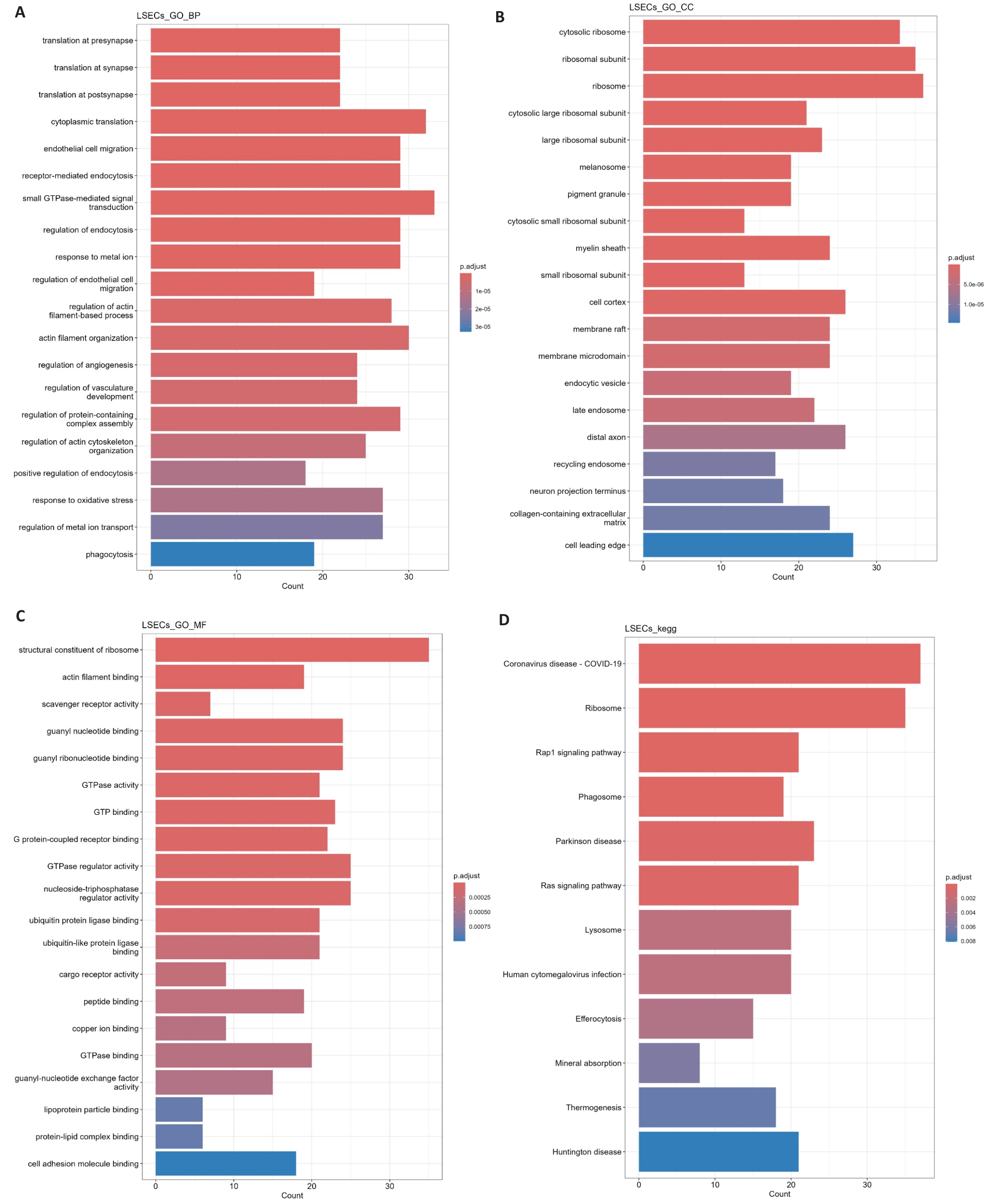
Fig.11 GO/KEGG enrichment analysis of differential genes in LSECs. A-C: Gene Ontology enrichment analysis of differentially expressed genes in LSECs, including biological process (A), cellular component (B), and molecular function (C). D: KEGG pathway enrichment analysis of differentially expressed genes in LSECs.
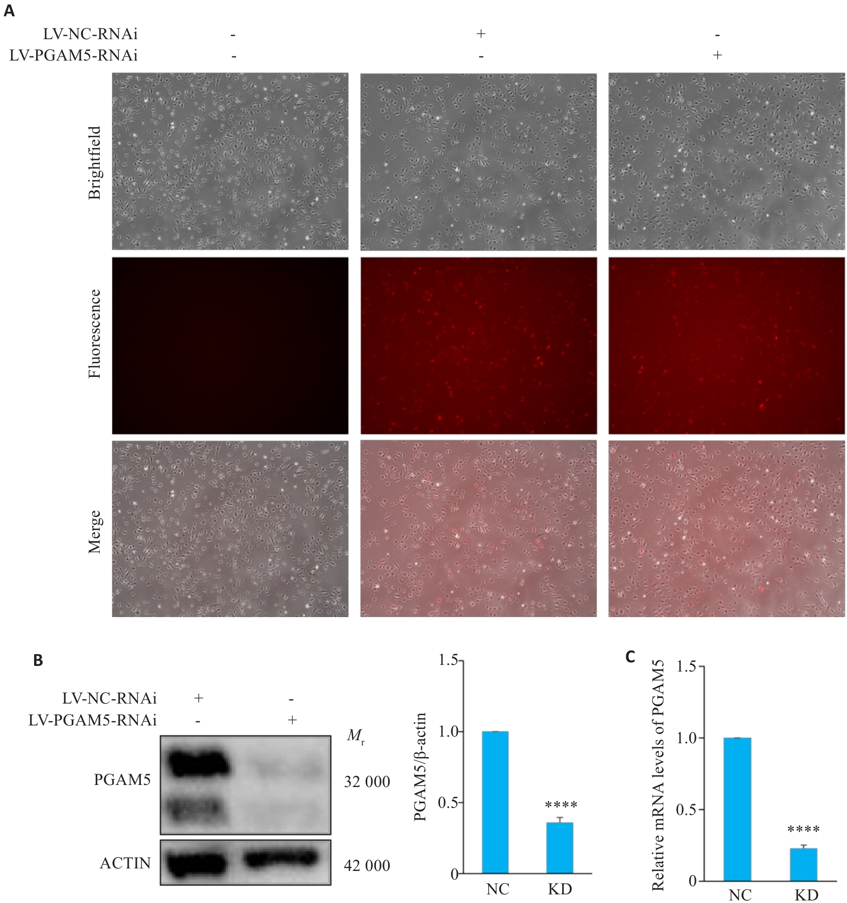
Fig.12 Validation of PGAM5 knockdown in HLSECs. A: Representative fluorescence images (×100) showing viral transduction efficiency in HLSECs. B: Western blotting of PGAM5 protein and quantitative analysis (n=3). C: Relative PGAM5 mRNA expression levels (n=3). ****P<0.0001 vs NC group.
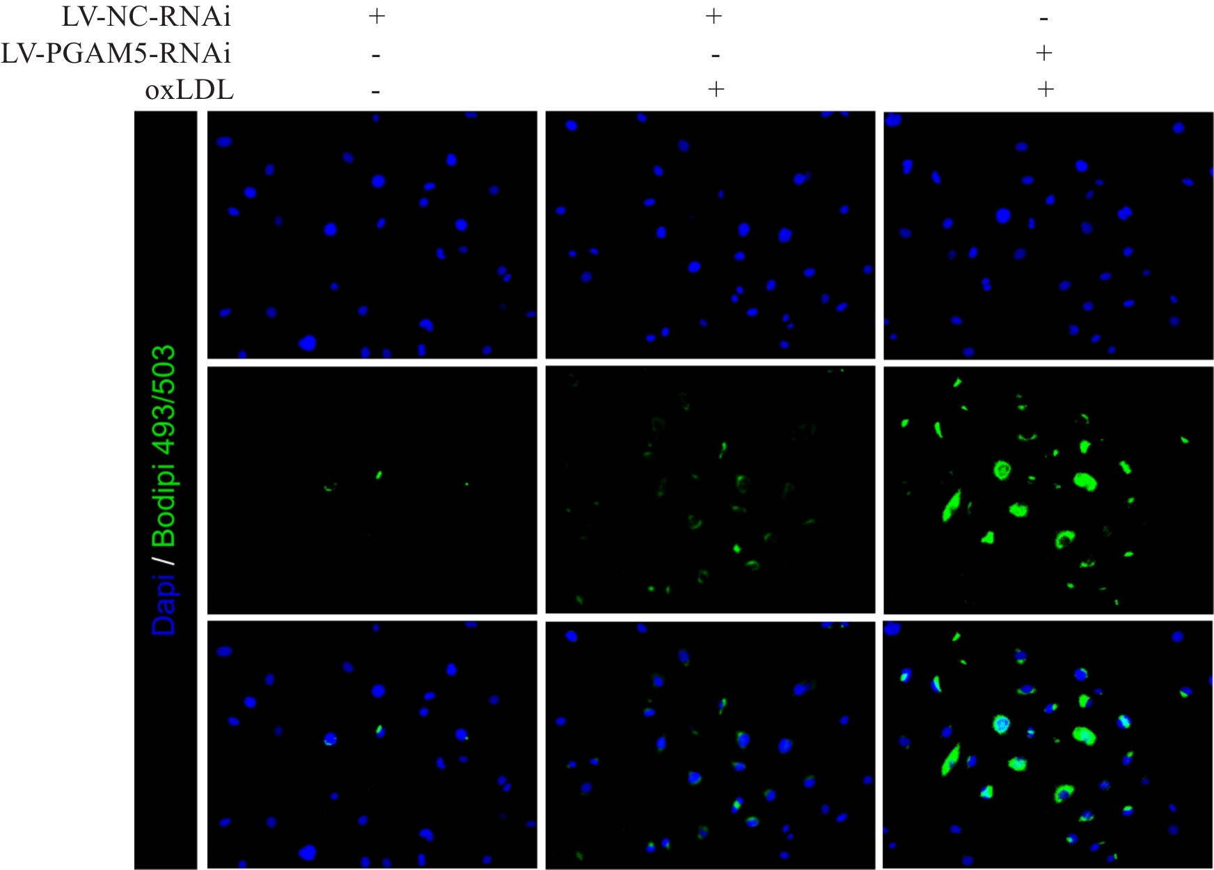
Fig.13 Bodipy staining of HLSECs after oxLDL treatment (×200).
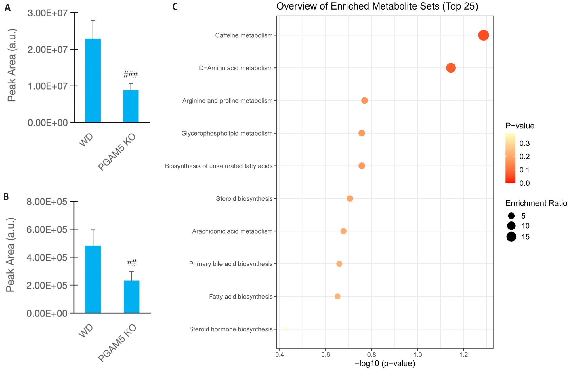
Fig.14 PGAM5 deficiency reshapes plasma metabolite profile in AS mice. A: Relative abundance of L-palmitoylcarnitine. B: Relative abundance of arachidonic acid. C: Pathway enrichment analysis of top differential metabolites (n=5). ##P<0.01, ###P<0.001 vs WD group.
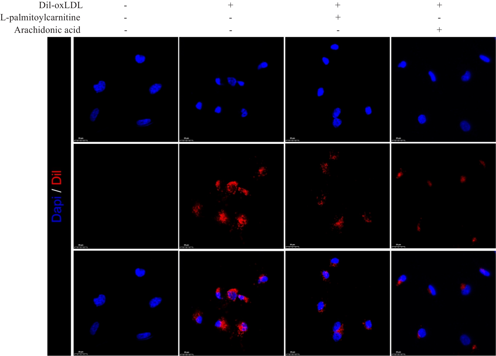
Fig.15 Dil-oxLDL uptake assay in HLSECs (×600).
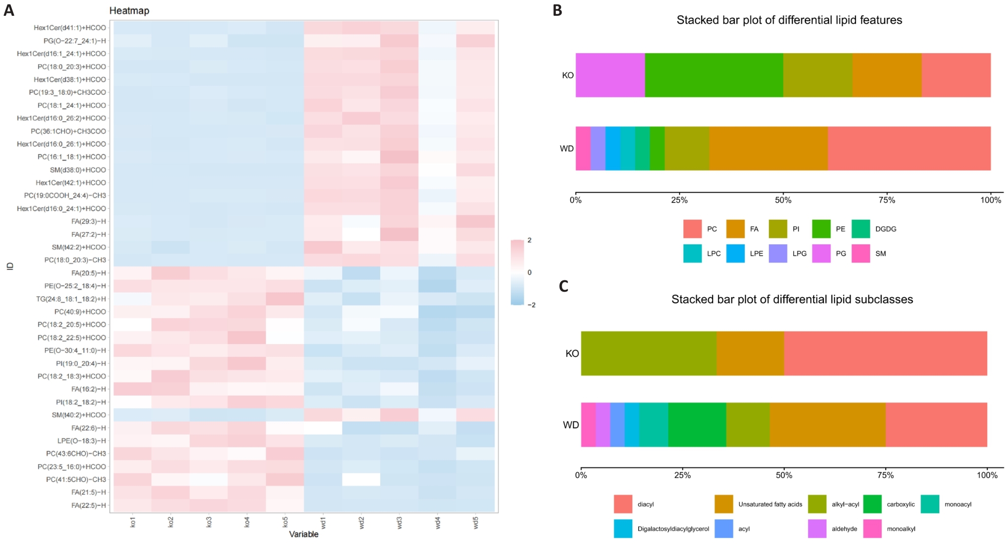
Fig.16 PGAM5 deficiency significantly alters the plasma metabolite profile in AS mice. A: Heatmap of the representative differential lipids. B: Distribution of differential lipid classes. C: Distribution of differential lipid subclasses.
| [1] | Temtem M, Mendonça MI, Santos M, et al. Validation of the SCORE2 risk prediction algorithm in a Portuguese population: a new model to estimate 10-year cardiovascular disease incidence in Europe[J]. Rev Portuguesa De Cardiol, 2024, 43(8): 437-44. doi:10.1016/j.repc.2023.10.011 |
| [2] | Fu JZ, Deng YH, Ma Y, et al. National and provincial-level prevalence and risk factors of carotid atherosclerosis in Chinese adults[J]. JAMA Netw Open, 2024, 7(1): e2351225. doi:10.1001/jamanetworkopen.2023.51225 |
| [3] | Gusev E, Sarapultsev A. Atherosclerosis and inflammation: insights from the theory of general pathological processes[J]. Int J Mol Sci, 2023, 24(9): 7910. doi:10.3390/ijms24097910 |
| [4] | Miano JM, Fisher EA, Majesky MW. Fate and state of vascular smooth muscle cells in atherosclerosis[J]. Circulation, 2021, 143(21): 2110-6. doi:10.1161/circulationaha.120.049922 |
| [5] | Liu MH, Chen RM, Zheng ZW, et al. Mechanisms of inflammatory microenvironment formation in cardiometabolic diseases: molecular and cellular perspectives[J]. Front Cardiovasc Med, 2025, 11: 1529903. doi:10.3389/fcvm.2024.1529903 |
| [6] | Mortensen MB, Dzaye O, Bøtker HE, et al. Low-density lipoprotein cholesterol is predominantly associated with atherosclerotic cardiovascular disease events in patients with evidence of coronary atherosclerosis: the western Denmark heart registry[J]. Circulation, 2023, 147(14): 1053-63. doi:10.1161/circulationaha.122.061010 |
| [7] | Zhang HL, Long XY, Niu GN, et al. Genetic insights into lipid traits and atherosclerosis risk: a Mendelian randomization and polygenic risk score analysis[J]. Int J Surg, 2025, 111(10): 6802-15. doi:10.1097/js9.0000000000002869 |
| [8] | Ference BA, Ginsberg HN, Graham I, et al. Low-density lipoproteins cause atherosclerotic cardiovascular disease. 1. Evidence from genetic, epidemiologic, and clinical studies. A consensus statement from the European Atherosclerosis Society Consensus Panel[J]. Eur Heart J, 2017, 38(32): 2459-72. doi:10.1093/eurheartj/ehx144 |
| [9] | Josefs T, Barrett TJ, Brown EJ, et al. Neutrophil extracellular traps promote macrophage inflammation and impair atherosclerosis resolution in diabetic mice[J]. JCI Insight, 2020, 5(7): e134796. doi:10.1172/jci.insight.134796 |
| [10] | Badmus OO, Hillhouse SA, Anderson CD, et al. Molecular mechanisms of metabolic associated fatty liver disease (MAFLD): functional analysis of lipid metabolism pathways[J]. Clin Sci, 2022, 136(18): 1347-66. doi:10.1042/cs20220572 |
| [11] | Zhang S, Hong FF, Ma C, et al. Hepatic lipid metabolism disorder and atherosclerosis[J]. Endocr Metab Immune Disord Drug Targets, 2022, 22(6): 590-600. doi:10.2174/1871530322666211220110810 |
| [12] | Zhu LY, Liu FZ, Shi CY, et al. Hepatic micropeptide modulates mitochondrial RNA processing machinery in hepatocellular carcinoma[J]. Mol Cell, 2025, 85(12): 2303-19.e7. doi:10.1016/j.molcel.2025.05.027 |
| [13] | Heeren J, Scheja L. Metabolic-associated fatty liver disease and lipoprotein metabolism[J]. Mol Metab, 2021, 50: 101238. doi:10.1016/j.molmet.2021.101238 |
| [14] | Rogge MM. The role of impaired mitochondrial lipid oxidation in obesity[J]. Biol Res Nurs, 2009, 10(4): 356-73. doi:10.1177/1099800408329408 |
| [15] | Mayr JA. Lipid metabolism in mitochondrial membranes[J]. J Inherit Metab Dis, 2015, 38(1): 137-44. doi:10.1007/s10545-014-9748-x |
| [16] | Ademowo OS, Dias HKI, Burton DGA, et al. Lipid (per) oxidation in mitochondria: an emerging target in the ageing process?[J]. Biogerontology, 2017, 18(6): 859-79. doi:10.1007/s10522-017-9710-z |
| [17] | Di L, Maiseyeu A. Low-density lipoprotein nanomedicines: mechanisms of targeting, biology, and theranostic potential[J]. Drug Deliv, 2021, 28(1): 408-21. doi:10.1080/10717544.2021.1886199 |
| [18] | Bruckdorfer KR. Antioxidants and CVD: symposium on ‘diet and CVD’[J]. Proc Nutr Soc, 2008, 67(2): 214-22. doi:10.1017/s0029665108007052 |
| [19] | Holte C, Szafranska K, Kruse L, et al. Highly oxidized albumin is cleared by liver sinusoidal endothelial cells via the receptors stabilin-1 and-2[J]. Sci Rep, 2023, 13(1): 19121. doi:10.1038/s41598-023-46462-9 |
| [20] | She L, Tu H, Zhang YZ, et al. Inhibition of phosphoglycerate mutase 5 reduces necroptosis in rat hearts following ischemia/reperfusion through suppression of dynamin-related protein 1[J]. Cardiovasc Drugs Ther, 2019, 33(1): 13-23. doi:10.1007/s10557-018-06848-8 |
| [21] | He WB, He WL, Sun ZC, et al. Physiological and pathological roles of PGAM5: an update and future trend[J]. iScience, 2025, 28(2): 111539. doi:10.1016/j.isci.2024.111539 |
| [22] | Qi YX, Rajbanshi B, Hao RH, et al. The dual role of PGAM5 in inflammation[J]. Exp Mol Med, 2025, 57(2): 298-311. doi:10.1038/s12276-025-01391-7 |
| [23] | Zhang Q, Zheng P, Pan Y, et al. Phosphoglycerate mutase 5 is important mediator for instigating arterial lipid accumulation and aggravating atherosclerosis[J]. JACC Basic Transl Sci, 2025, 10(5): 652-73. doi:10.1016/j.jacbts.2024.12.007 |
| [24] | Xu JS, Guo PC, Hao SJ, et al. A spatiotemporal atlas of mouse liver homeostasis and regeneration[J]. Nat Genet, 2024, 56(5): 953-69. doi:10.1038/s41588-024-01709-7 |
| [25] | Du YY, Wu SS, Xi SY, et al. ASH1L in hepatoma cells and hepatic stellate cells promotes fibrosis-associated hepatocellular carcinoma by modulating tumor-associated macrophages[J]. Adv Sci, 2024, 11(45): 2404756. doi:10.1002/advs.202404756 |
| [26] | Sinha S, Ali Q, Zhang T, et al. Aging disrupts hepatocyte zonation homeostasis in mice and humans[J]. Hepatology, 2026, 83(5): 1143-57. doi:10.1097/hep.0000000000001451 |
| [27] | Dai CY, Tsai YM, Chang CY, et al. Reconstruction of the hepatic microenvironment and pathological changes underlying type II diabetes through single-cell RNA sequencing[J]. Int J Biol Sci, 2024, 20(14): 5531-47. doi:10.7150/ijbs.99176 |
| [28] | Yang W, He H, Wang TT, et al. Single-cell transcriptomic analysis reveals a hepatic stellate cell-activation roadmap and myofibroblast origin during liver fibrosis in mice[J]. Hepatology, 2021, 74(5): 2774-90. doi:10.1002/hep.31987 |
| [29] | Rocha AS, Vidal V, Mertz M, et al. The angiocrine factor Rspondin3 is a key determinant of liver zonation[J]. Cell Rep, 2015, 13(9): 1757-64. doi:10.1016/j.celrep.2015.10.049 |
| [30] | Wang B, Zhao LD, Fish M, et al. Self-renewing diploid Axin2+ cells fuel homeostatic renewal of the liver[J]. Nature, 2015, 524(7564): 180-5. doi:10.1038/nature14863 |
| [31] | Halpern KB, Shenhav R, Massalha H, et al. Paired-cell sequencing enables spatial gene expression mapping of liver endothelial cells[J]. Nat Biotechnol, 2018, 36(10): 962-70. doi:10.1038/nbt.4231 |
| [32] | Su TT, Yang YL, Lai SC, et al. Single-cell transcriptomics reveals zone-specific alterations of liver sinusoidal endothelial cells in cirrhosis[J]. Cell Mol Gastroenterol Hepatol, 2021, 11(4): 1139-61. doi:10.1016/j.jcmgh.2020.12.007 |
| [33] | Lin ZY, Liu F, Shi PL, et al. Fatty acid oxidation promotes reprogramming by enhancing oxidative phosphorylation and inhibiting protein kinase C[J]. Stem Cell Res Ther, 2018, 9(1): 47. doi:10.1186/s13287-018-0792-6 |
| [34] | Berra-Romani R, Faris P, Negri S, et al. Arachidonic acid evokes an increase in intracellular Ca2+ concentration and nitric oxide production in endothelial cells from human brain microcirculation[J]. Cells, 2019, 8(7): 689. doi:10.3390/cells8070689 |
| [35] | Vial G, Dubouchaud H, Couturier K, et al. Effects of a high-fat diet on energy metabolism and ROS production in rat liver[J]. J Hepatol, 2011, 54(2): 348-56. doi:10.1016/j.jhep.2010.06.044 |
| [36] | Patten DA, Wilkinson AL, O'Keeffe A, et al. Scavenger receptors: novel roles in the pathogenesis of liver inflammation and cancer[J]. Semin Liver Dis, 2022, 42(1): 61-76. doi:10.1055/s-0041-1733876 |
| [37] | Shetty S, Lalor PF, Adams DH. Liver sinusoidal endothelial cells: gatekeepers of hepatic immunity[J]. Nat Rev Gastroenterol Hepatol, 2018, 15(9): 555-67. doi:10.1038/s41575-018-0020-y |
| [38] | Wilkinson AL, Qurashi M, Shetty S. The role of sinusoidal endothelial cells in the axis of inflammation and cancer within the liver[J]. Front Physiol, 2020, 11: 990. doi:10.3389/fphys.2020.00990 |
| [39] | Smedsrød B, Melkko J, Araki N, et al. Advanced glycation end products are eliminated by scavenger-receptor-mediated endocytosis in hepatic sinusoidal Kupffer and endothelial cells[J]. Biochem J, 1997, 322(2): 567-73. doi:10.1042/bj3220567 |
| [40] | Lee SJ, Evers S, Roeder D, et al. Mannose receptor-mediated regulation of serum glycoprotein homeostasis[J]. Science, 2002, 295(5561): 1898-901. doi:10.1126/science.1069540 |
| [41] | Lalor PF, Lai WK, Curbishley SM, et al. Human hepatic sinusoidal endothelial cells can be distinguished by expression of phenotypic markers related to their specialised functions in vivo [J]. World J Gastroenterol, 2006, 12(34): 5429-39. doi:10.3748/wjg.v12.i34.5429 |
| [42] | Hu YF, Li W, Cheng X, et al. Emerging roles and therapeutic applications of arachidonic acid pathways in cardiometabolic diseases[J]. Circ Res, 2024, 135(1): 222-60. doi:10.1161/circresaha.124.324383 |
| [43] | Badimon L, Vilahur G, Rocca B, et al. The key contribution of platelet and vascular arachidonic acid metabolism to the pathophysiology of atherothrombosis[J]. Cardiovasc Res, 2021, 117(9): 2001-15. doi:10.1093/cvr/cvab003 |
| [44] | Gong X, Qiu T, Zhou JY, et al. TGF-β1 promotes angiogenesis via endothelial-to-mesenchymal transition in infantile hemangioma[J]. Arterioscler Thromb Vasc Biol, 2025, 45(10): 1912-27. doi:10.1161/atvbaha.125.322793 |
| [45] | Ba HX, Guo YF, Jiang YJ, et al. Unveiling the metabolic landscape of pulmonary hypertension: insights from metabolomics[J]. Respir Res, 2024, 25(1): 221. doi:10.1186/s12931-024-02775-5 |
| [46] | Taiwo A, Merrill RA, Wendt L, et al. Metabolite perturbations in type 1 diabetes associated with metabolic dysfunction-associated steatotic liver disease[J]. Front Endocrinol, 2025, 16: 1500242. doi:10.3389/fendo.2025.1500242 |
| [47] | Chong SY, Wang XY, van Bloois L, et al. Injectable liposomal docosahexaenoic acid alleviates atherosclerosis progression and enhances plaque stability[J]. J Control Release, 2023, 360: 344-64. doi:10.1016/j.atherosclerosis.2023.06.702 |
| [48] | Djuricic I, Calder PC. N-3 fatty acids (EPA and DHA) and cardiovascular health-updated review of mechanisms and clinical outcomes[J]. Curr Atheroscler Rep, 2025, 27(1): 116. doi:10.1007/s11883-025-01363-2 |
| Viewed | ||||||
|
Full text |
|
|||||
|
Abstract |
|
|||||
