Journal of Southern Medical University ›› 2024, Vol. 44 ›› Issue (6): 1079-1087.doi: 10.12122/j.issn.1673-4254.2024.06.08
Previous Articles Next Articles
Jie CHEN1,2( ), Chenxu LIU1,2, Chun WANG2,3, Li LI1,2, Weiting TAO1,2, Jingru XUN4, Honghui TANG4, Li HUANG1,2()
Received:2023-07-14
Online:2024-06-20
Published:2024-07-01
Contact:
Li HUANG
E-mail:cj332022@163.com;zi05@163.com
Jie CHEN, Chenxu LIU, Chun WANG, Li LI, Weiting TAO, Jingru XUN, Honghui TANG, Li HUANG. Exogenous leptin improves cerebral ischemia-reperfusion-induced glutamate excitotoxic injury in mice by up-regulating GLT-1 and GLAST expression in astrocytes[J]. Journal of Southern Medical University, 2024, 44(6): 1079-1087.
Add to citation manager EndNote|Ris|BibTeX
URL: https://www.j-smu.com/EN/10.12122/j.issn.1673-4254.2024.06.08
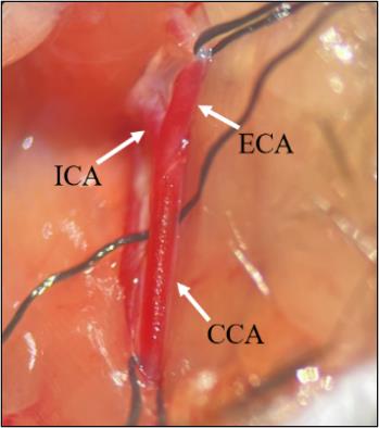
Fig.1 Microscopic observation of mouse right common carotid artery (CCA) and its branches.
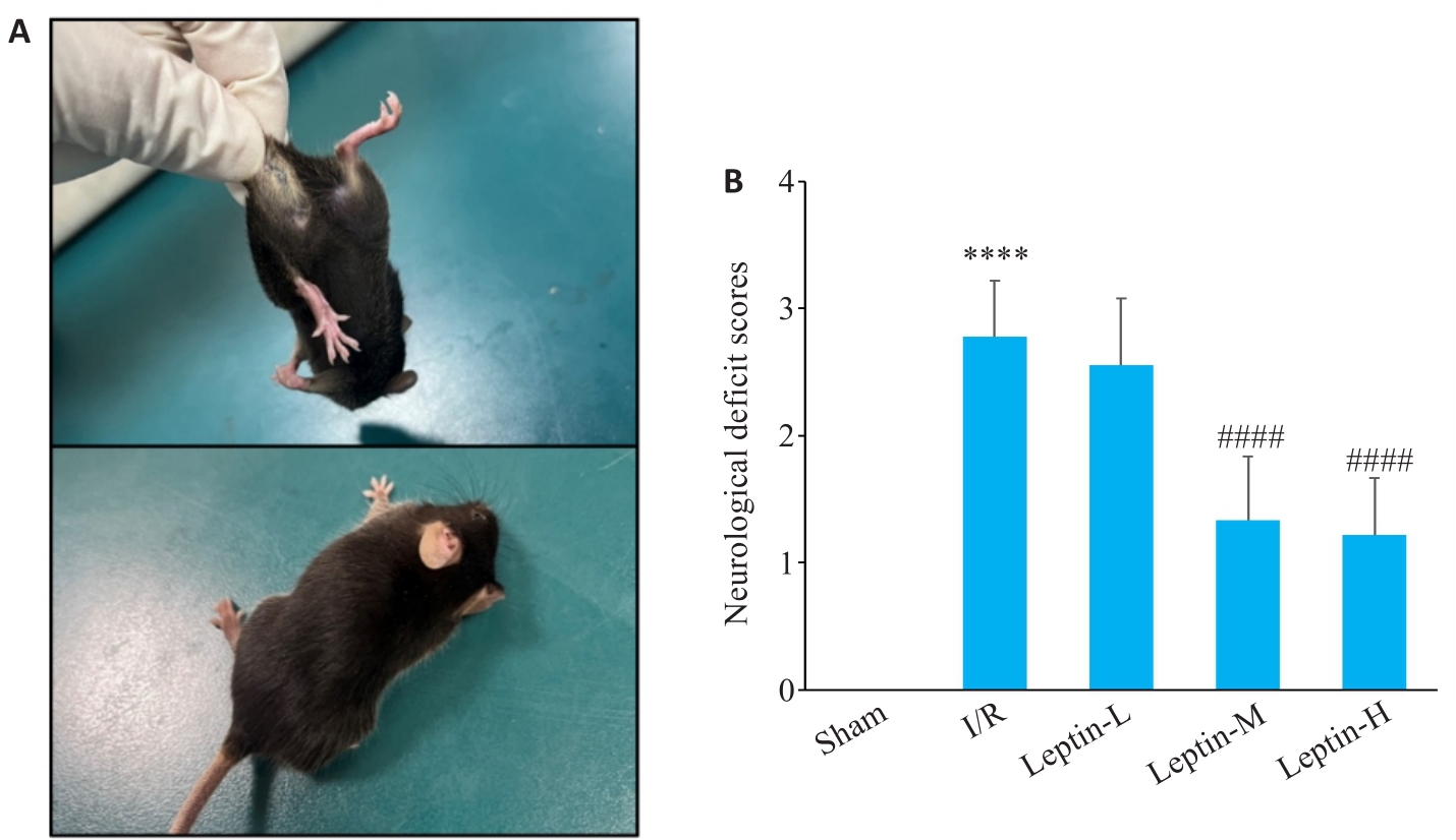
Fig.2 Posture of the mice following brain I/R modeling (A) and neurological deficit scores of the mice in different groups (B), n=9. ****P<0.0001 vs Sham group; ####P<0.0001 vs I/R group.
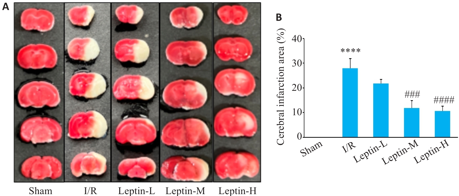
Fig.3 TTC staining for determining the size of cerebral infarct area of the mice. A: TTC staining of the brain tissue sections. B: Quantitative analysis of brain infarct area (n=3). ****P<0.0001 vs Sham group; ###P<0.001, ####P<0.0001 vs I/R group.
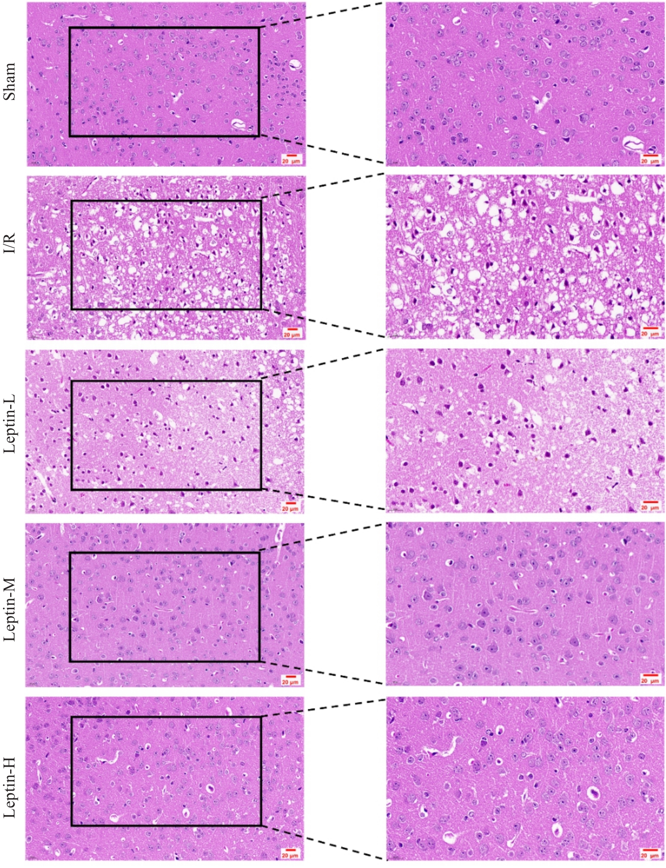
Fig.4 HE staining for examining histopathological changes in the cortical brain tissues of the mice (scale bar=20 μm).
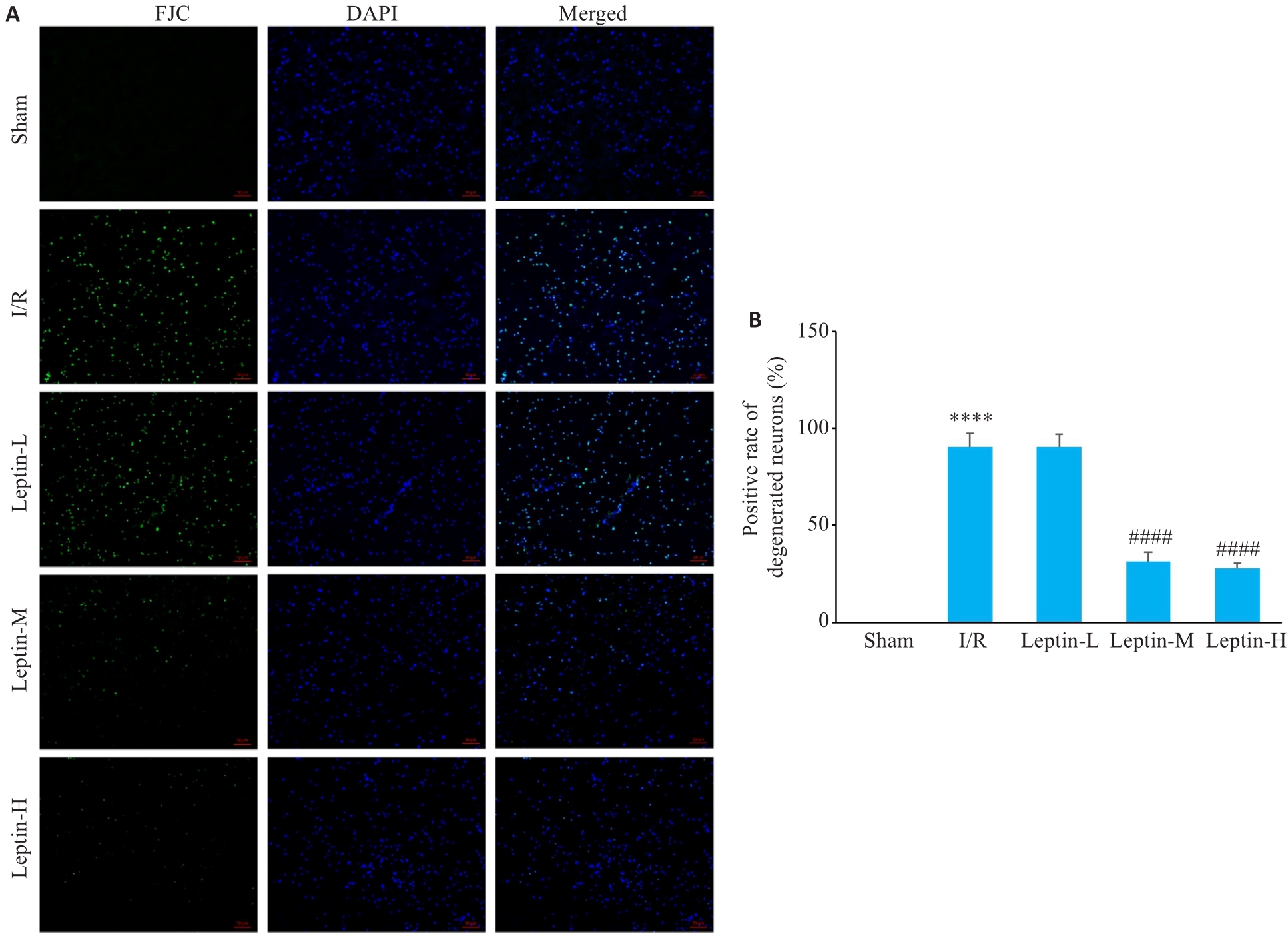
Fig.5 FJC staining for assessing cortical neuronal degenerative damage. A: FJC staining iamges (scale bar=20 μm). B: Positive rate of the degenerated neurons. ****P<0.0001 vs Sham group; ####P<0.0001 vs I/R group.
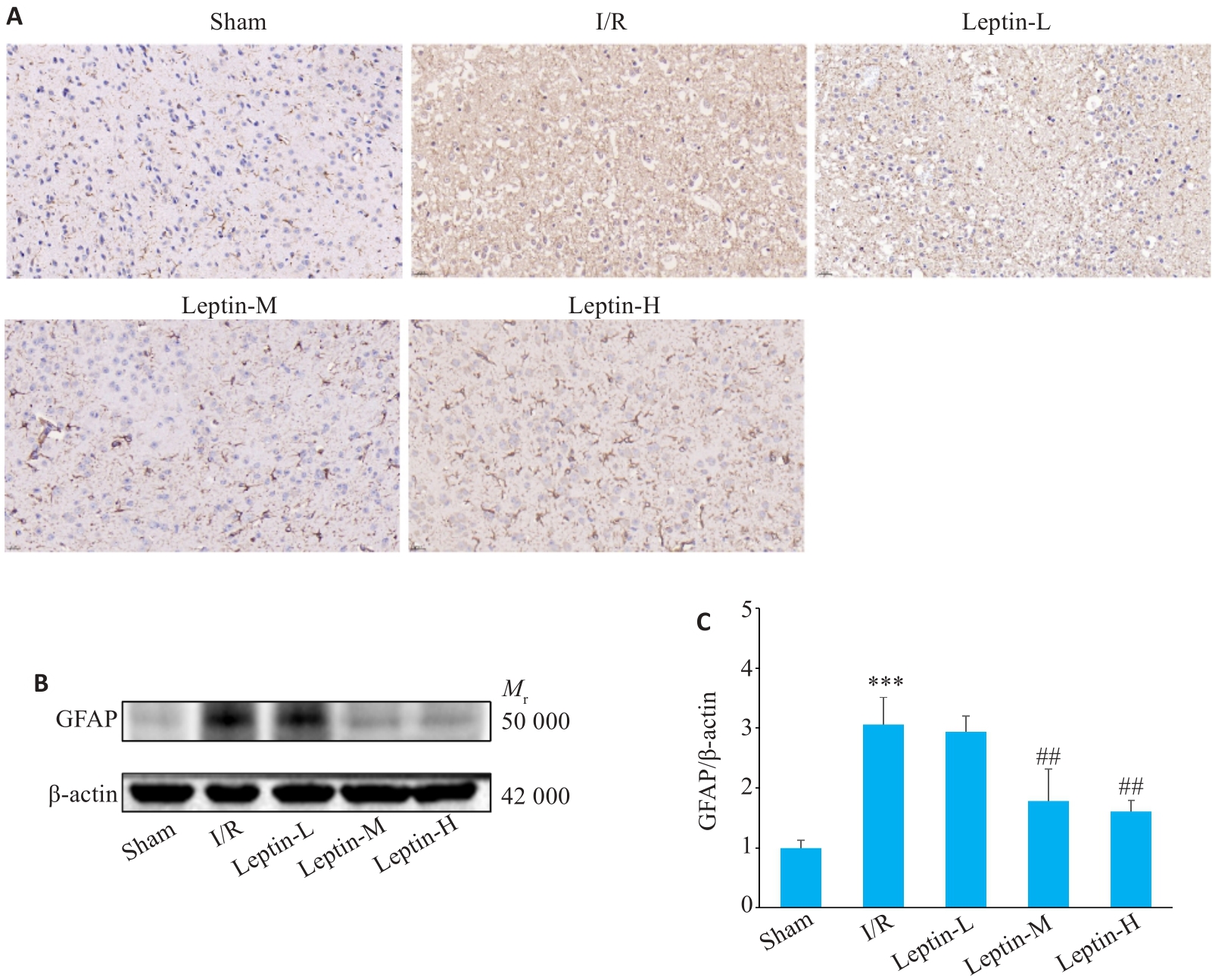
Fig.6 Expression of GFAP protein in mouse cortical brain tissue. A: Expression of GFAP detected by immunohistochemistry (Original magnification: ×40). B: Expression of GFAP detected by Western blotting (n=3). C: Quantitative analysis of GFAP expression. ***P<0.001 vs Sham group; ##P<0.01 vs I/R group.
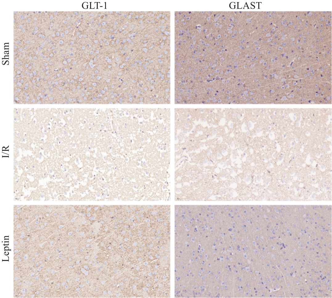
Fig.7 Expression of GLT-1 and GLAST proteins in mouse cortical brain tissue (×40).
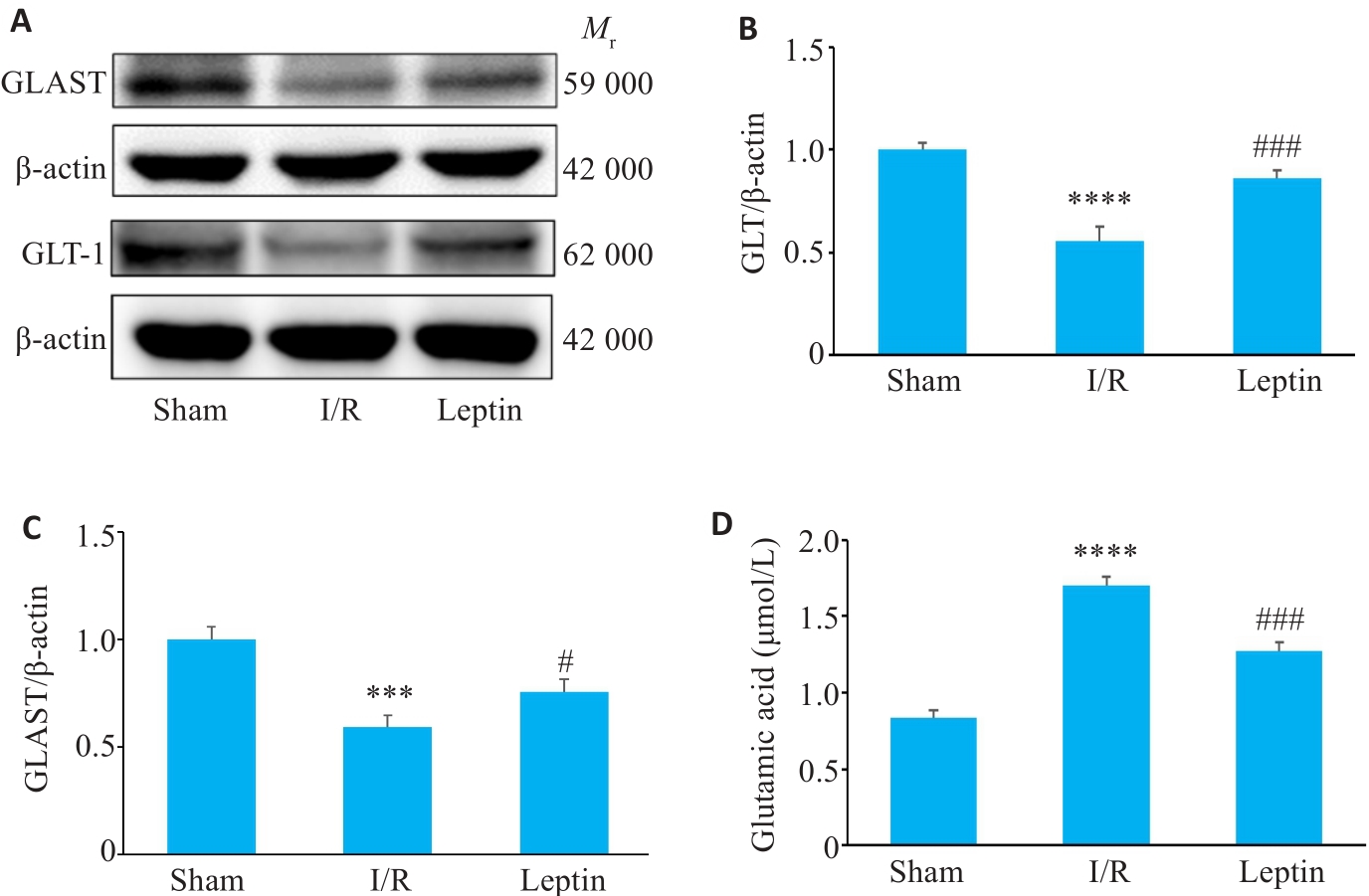
Fig.8 Changes in the expression of GLT-1 and GLAST proteins and Glu content in mouse cortical brain tissue. A: Expression of GLT-1 and GLAST detected by Western blotting (n=3). B: Quantitative analysis of GLT-1 expression. C: Quantitative analysis of GLAST expression; D: Content of Glu (n=3). ***P<0.001, ****P<0.0001 vs Sham group; #P<0.01, ###P<0.001 vs I/R group.
| 1 | Collaborators G 2 N. Global, regional, and national burden of neurological disorders, 1990-2016: a systematic analysis for the Global Burden of Disease Study 2016[J]. Lancet Neurol, 2019, 18(5): 459-80. |
| 2 | Campbell BCV, Khatri P. Stroke[J]. Lancet, 2020, 396(10244): 129-42. |
| 3 | Ghosh M, Lane M, Krizman E, et al. The transcription factor Pax6 contributes to the induction of GLT-1 expression in astrocytes through an interaction with a distal enhancer element[J]. J Neurochem, 2016, 136(2): 262-75. |
| 4 | Kim K, Lee SG, Kegelman TP, et al. Role of excitatory amino acid transporter-2 (EAAT2) and glutamate in neurodegeneration: opportunities for developing novel therapeutics[J]. J Cell Physiol, 2011, 226(10): 2484-93. |
| 5 | Murphy-Royal C, Dupuis J, Groc L, et al. Astroglial glutamate transporters in the brain: regulating neurotransmitter homeostasis and synaptic transmission[J]. J Neurosci Res, 2017, 95(11): 2140-51. |
| 6 | Verkhratsky A, Nedergaard M. Physiology of astroglia[J]. Physiol Rev, 2018, 98(1): 239-389. |
| 7 | Danbolt NC, Furness DN, Zhou Y. Neuronal vs glial glutamate uptake: resolving the conundrum[J]. Neurochem Int, 2016, 98: 29-45. |
| 8 | Malik AR, Willnow TE. Excitatory amino acid transporters in physiology and disorders of the central nervous system[J]. Int J Mol Sci, 2019, 20(22): 5671. |
| 9 | Melone M, Ciriachi C, Pietrobon D, et al. Heterogeneity of astrocytic and neuronal GLT-1 at cortical excitatory synapses, as revealed by its colocalization with Na+/K+-ATPase α isoforms[J]. Cereb Cortex, 2019, 29(8): 3331-50. |
| 10 | Roberts RC, Roche JK, McCullumsmith RE. Localization of excitatory amino acid transporters EAAT1 and EAAT2 in human postmortem cortex: a light and electron microscopic study[J]. Neuroscience, 2014, 277: 522-40. |
| 11 | Kashem MA, Sultana N, Pow DV, et al. GLAST (GLutamate and ASpartate Transporter) in human prefrontal cortex; interactome in healthy brains and the expression of GLAST in brains of chronic alcoholics[J]. Neurochem Int, 2019, 125: 111-6. |
| 12 | Danbolt NC. Glutamate uptake[J]. Prog Neurobiol, 2001, 65(1): 1-105. |
| 13 | Suárez-Pozos E, Thomason EJ, Fuss B. Glutamate transporters: expression and function in oligodendrocytes[J]. Neurochem Res, 2020, 45(3): 551-60. |
| 14 | Pajarillo E, Rizor A, Lee J, et al. The role of astrocytic glutamate transporters GLT-1 and GLAST in neurological disorders: potential targets for neurotherapeutics[J]. Neuropharmacology, 2019, 161: 107559. |
| 15 | Banks WA, Kastin AJ, Huang W, et al. Leptin enters the brain by a saturable system independent of insulin[J]. Peptides, 1996, 17(2): 305-11. |
| 16 | Hu SJ, Cheng DB, Peng DT, et al. Leptin attenuates cerebral ischemic injury in rats by modulating the mitochondrial electron transport chain via the mitochondrial STAT3 pathway[J]. Brain Behav, 2019, 9(2): e01200. |
| 17 | Zhang WF, Jin YC, Wang D, et al. Neuroprotective effects of leptin on cerebral ischemia through JAK2/STAT3/PGC-1-mediated mitochondrial function modulation[J]. Brain Res Bull, 2020, 156: 118-30. |
| 18 | Zhang Y, Cheng DB, Jie CX, et al. Leptin alleviates endoplasmic reticulum stress induced by cerebral ischemia/reperfusion injury via the PI3K/Akt signaling pathway[J]. Biosci Rep, 2022, 42(12): BSR20221443. |
| 19 | Longa EZ, Weinstein PR, Carlson S, et al. Reversible middle cerebral artery occlusion without craniectomy in rats[J]. Stroke, 1989, 20(1): 84-91. |
| 20 | Jiwaji Z, Hardingham GE. Good, bad, and neglectful: Astrocyte changes in neurodegenerative disease[J]. Free Radic Biol Med, 2022, 182: 93-9. |
| 21 | Liu ZW, AstrocytesChopp M., therapeutic targets for neuroprotection and neurorestoration in ischemic stroke[J]. Prog Neurobiol, 2016, 144: 103-20. |
| 22 | Pekny M, Wilhelmsson U, Pekna M. The dual role of astrocyte activation and reactive gliosis[J]. Neurosci Lett, 2014, 565: 30-8. |
| 23 | Michinaga S, Koyama Y. Pathophysiological responses and roles of astrocytes in traumatic brain injury[J]. Int J Mol Sci, 2021, 22(12): 6418. |
| 24 | Zhang SF, Shang DS, Shi H, et al. Function of astrocytes in neuroprotection and repair after ischemic stroke[J]. Eur Neurol, 2021, 84(6): 426-34. |
| 25 | Mahmoud S, Gharagozloo M, Simard C, et al. Astrocytes maintain glutamate homeostasis in the CNS by controlling the balance between glutamate uptake and release[J]. Cells, 2019, 8(2): 184. |
| 26 | Yeh TH, Hwang HM, Chen JJ, et al. Glutamate transporter function of rat hippocampal astrocytes is impaired following the global ischemia[J]. Neurobiol Dis, 2005, 18(3): 476-83. |
| 27 | Fontana AC. Current approaches to enhance glutamate transporter function and expression[J]. J Neurochem, 2015, 134(6): 982-1007. |
| 28 | Roettger V, Lipton P. Mechanism of glutamate release from rat hippocampal slices during in vitro ischemia[J]. Neuroscience, 1996, 75(3): 677-85. |
| 29 | Rossi DJ, Oshima T, Attwell D. Glutamate release in severe brain ischaemia is mainly by reversed uptake[J]. Nature, 2000, 403(6767): 316-21. |
| 30 | Shen YL, Lu HL, Xu RN, et al. The expression of GLAST and GLT1 in a transient cerebral ischemia Mongolian gerbil model[J]. Neuropsychiatr Dis Treat, 2020, 16: 789-800. |
| 31 | Rao VL, Dogan A, Bowen KK, et al. Antisense knockdown of the glial glutamate transporter GLT-1 exacerbates hippocampal neuronal damage following traumatic injury to rat brain[J]. Eur J Neurosci, 2001, 13(1): 119-28. |
| 32 | Naranjo V, Contreras A, Merino B, et al. Specific deletion of the astrocyte leptin receptor induces changes in hippocampus glutamate metabolism, synaptic transmission and plasticity[J]. Neuroscience, 2020, 447: 182-90. |
| 33 | Zhang J, Deng Z, Liao J, et al. Leptin attenuates cerebral ischemia injury through the promotion of energy metabolism via the PI3K/Akt pathway[J]. J Cereb Blood Flow Metab, 2013, 33(4): 567-74. |
| Viewed | ||||||
|
Full text |
|
|||||
|
Abstract |
|
|||||