
Journal of Southern Medical University ›› 2026, Vol. 46 ›› Issue (10): 2372-2383.doi: 10.12122/j.issn.1673-4254.2026.10.07
Zebin ZHOU1( ), Yulin YANG1, Huanchang YAN2, Xin WAN1, Zhirou LEI1, Haiying WANG1, Shixing TANG1,3()
Received:2026-03-20
Online:2026-10-20
Published:2026-10-26
Contact:
Shixing TANG
E-mail:smuzebin@163.com;tamgshixing@smu.edu.cn
Supported by:Zebin ZHOU, Yulin YANG, Huanchang YAN, Xin WAN, Zhirou LEI, Haiying WANG, Shixing TANG. Gagp17 protein KSKK→KSQQ substitution and ΔKSKK deletion mutations promote HIV-1 release and Gagp17 maturation in HIV-1 CRF55_01B and CRF59_01B strains[J]. Journal of Southern Medical University, 2026, 46(10): 2372-2383.
Add to citation manager EndNote|Ris|BibTeX
URL: https://www.j-smu.com/EN/10.12122/j.issn.1673-4254.2026.10.07
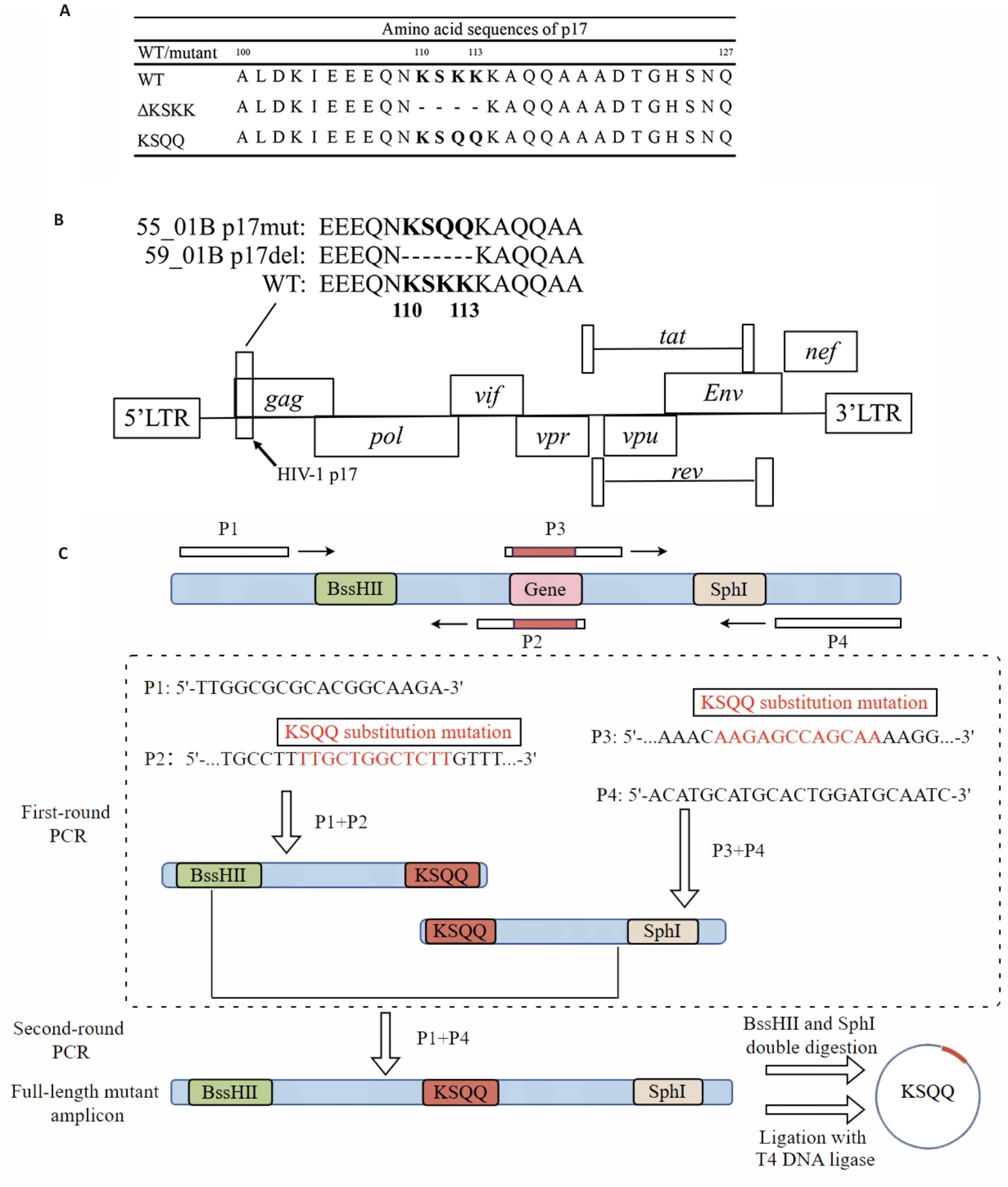
Fig.1 Schematic diagram of the HIV-1 genome, p17 mutants, and overlap PCR extension. A: HIV-1 genome is shown with a detailed view of p17 mutations, highlighting the KSQQ mutation and the KSKK deletion within Gagp17. B: Wild-type (WT) HIV-1 and Gagp17 mutants. The KSQQ mutation and KSKK deletion in Gagp17 are highlighted and compared with WT HIV-1. C: Construction of a KSQQ mutant gene fragment by overlap extension PCR.
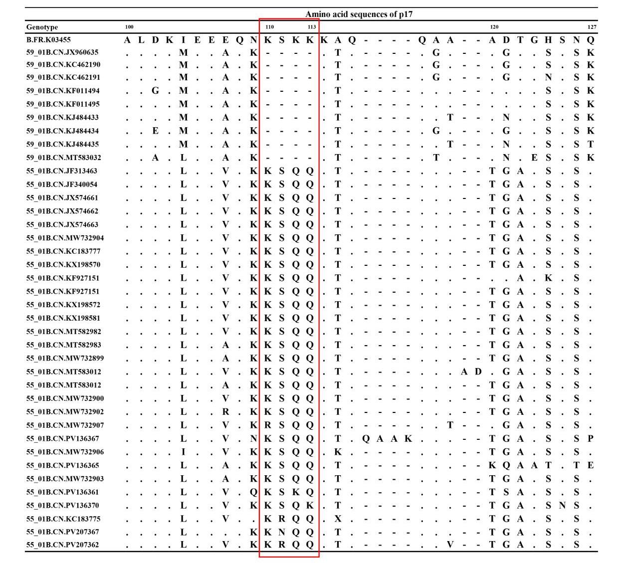
Fig.2 Alignment of HIV-1 Gagp17 amino acid sequences. The sequences were retrieved from the LANL HIV Sequence Database (https://www.hiv.lanl.gov/). The alignment was generated using MEGA6.014 and BioEdit7.1.9; numbering is based on HXB2 (B.FR.K03455). Dots (.) indicate residues identical to the HXB2 reference; dashes (-) denote amino acid deletions.
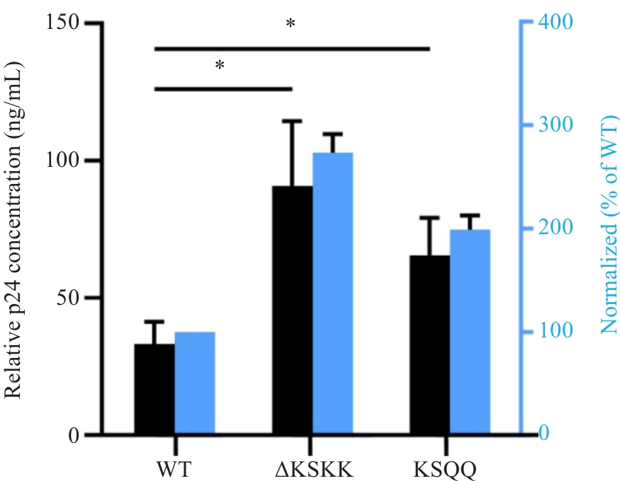
Fig.3 Detection of HIV-1 p24 concentration in the supernatant of cells transfected with HIV-1 p17 WT and mutants by using ELISA. Left y axis and black are the concentration of HIV-1 p24 while right y axis and blue are HIV-1 p24 level relative of that of WT (100%). WT: Wild type of pNL-4-3/KFS with KSKK served as the control. *P<0.05 (n=3).
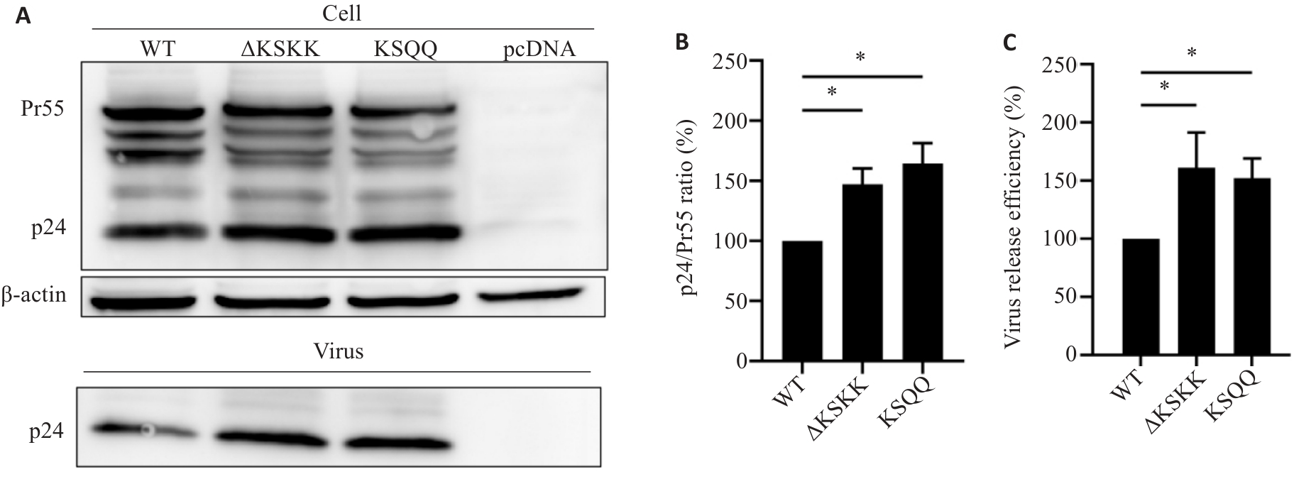
Fig.4 Evaluation of HIV-1 p17 mutant virus release and Gag protein processing efficiency by Western blotting. A: Western blotting for detecting cellular and viral proteins. B: Gag protein processing efficiency. C: Virus release efficiency. WT: pNL-4-3/KFS with KSKK served as the control, with its release and protein processing efficiencies as 100%. *P<0.05 (n=3).
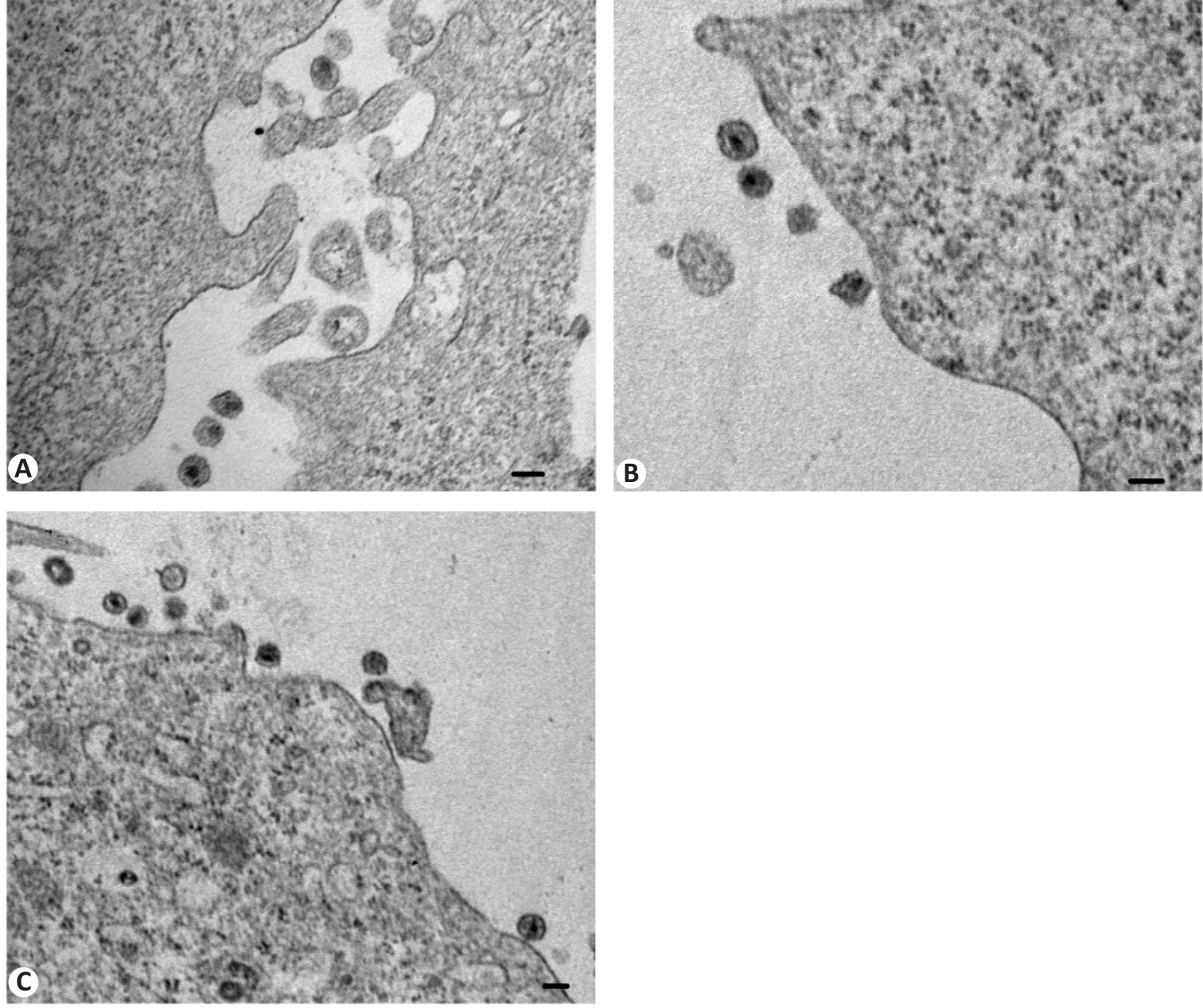
Fig.5 Electron micrographs of wild-type (WT) and Gagp17 mutant viral particles. A: WT. B: ΔKSKK deletion mutant. C: KSQQ substitution mutant. Scale bar=100 nm.
| Virus | Cell type | Number of particles (n) | Number of particles with different core shapes [n (%)] | ||
|---|---|---|---|---|---|
| Cone shaped | Centric | Acentric | |||
| WT | Hela | 33 | 6 (18.2)a | 12 (36.4) | 15 (45.4) |
| ΔKSKK | Hela | 55 | 15 (27.2) | 20 (36.4) | 20 (36.4) |
| KSQQ | Hela | 48 | 12 (25.0) | 19 (50.0) | 12 (25.0) |
Tab.1 Electron microscopy analysis of wild-type HIV-1 and Gagp17 mutant viral particles
| Virus | Cell type | Number of particles (n) | Number of particles with different core shapes [n (%)] | ||
|---|---|---|---|---|---|
| Cone shaped | Centric | Acentric | |||
| WT | Hela | 33 | 6 (18.2)a | 12 (36.4) | 15 (45.4) |
| ΔKSKK | Hela | 55 | 15 (27.2) | 20 (36.4) | 20 (36.4) |
| KSQQ | Hela | 48 | 12 (25.0) | 19 (50.0) | 12 (25.0) |
| Subtype/CRF | Total (n) | ΔKSKK [n (%)] | KSQQ [n (%)]** |
|---|---|---|---|
| C | 2996 | 38 (20.4) | 2958 (54.2) |
| 01_AE | 1670 | 17 (9.1) | 1653 (30.3) |
| 07_BC | 135 | 42 (22.6) | 93 (1.7) |
| F1 | 92 | 0 (0.0) | 92 (1.7) |
| BC | 87 | 25 (13.4) | 62 (1.1) |
| 01B | 69 | 1 (0.5) | 68 (1.2) |
| 01BC | 54 | 1 (0.5) | 53 (1.0) |
| G | 53 | 4 (2.2) | 49 (0.9) |
| 107 | 47 | 1 (0.5) | 46 (0.8) |
| 55_01B | 23 | 0 (0.0) | 23 (0.4) |
| BF1 | 21 | 1 (0.5) | 20 (0.4) |
| 01C | 18 | 2 (1.1) | 16 (0.3) |
| A1 | 16 | 6 (3.2) | 10 (0.2) |
| A1C | 12 | 2 (1.1) | 10 (0.2) |
| H | 10 | 3 (1.6) | 7 (0.1) |
| 07C | 10 | 3 (1.6) | 7 (0.1) |
| 145_0755 | 10 | 0 (0.0) | 10 (0.2) |
| 59_01B | 9 | 9 (4.8) | 0 (0.0) |
| 24_BG | 9 | 0 (0.0) | 9 (0.2) |
| 33_01B | 9 | 0 (0.0) | 9 (0.2) |
| F2 | 8 | 1 (0.5) | 7 (0.1) |
| 12_BF | 8 | 0 (0.0) | 8 (0.1) |
| 137_0107 | 8 | 0 (0.0) | 8 (0.1) |
| 02_AG | 7 | 1 (0.5) | 6 (0.1) |
| 85_BC | 7 | 0 (0.0) | 7 (0.1) |
| 119_0107 | 7 | 0 (0.0) | 7 (0.1) |
| 89_BF1 | 7 | 0 (0.0) | 7 (0.1) |
| B | 4 | 4 (2.2) | 0 (0.0) |
| 62_BC | 3 | 3 (1.6) | 0 (0.0) |
| 152_DG | 3 | 3 (1.6) | 0 (0.0) |
| Othersa | 233 | 19 (10.2) | 214 (3.9) |
| Total | 5645 | 186 | 5459 |
Tab.2 HIV-1 subtype distribution of the mutant viruses with ΔKSKK deletion mutation or KSKK→KSQQ substitution mutation in Gagp17 amino acids
| Subtype/CRF | Total (n) | ΔKSKK [n (%)] | KSQQ [n (%)]** |
|---|---|---|---|
| C | 2996 | 38 (20.4) | 2958 (54.2) |
| 01_AE | 1670 | 17 (9.1) | 1653 (30.3) |
| 07_BC | 135 | 42 (22.6) | 93 (1.7) |
| F1 | 92 | 0 (0.0) | 92 (1.7) |
| BC | 87 | 25 (13.4) | 62 (1.1) |
| 01B | 69 | 1 (0.5) | 68 (1.2) |
| 01BC | 54 | 1 (0.5) | 53 (1.0) |
| G | 53 | 4 (2.2) | 49 (0.9) |
| 107 | 47 | 1 (0.5) | 46 (0.8) |
| 55_01B | 23 | 0 (0.0) | 23 (0.4) |
| BF1 | 21 | 1 (0.5) | 20 (0.4) |
| 01C | 18 | 2 (1.1) | 16 (0.3) |
| A1 | 16 | 6 (3.2) | 10 (0.2) |
| A1C | 12 | 2 (1.1) | 10 (0.2) |
| H | 10 | 3 (1.6) | 7 (0.1) |
| 07C | 10 | 3 (1.6) | 7 (0.1) |
| 145_0755 | 10 | 0 (0.0) | 10 (0.2) |
| 59_01B | 9 | 9 (4.8) | 0 (0.0) |
| 24_BG | 9 | 0 (0.0) | 9 (0.2) |
| 33_01B | 9 | 0 (0.0) | 9 (0.2) |
| F2 | 8 | 1 (0.5) | 7 (0.1) |
| 12_BF | 8 | 0 (0.0) | 8 (0.1) |
| 137_0107 | 8 | 0 (0.0) | 8 (0.1) |
| 02_AG | 7 | 1 (0.5) | 6 (0.1) |
| 85_BC | 7 | 0 (0.0) | 7 (0.1) |
| 119_0107 | 7 | 0 (0.0) | 7 (0.1) |
| 89_BF1 | 7 | 0 (0.0) | 7 (0.1) |
| B | 4 | 4 (2.2) | 0 (0.0) |
| 62_BC | 3 | 3 (1.6) | 0 (0.0) |
| 152_DG | 3 | 3 (1.6) | 0 (0.0) |
| Othersa | 233 | 19 (10.2) | 214 (3.9) |
| Total | 5645 | 186 | 5459 |
| Time period | Total (n)b | ΔKSKK [n (%)] | KSQQ [n (%)]** |
|---|---|---|---|
| Before 2000 | 142 | 8 (4.3) | 134 (2.5) |
| 2000-2010 | 2102 | 117 (62.9) | 1985 (36.4) |
| After 2010 | 3401 | 61 (32.8) | 3340 (61.1) |
| Total | 5645 | 186 | 5459 |
Tab.3 Temporal distribution of the mutant viruses with ΔKSKK deletion mutation or KSKK→KSQQ substitution in Gagp17 amino acids
| Time period | Total (n)b | ΔKSKK [n (%)] | KSQQ [n (%)]** |
|---|---|---|---|
| Before 2000 | 142 | 8 (4.3) | 134 (2.5) |
| 2000-2010 | 2102 | 117 (62.9) | 1985 (36.4) |
| After 2010 | 3401 | 61 (32.8) | 3340 (61.1) |
| Total | 5645 | 186 | 5459 |
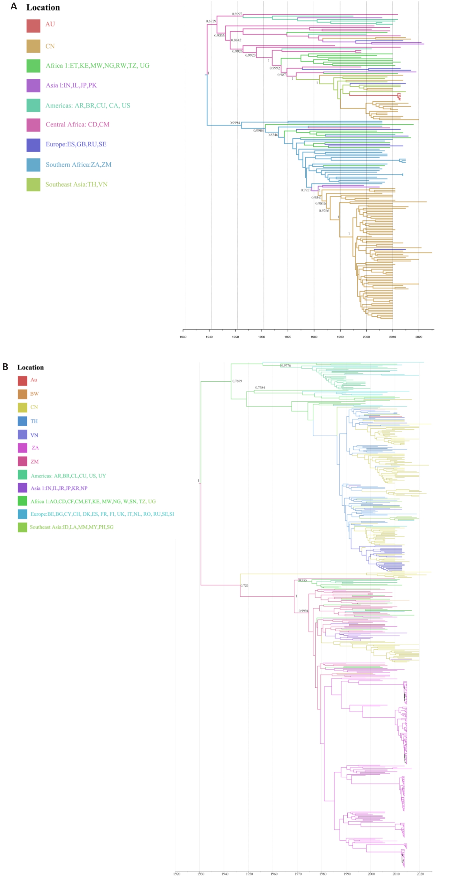
Fig.6 Spatiotemporal distribution of mutations in HIV-1 Gagp17. A: ΔKSKK deletion mutation. B: KSKK→KSQQ substitution mutation. Location: Country composition within each block.
| Region | Total outflow ∑PP | Total inflow ∑PP | Net flow: out-in | Regional role |
|---|---|---|---|---|
| Australia | 2.7619 | 0.6334 | 2.1285 | Super-hub |
| Southern africa | 2.1524 | 0 | 2.1524 | Primary source |
| Southeast asia | 1.7959 | 0.5807 | 1.2152 | Secondary source |
| Asia1 | 1.522 | 1.7731 | -0.2511 | Intermediate hub |
| China | 0.9626 | 1.4752 | -0.5126 | Receiver |
| Europe | 0.6948 | 1.3893 | -0.6945 | Receiver |
| Africa1 | 0.6039 | 1.833 | -1.2291 | Receiver |
| Central africa | 0 | 1.8842 | -1.8842 | Primary sink |
| Americas | 0 | 0.9246 | -0.9246 | Sink |
Tab.5 Net flow direction of transmission for HIV-1 ΔKSKK mutant virus
| Region | Total outflow ∑PP | Total inflow ∑PP | Net flow: out-in | Regional role |
|---|---|---|---|---|
| Australia | 2.7619 | 0.6334 | 2.1285 | Super-hub |
| Southern africa | 2.1524 | 0 | 2.1524 | Primary source |
| Southeast asia | 1.7959 | 0.5807 | 1.2152 | Secondary source |
| Asia1 | 1.522 | 1.7731 | -0.2511 | Intermediate hub |
| China | 0.9626 | 1.4752 | -0.5126 | Receiver |
| Europe | 0.6948 | 1.3893 | -0.6945 | Receiver |
| Africa1 | 0.6039 | 1.833 | -1.2291 | Receiver |
| Central africa | 0 | 1.8842 | -1.8842 | Primary sink |
| Americas | 0 | 0.9246 | -0.9246 | Sink |
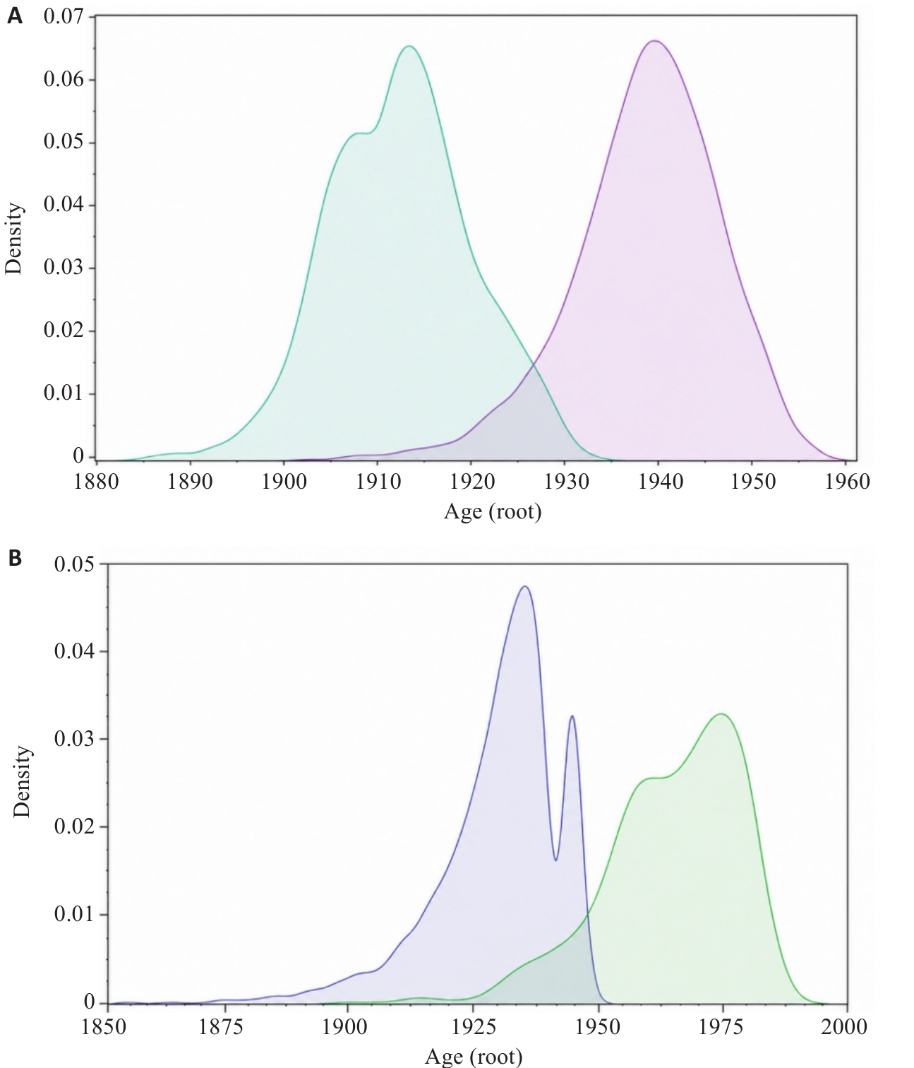
Fig.7 Comparison of prior and posterior density distributions for root node age (sensitivity analysis). Green line: prior density curve obtained from sampling the prior only (without data); Purple and pink line: posterior density curve obtained from the main analysis (including data). A: ΔKSKK deletion mutation. B: KSKK→KSQQ substitution mutation. The overlapping area between the prior and posterior curves is less than 20%. This figure is used to assess the sensitivity of phylogeographic inference to prior assumptions.
| [1] | Wang D, Feng Y, Hao JJ, et al. National and regional molecular epidemiology of HIV-1: China, 2004-2023[J]. China CDC Week, 2024, 6: 1257-63. |
| [2] | Gui T, Zhao J, Sun CR, et al. Genetic characterization of a unique recombinant originating from CRF55_01B, CRF01_AE, and CRF07_BC in Shenzhen, China[J]. AIDS Res Hum Retroviruses, 2015, 31(5): 559-63. doi:10.1089/aid.2014.0337 |
| [3] | 唐 虎, 杨梓杰, 郑陈丽, 等. 深圳市2022—2023年新报告HIV/AIDS患者HIV-1分子传播网络特征[J]. 中国艾滋病性病, 2025, 31(3): 254-60. |
| [4] | Luo YF, Wu H, Liang CY, et al. Molecular cluster, transmission characteristics, origin and dynamics analysis of HIV-1 CRF59_01B in China: a molecular epidemiology study[J]. Acta Trop, 2024, 260: 107396. doi:10.1016/j.actatropica.2024.107396 |
| [5] | Yan HC, Wu H, Li SM, et al. The origin and spread of HIV-1 CRF59_01B epidemic in China: a molecular network and phylogeographic analysis[J]. J Med Virol, 2024, 96(7): e29799. doi:10.1002/jmv.29799 |
| [6] | Qu K, Ke ZL, Zila V, et al. Maturation of the matrix and viral membrane of HIV-1[J]. Science, 2021, 373(6555): 700-4. doi:10.1126/science.abe6821 |
| [7] | Wen Y, Feigenson GW, Vogt VM, et al. Mechanisms of PI(4, 5)P2 enrichment in HIV-1 viral membranes[J]. J Mol Biol, 2020, 432(19): 5343-64. doi:10.1016/j.jmb.2020.07.018 |
| [8] | Zhang YX, Li MZ, Lin FY, et al. HIV-1 matrix protein p17: a key factor in HIV-associated cancers[J]. Front Virol, 2025, 5: 1584507. doi:10.3389/fviro.2025.1584507 |
| [9] | Cabrera-Rodríguez R, Pérez-Yanes S, Lorenzo-Sánchez I, et al. HIV infection: shaping the complex, dynamic, and interconnected network of the cytoskeleton[J]. Int J Mol Sci, 2023, 24(17): 13104. doi:10.3390/ijms241713104 |
| [10] | Stacey JCV, Hrebík D, Nand E, et al. The conserved HIV-1 spacer peptide 2 triggers matrix lattice maturation[J]. Nature, 2025, 640(8057): 258-64. doi:10.1038/s41586-025-08624-9 |
| [11] | Zhang LF, Li S, Xu XY, et al. HIV-1 p17 matrix protein enhances type I interferon responses through the p17-OLA1-STING axis[J]. J Cell Sci, 2024, 137(2): jcs261500. doi:10.1242/jcs.261500 |
| [12] | Samal AB, Green TJ, Saad JS. Atomic view of the HIV-1 matrix lattice; implications on virus assembly and envelope incorporation[J]. Proc Natl Acad Sci U S A, 2022, 119(23): e2200794119. doi:10.1073/pnas.2200794119 |
| [13] | Bukrinskaya A. HIV-1 matrix protein: a mysterious regulator of the viral life cycle[J]. Virus Res, 2007, 124(1/2): 1-11. doi:10.1016/j.virusres.2006.07.001 |
| [14] | Morikawa Y, Kishi T, Zhang WH, et al. A molecular determinant of human immunodeficiency virus particle assembly located in matrix antigen p17[J]. J Virol, 1995, 69(7): 4519-23. doi:10.1128/jvi.69.7.4519-4523.1995 |
| [15] | Bielejec F, Baele G, Vrancken B, et al. SpreaD3: interactive visualization of spatiotemporal history and trait evolutionary processes[J]. Mol Biol Evol, 2016, 33(8): 2167-9. doi:10.1093/molbev/msw082 |
| [16] | Tee KK, Thomson MM, Hemelaar J. Editorial: HIV-1 genetic diversity, volume II[J]. Front Microbiol, 2022, 13: 1007037. doi:10.3389/fmicb.2022.1007037 |
| [17] | Yan HC, Luo YF, Wu H, et al. Evolving molecular HIV clusters revealed genotype-specific dynamics in Guangzhou, China (2008-2020)[J]. Int J Infect Dis, 2024, 148: 107218. doi:10.1016/j.ijid.2024.107218 |
| [18] | Lacouture C, Carrio B, Favard C, et al. HIV-1 assembly–when virology meets biophysics[J]. J Cell Sci, 2024, 137(19): jcs262064. doi:10.1242/jcs.262064 |
| [19] | Sumner C, Ono A. Relationship between HIV-1 gag multimerization and membrane binding[J]. Viruses, 2022, 14(3): 622. doi:10.3390/v14030622 |
| [20] | Freed EO. HIV-1 assembly, release and maturation[J]. Nat Rev Microbiol, 2015, 13(8): 484-96. doi:10.1038/nrmicro3490 |
| [21] | Chukkapalli V, Hogue IB, Boyko V, et al. Interaction between the human immunodeficiency virus type 1 Gag matrix domain and phosphatidylinositol-(4, 5)‑bisphosphate is essential for efficient gag membrane binding[J]. J Virol, 2008, 82(5): 2405-17. doi:10.1128/jvi.01614-07 |
| [22] | Breitschwerdt S, Grandel B, Asbach B, et al. Consensus sequences for gag and pol introduced into HIV-1 clade B laboratory strains differentially influence the impact of point mutations associated with immune escape and with drug resistance on viral replicative capacity[J]. Viruses, 2025, 17(6): 842. doi:10.3390/v17060842 |
| [23] | Sonawane A, Selvam D, Yue L, et al. Virulence and replicative fitness of HIV-1 transmitted/founder (T/F) viruses harbouring drug resistance-associated mutation[J]. Viruses, 2024, 16(12): 1854. doi:10.3390/v16121854 |
| [24] | Mattei S, Glass B, Hagen WJH, et al. The structure and flexibility of conical HIV-1 capsids determined within intact virions[J]. Science, 2016, 354(6318): 1434-7. doi:10.1126/science.aah4972 |
| [25] | Worobey M, Gemmel M, Teuwen DE, et al. Direct evidence of extensive diversity of HIV-1 in Kinshasa by 1960[J]. Nature, 2008, 455(7213): 661-4. doi:10.1038/nature07390 |
| [26] | Zhang WQ, Han XX, An MH, et al. Identification and characterization of a novel HIV-1 circulating recombinant form (CRF59_01B) identified among men-who-have-sex-with-men in China[J]. PLoS One, 2014, 9(6): e99693. doi:10.1371/journal.pone.0099693 |
| [27] | Han XX, An MH, Zhang WQ, et al. Genome sequences of a novel HIV-1 circulating recombinant form, CRF55_01B, identified in China[J]. Genome Announc, 2013, 1(1): e00050-12. doi:10.1128/genomea.00050-12 |
| Viewed | ||||||
|
Full text |
|
|||||
|
Abstract |
|
|||||
