Journal of Southern Medical University ›› 2025, Vol. 45 ›› Issue (8): 1599-1607.doi: 10.12122/j.issn.1673-4254.2025.08.04
Kun WANG1,2( ), Haiyan ZUO2, Jiaojiao ZHANG2, Xin WU2, Wenhui WANG2, Shengbing WU2, Meiqi ZHOU2()
Received:2025-04-01
Online:2025-08-20
Published:2025-09-05
Contact:
Meiqi ZHOU
E-mail:ahwk0819@ahtcm.edu.cn;meiqizhou@163.com
Supported by:Kun WANG, Haiyan ZUO, Jiaojiao ZHANG, Xin WU, Wenhui WANG, Shengbing WU, Meiqi ZHOU. Electroacupuncture improves myocardial injury in rats with acute myocardial ischemia by inhibiting HPA axis hyperactivity via modulating hippocampal glutamatergic system[J]. Journal of Southern Medical University, 2025, 45(8): 1599-1607.
Add to citation manager EndNote|Ris|BibTeX
URL: https://www.j-smu.com/EN/10.12122/j.issn.1673-4254.2025.08.04
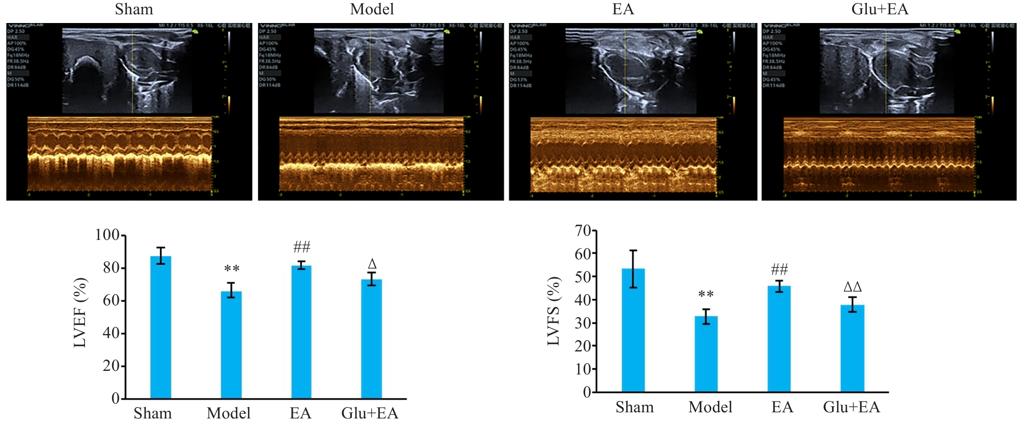
Fig.1 Echocardiography of the rats in the 4 groups. **P<0.01 vs Sham group; ##P<0.01 vs Model group; ∆P<0.05, ∆∆P<0.01 vs EA group.
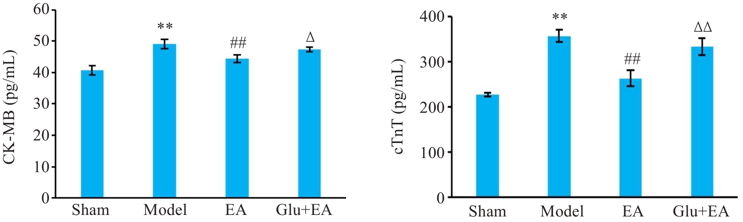
Fig.2 Serum levels of CK-MB and cTnT of the rats. **P<0.01 vs Sham group; ##P<0.01 vs Model group; ∆P<0.05, ∆∆P<0.01 vs EA group.

Fig.3 Pathological changes in the myocardial tissue of the rats (HE staining, scale bar=50 μm).
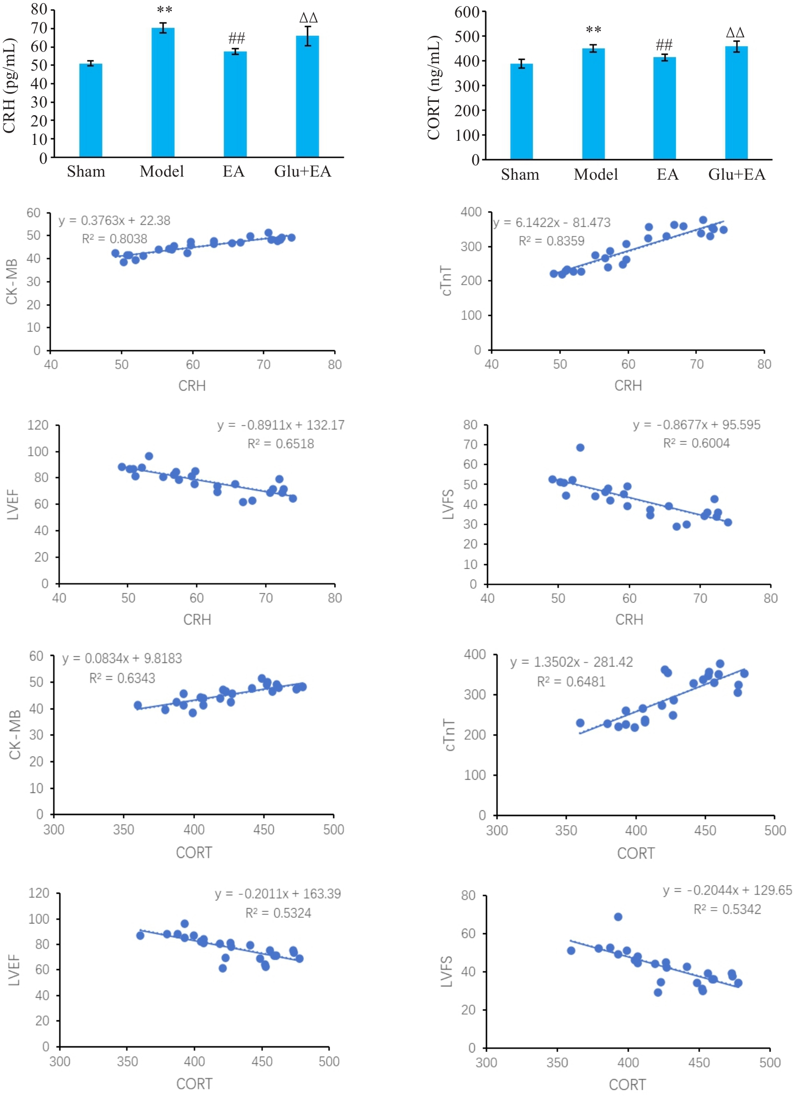
Fig.4 Serum levels of CRH and CORT of the rats. **P<0.01 vs Sham group; ##P<0.01 vs Model group; ∆∆P<0.01 vs EA group.
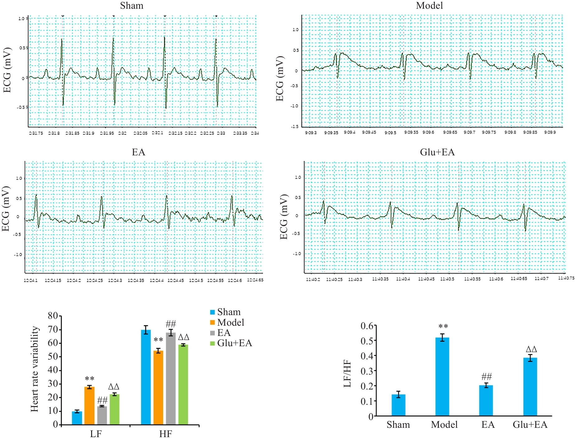
Fig.5 Heart rate variability of the rats. **P<0.01 vs Sham group; ##P<0.01 vs Model group; ∆∆P<0.01 vs EA group. LF: Low frequency; HF: High frequency.

Fig.6 Expressions of TH and GAP 43 in the myocardial tissue of the rats (Immunofluorescence staining, scale bar=20 μm). **P<0.01 vs Sham group; ##P<0.01 vs Model group; ∆∆P<0.01 vs EA group.
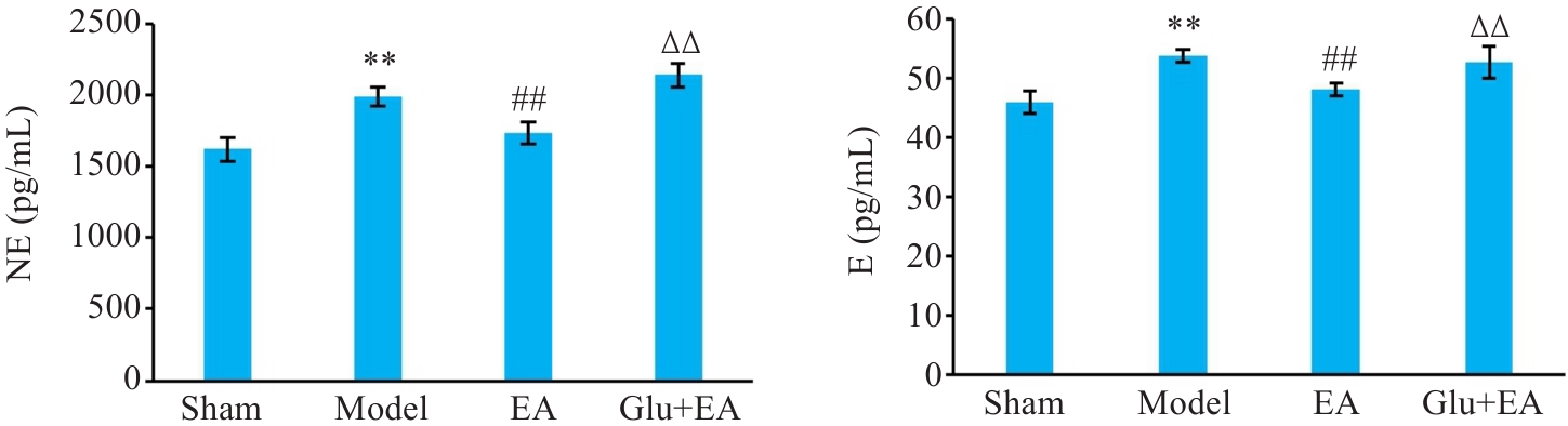
Fig.7 Serum levels of NE and E of the rats. **P<0.01 vs Sham group; ##P<0.01 vs Model group; ∆∆P<0.01 vs EA group.
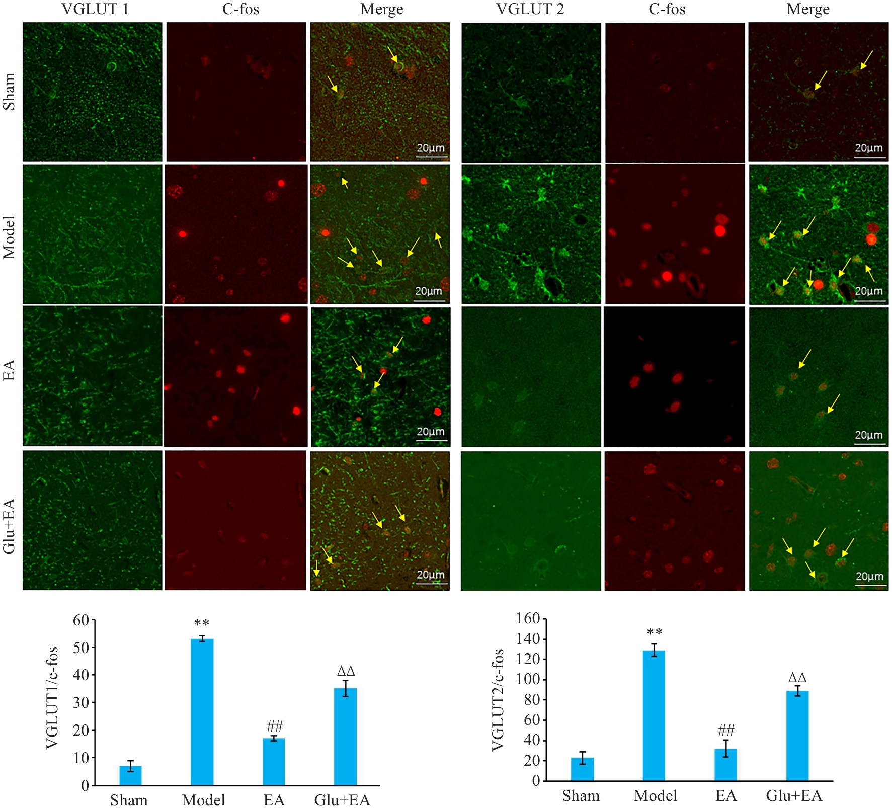
Fig.8 Colocalization of VGLUT1, VGLUT2, and c-fos in rat hippocampus in different groups (immunofluorescence staining, scale bar=20 μm). **P<0.01 vs Sham group; ##P<0.01 vs Model group; ∆∆P<0.01 vs EA group.
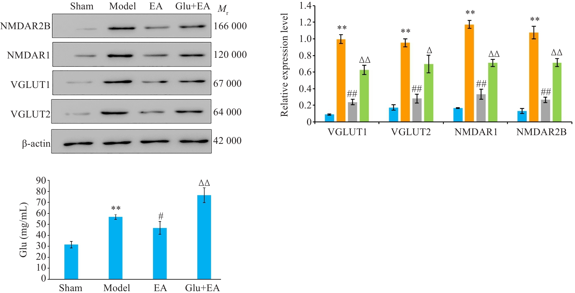
Fig.9 Expressions of VGLUT1, VGLUT2, NMDAR1, NMDAR2B and Glu in the hippocampus of the rats. **P<0.01 vs Sham group; #P<0.05, ##P<0.01 vs Model group; ∆∆P<0.01, ∆P<0.05 vs EA group.
| [1] | Rong W, Li J, Pan D, et al. Cardioprotective mechanism of leonurine against myocardial ischemia through a liver-cardiac crosstalk metabolomics study[J]. Biomolecules, 2022, 12(10): 1512. doi:10.3390/biom12101512 |
| [2] | Oliveira JB, Soares AASM, Sposito AC. Chapter two inflammatory response during myocardial infarction[J]. Adv Clin Chem, 2018, 84: 39-79. doi:10.1016/bs.acc.2017.12.002 |
| [3] | 王 阶, 陈 光. 冠心病稳定型心绞痛中医诊疗专家共识[J]. 中医杂志, 2018, 59(5): 447-50. |
| [4] | DeVon HA, Uwizeye G, Cai HY, et al. Feasibility and preliminary efficacy of acupuncture for angina in an underserved diverse population[J]. Acupunct Med, 2022, 40(2): 152-9. doi:10.1177/09645284211055754 |
| [5] | Wang N, Ma J, Ma Y, et al. Electroacupuncture pretreatment mitigates myocardial ischemia/reperfusion injury via XBP1/GRP78/Akt pathway[J]. Front Cardiovasc Med, 2021, 8: 629547. doi:10.3389/fcvm.2021.629547 |
| [6] | Qianhui S, Kai C, Xingye D, et al. Effect of electroacupuncture at Neiguan (PC6) at different time points on myocardial ischemia reperfusion arrhythmia in rats[J]. J Tradit Chin Med, 2024, 44(1): 113-21. doi:10.19852/j.cnki.jtcm.20231110.004 |
| [7] | 左海燕, 吴生兵, 吴 欣, 等. 基于VEGF-C/VEGFR-3通路探讨电针对急性心肌缺血小鼠心肌炎性损伤及细胞凋亡的影响[J]. 中国针灸, 2022, 42(11): 1269-77. |
| [8] | Zhang T, Yang WX, Wang YL, et al. Electroacupuncture preconditioning attenuates acute myocardial ischemia injury through inhibiting NLRP3 inflammasome activation in mice[J]. Life Sci, 2020, 248: 117451. doi:10.1016/j.lfs.2020.117451 |
| [9] | Zhang F, Wang QY, Zhou J, et al. Electroacupuncture attenuates myocardial ischemia-reperfusion injury by inhibiting microglial engulfment of dendritic spines[J]. iScience, 2023, 26(9): 107645. doi:10.1016/j.isci.2023.107645 |
| [10] | Wang M, Pan W, Xu Y, et al. Microglia-mediated neuro-inflammation: a potential target for the treatment of cardiovascular diseases[J]. J Inflamm Res, 2022, 15: 3083-94. doi:10.2147/jir.s350109 |
| [11] | 崔 翔, 曹洪垒, 刘 群, 等. 电针“内关” 穴对慢性心肌缺血大鼠心交感神经活动及心功能的影响[J]. 针刺研究, 2020, 45(4): 264-8. doi:10.13702/j.1000-0607.190942 |
| [12] | Burford NG, Webster NA, Cruz-Topete D. Hypothalamic-pituitary-adrenal axis modulation of glucocorticoids in the cardiovascular system[J]. Int J Mol Sci, 2017, 18(10): E2150. doi:10.3390/ijms18102150 |
| [13] | Li NC, Li MY, Chen B, et al. A new perspective of acupuncture: the interaction among three networks leads to neutralization[J]. Evid Based Complement Alternat Med, 2019, 2019: 2326867. doi:10.1155/2019/2326867 |
| [14] | Li YC, Zheng XX, Xia SZ, et al. Paeoniflorin ameliorates depressive-like behavior in prenatally stressed offspring by restoring the HPA axis- and glucocorticoid receptor- associated dysfunction[J]. J Affect Disord, 2020, 274: 471-81. doi:10.1016/j.jad.2020.05.078 |
| [15] | Zheng JY, Zhu J, Wang Y, et al. Effects of acupuncture on hypothalamic-pituitary-adrenal axis: Current status and future perspectives[J]. J Integr Med, 2024, 22(4): 445-58. doi:10.1016/j.joim.2024.06.004 |
| [16] | Kun W, Jie Z, Shuai C, et al. Electroacupuncture ameliorates cardiac dysfunction in myocardial ischemia model rats: a potential role of the hypothalamic-pituitary-adrenal axis[J]. J Tradit Chin Med, 2023, 43(5): 944-54. doi:10.19852/j.cnki.jtcm.20230727.001 |
| [17] | 郭 鹞. 人类疾病的动物模型[M]. 北京: 人民卫生出版社, 1982. |
| [18] | Olajide OJ, Gbadamosi IT, Yawson EO, et al. Hippocampal degeneration and behavioral impairment during Alzheimer-like pathogenesis involves glutamate excitotoxicity[J]. J Mol Neurosci, 2021, 71(6): 1205-20. doi:10.1007/s12031-020-01747-w |
| [19] | Zacchigna S, Paldino A, Falcão-Pires I, et al. Towards standardization of echocardiography for the evaluation of left ventricular function in adult rodents: a position paper of the ESC Working Group on Myocardial Function[J]. Cardiovasc Res, 2021, 117(1): 43-59. doi:10.1093/cvr/cvaa110 |
| [20] | 李思成, 王 华, 吴 松, 等. “心与小肠相表里” 理论在心肌缺血疾病中的运用[J]. 时珍国医国药, 2020, 31(1): 153-5. doi:10.3969/j.issn.1008-0805.2020.01.051 |
| [21] | 石文英, 严 洁, 常小荣, 等. 基于心脑相关理论探讨电针心经、心包经穴对大脑中动脉梗阻大鼠心、脑细胞凋亡的影响[J]. 针刺研究, 2019, 44(2): 107-12. |
| [22] | 张 倩, 周美启. 试析经脉脏腑相关理论的文化因素[J]. 中国针灸, 2019, 39(5): 535-9. |
| [23] | Fan X, Cao J, Li M, et al. Stroke related brain-heart crosstalk: pathophysiology, clinical implications, and underlying mechanisms[J]. Adv Sci: Weinh, 2024, 11(14): e2307698. doi:10.1002/advs.202307698 |
| [24] | 李云峰, 罗质璞. 应激诱发抑郁症机制的研究进展[J]. 生理科学进展, 2002, 33(2): 142-4. |
| [25] | Tseng YT, Schaefke B, Wei P, et al. Defensive responses: behaviour, the brain and the body[J]. Nat Rev Neurosci, 2023, 24(11): 655-71. doi:10.1038/s41583-023-00736-3 |
| [26] | Wu M, Li A, Guo YX, et al. GABAergic neurons in the nucleus accumbens core mediate the antidepressant effects of sevoflurane[J]. Eur J Pharmacol, 2023, 946: 175627. doi:10.1016/j.ejphar.2023.175627 |
| [27] | Yang JW, Ye Y, Wang XR, et al. Acupuncture attenuates renal sympathetic activity and blood pressure via beta-adrenergic receptors in spontaneously hypertensive rats[J]. Neural Plast, 2017, 2017: 8696402. doi:10.1155/2017/8696402 |
| [28] | Hou LW, Fang JL, Zhang JL, et al. Auricular vagus nerve stimulation ameliorates functional dyspepsia with depressive-like behavior and inhibits the hypothalamus-pituitary-adrenal axis in a rat model[J]. Dig Dis Sci, 2022, 67(10): 4719-31. doi:10.1007/s10620-021-07332-4 |
| [29] | 袁 璟, 王军蒙, 蔡 云, 等. 电针对心肌缺血小鼠心肌组织交感神经相关物质的影响[J]. 中国针灸, 2019, 39(5): 501-6. doi:10.13703/j.0255-2930.2019.05.011 |
| [30] | 王帅亚, 舒 琪, 陈翩翩, 等. 电针预处理对心肌缺血再灌注损伤大鼠小脑顶核GABAA受体及交感神经活动的影响[J]. 中国针灸, 2023, 43(6): 669-78. |
| [31] | Gold PW, Wong ML. Re-assessing the catecholamine hypothesis of depression: the case of melancholic depression[J]. Mol Psychiatry, 2021, 26(11): 6121-4. doi:10.1038/s41380-021-01133-x |
| [32] | 贺文彬, 张俊龙, 陈乃宏. 基于海马-HPA轴负反馈调控机制对中医肾脑关系的理论分析[J]. 中华中医药杂志, 2016, 31(9): 3426-8. |
| [33] | 陈淑媛, 贺 薇, 石玉秀. 海马-HPA轴-PTSD的相关性[J]. 中外医疗, 2010, 29(25): 187, 189. |
| [34] | 王 洁, 吴子建, 蔡荣林, 等. 电针“心俞-神门”对急性心肌缺血模型大鼠心肌与海马组织Glu、Asp和NR1表达的影响[J]. 湖南中医药大学学报, 2021, 41(1): 79-84. |
| [35] | 陈振星, 何淑芳, 许士进, 等. 特定药物激活受体介导PVN谷氨酸能神经元抑制对小鼠心肌缺血/再灌注损伤的影响[J]. 中国药理学通报, 2022, 38(1): 47-53. |
| [36] | Shu Q, Wang SY, Chen PP, et al. Glutamatergic neurons in lateral hypothalamus play a vital role in acupuncture preconditioning to alleviate MIRI[J]. J Neurophysiol, 2023, 129(2): 320-32. doi:10.1152/jn.00424.2022 |
| [37] | 童思佳, 王 堃, 吴生兵, 等. 电针对急性心肌缺血大鼠海马谷氨酸系统的影响[J]. 中国中医药信息杂志, 2024, 31(3): 92-7. |
| Viewed | ||||||
|
Full text |
|
|||||
|
Abstract |
|
|||||