Journal of Southern Medical University ›› 2025, Vol. 45 ›› Issue (1): 197-205.doi: 10.12122/j.issn.1673-4254.2025.01.23
Peishan ZHOU1,2( ), Wei YANG1, Qingyuan LI2, Xiaofang GUO3, Rong FU1(), Side LIU2()
Received:2024-09-30
Online:2025-01-20
Published:2025-01-20
Contact:
Rong FU, Side LIU
E-mail:2279626154@qq.com;furong@smu.edu.cn;liuside2011@163.com
Peishan ZHOU, Wei YANG, Qingyuan LI, Xiaofang GUO, Rong FU, Side LIU. A fusion model of manually extracted visual features and deep learning features for rebleeding risk stratification in peptic ulcers[J]. Journal of Southern Medical University, 2025, 45(1): 197-205.
Add to citation manager EndNote|Ris|BibTeX
URL: https://www.j-smu.com/EN/10.12122/j.issn.1673-4254.2025.01.23
| Ulcer grade | Total cases | Total images | Location | Male | Female | Average age (year) |
|---|---|---|---|---|---|---|
| Ia | 26 | 78 | Duodenum (bulb, descending part), Stomach (antrum) | 22 | 4 | 51.89±16.85 |
| Ib | 194 | 1085 | Duodenum (bulb) | 155 | 40 | 50.98±17.33 |
| IIa | 196 | 1055 | Duodenum (bulb), Stomach (antrum, gastric angle) | 160 | 36 | 50.73±17.89 |
| IIb | 80 | 435 | Duodenum (bulb, descending part), Stomach (antrum) | 64 | 16 | 47.88±16.83 |
| IIc | 69 | 295 | Duodenum (bulb), Stomach (antrum, body) | 59 | 10 | 49.70±16.06 |
| III | 142 | 625 | Duodenum, Stomach (antrum, gastric angle) | 106 | 36 | 49.31±17.61 |
Tab.1 Ulcer grade, total cases, total images, and patient demographic data in the Forrest dataset
| Ulcer grade | Total cases | Total images | Location | Male | Female | Average age (year) |
|---|---|---|---|---|---|---|
| Ia | 26 | 78 | Duodenum (bulb, descending part), Stomach (antrum) | 22 | 4 | 51.89±16.85 |
| Ib | 194 | 1085 | Duodenum (bulb) | 155 | 40 | 50.98±17.33 |
| IIa | 196 | 1055 | Duodenum (bulb), Stomach (antrum, gastric angle) | 160 | 36 | 50.73±17.89 |
| IIb | 80 | 435 | Duodenum (bulb, descending part), Stomach (antrum) | 64 | 16 | 47.88±16.83 |
| IIc | 69 | 295 | Duodenum (bulb), Stomach (antrum, body) | 59 | 10 | 49.70±16.06 |
| III | 142 | 625 | Duodenum, Stomach (antrum, gastric angle) | 106 | 36 | 49.31±17.61 |
| Forrest classification | Endoscopic appearance of ulcer lesions | Risk assessment |
|---|---|---|
| Forrest I active bleeding lesion | Ia: Spurting bleeding Ib: Oozing bleeding | High risk |
| Forrest II signs of recent hemorrhage | IIa: Visible vessel IIb: Adherent clot IIc: Black base | Low risk |
| Forrest III no signs of recent bleeding | III: Clean base | No endoscopic treatment required |
Tab.2 Endoscopic manifestations and risk assessment scale of peptic ulcers
| Forrest classification | Endoscopic appearance of ulcer lesions | Risk assessment |
|---|---|---|
| Forrest I active bleeding lesion | Ia: Spurting bleeding Ib: Oozing bleeding | High risk |
| Forrest II signs of recent hemorrhage | IIa: Visible vessel IIb: Adherent clot IIc: Black base | Low risk |
| Forrest III no signs of recent bleeding | III: Clean base | No endoscopic treatment required |
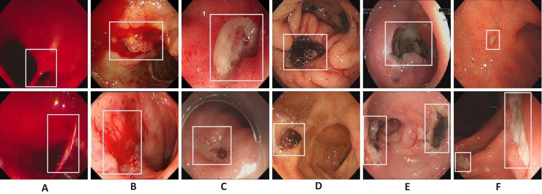
Fig.1 Representative endoscopic images of peptic ulcers in the 6 grades of rebleeding risks. A: Images of grade Ia ulcer showing jet-like bleeding. B: Grade Ib ulcer images showing diffuse bleeding. C: Grade IIa ulcer images showing exposure of the vascular head. D: Grade IIb ulcer images showing an attached blood clot. E: Grade IIc ulcer images with black substrate. F: Grade III ulcer images showing a clean base.
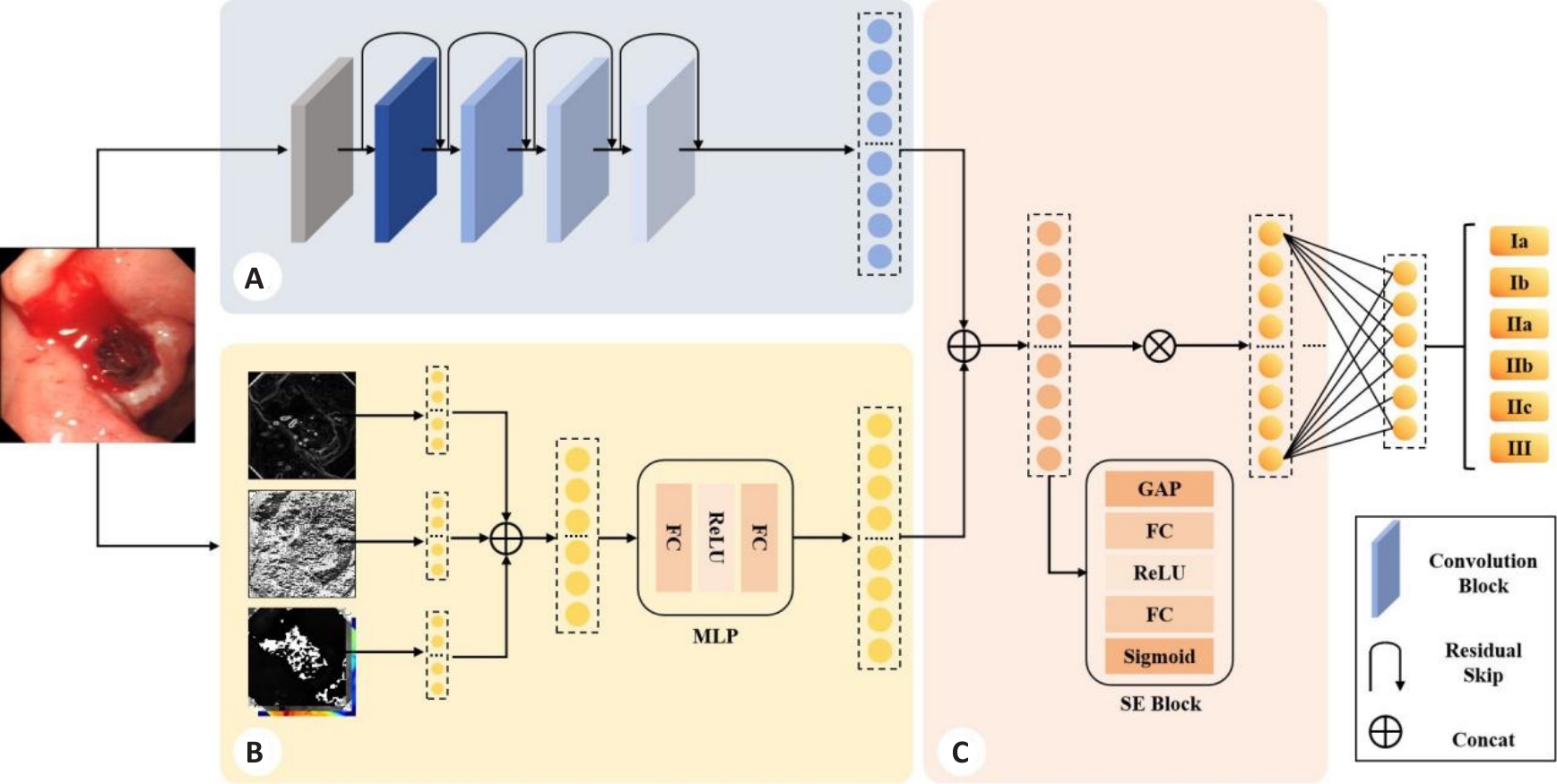
Fig.2 Framework of the proposed method. A: Deep feature extraction module (DFM). B: Handcrafted feature extraction module (HFM). C: Feature fusion module (FFM).
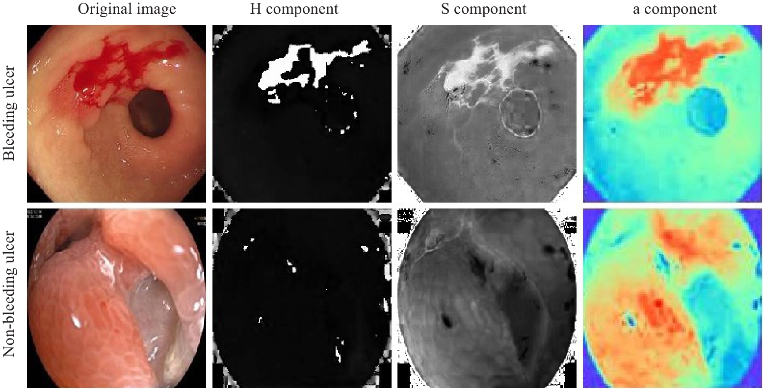
Fig.3 Representative plots of color features for active bleeding versus nonbleeding ulcers.
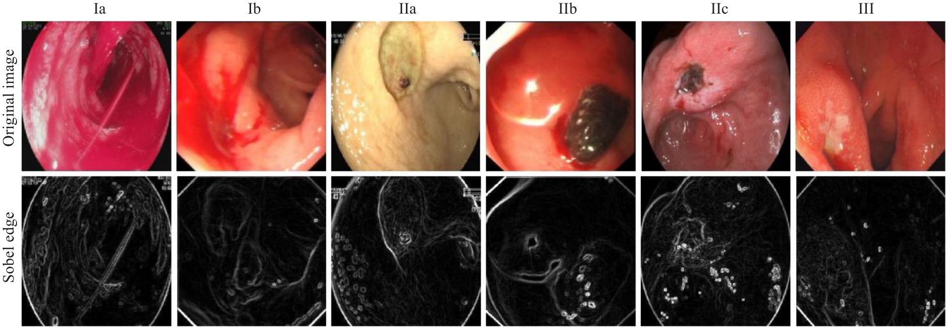
Fig.4 Representative plots of sobel margin of an ulcer with grade 6 rebleeding risk.
| Methods | Accuracy (%) | Precision (%) | Recall (%) | F1-score (%) |
|---|---|---|---|---|
| Simonyan K[ | 57.13±3.83 | 55.48±4.17 | 57.13±3.83 | 55.08±4.81 |
| Tan M[ | 71.35±1.88 | 72.00±1.76 | 71.35±1.88 | 71.07±2.10 |
| Yen HH[ | 70.50±1.86 | 70.77±1.73 | 70.50±1.86 | 69.99±2.10 |
| Cao W[ | 66.96±1.51 | 67.36±2.00 | 66.96±1.51 | 66.08±1.96 |
| Z. Niu[ | 69.75±1.72 | 70.03±1.27 | 69.75±1.72 | 69.08±1.78 |
| Polat, G[ | 71.41±2.27 | 71.60±1.71 | 71.41±2.27 | 70.97±2.01 |
| MFFM | 74.94±0.73 | 74.45±1.00 | 74.94±0.73 | 74.27±0.66 |
Tab.3 Performance comparison of different methods in peptic ulcer rebleeding risk assessment
| Methods | Accuracy (%) | Precision (%) | Recall (%) | F1-score (%) |
|---|---|---|---|---|
| Simonyan K[ | 57.13±3.83 | 55.48±4.17 | 57.13±3.83 | 55.08±4.81 |
| Tan M[ | 71.35±1.88 | 72.00±1.76 | 71.35±1.88 | 71.07±2.10 |
| Yen HH[ | 70.50±1.86 | 70.77±1.73 | 70.50±1.86 | 69.99±2.10 |
| Cao W[ | 66.96±1.51 | 67.36±2.00 | 66.96±1.51 | 66.08±1.96 |
| Z. Niu[ | 69.75±1.72 | 70.03±1.27 | 69.75±1.72 | 69.08±1.78 |
| Polat, G[ | 71.41±2.27 | 71.60±1.71 | 71.41±2.27 | 70.97±2.01 |
| MFFM | 74.94±0.73 | 74.45±1.00 | 74.94±0.73 | 74.27±0.66 |
| Methods | F1-score (%) | |||||
|---|---|---|---|---|---|---|
| Ia | Ib | IIa | IIb | IIc | III | |
| Simonyan K[ | 37.48±12.05 | 78.20±1.48 | 55.40±8.52 | 29.50±11.19 | 18.03±5.53 | 53.63±3.76 |
| Tan M[ | 45.43±7.35 | 88.41±0.76 | 68.32±3.37 | 55.27±4.06 | 44.48±7.86 | 74.83±2.58 |
| Yen HH[ | 35.93±14.47 | 87.05±1.13 | 68.86±2.56 | 53.44±3.69 | 43.49±5.82 | 72.74±2.51 |
| Cao W[ | 14.21±11.31 | 85.86±1.36 | 64.45±2.08 | 45.94±4.09 | 35.77±12.37 | 72.24±1.43 |
| Z. Niu[ | 36.55±7.50 | 87.99±1.22 | 66.85±3.60 | 51.58±2.65 | 38.73±3.83 | 74.61±2.59 |
| Polat, G[ | 27.74±17.36 | 86.62±1.77 | 71.31±4.00 | 57.02±4.19 | 44.97±3.78 | 72.54±3.87 |
| MFFM | 40.07±5.43 | 90.16±0.86 | 75.44±1.27 | 53.97±2.81 | 48.84±5.43 | 77.13±1.64 |
Tab.4 Comparison of F1 scores of different methods for assessing rebleeding risk levels of the ulcers
| Methods | F1-score (%) | |||||
|---|---|---|---|---|---|---|
| Ia | Ib | IIa | IIb | IIc | III | |
| Simonyan K[ | 37.48±12.05 | 78.20±1.48 | 55.40±8.52 | 29.50±11.19 | 18.03±5.53 | 53.63±3.76 |
| Tan M[ | 45.43±7.35 | 88.41±0.76 | 68.32±3.37 | 55.27±4.06 | 44.48±7.86 | 74.83±2.58 |
| Yen HH[ | 35.93±14.47 | 87.05±1.13 | 68.86±2.56 | 53.44±3.69 | 43.49±5.82 | 72.74±2.51 |
| Cao W[ | 14.21±11.31 | 85.86±1.36 | 64.45±2.08 | 45.94±4.09 | 35.77±12.37 | 72.24±1.43 |
| Z. Niu[ | 36.55±7.50 | 87.99±1.22 | 66.85±3.60 | 51.58±2.65 | 38.73±3.83 | 74.61±2.59 |
| Polat, G[ | 27.74±17.36 | 86.62±1.77 | 71.31±4.00 | 57.02±4.19 | 44.97±3.78 | 72.54±3.87 |
| MFFM | 40.07±5.43 | 90.16±0.86 | 75.44±1.27 | 53.97±2.81 | 48.84±5.43 | 77.13±1.64 |
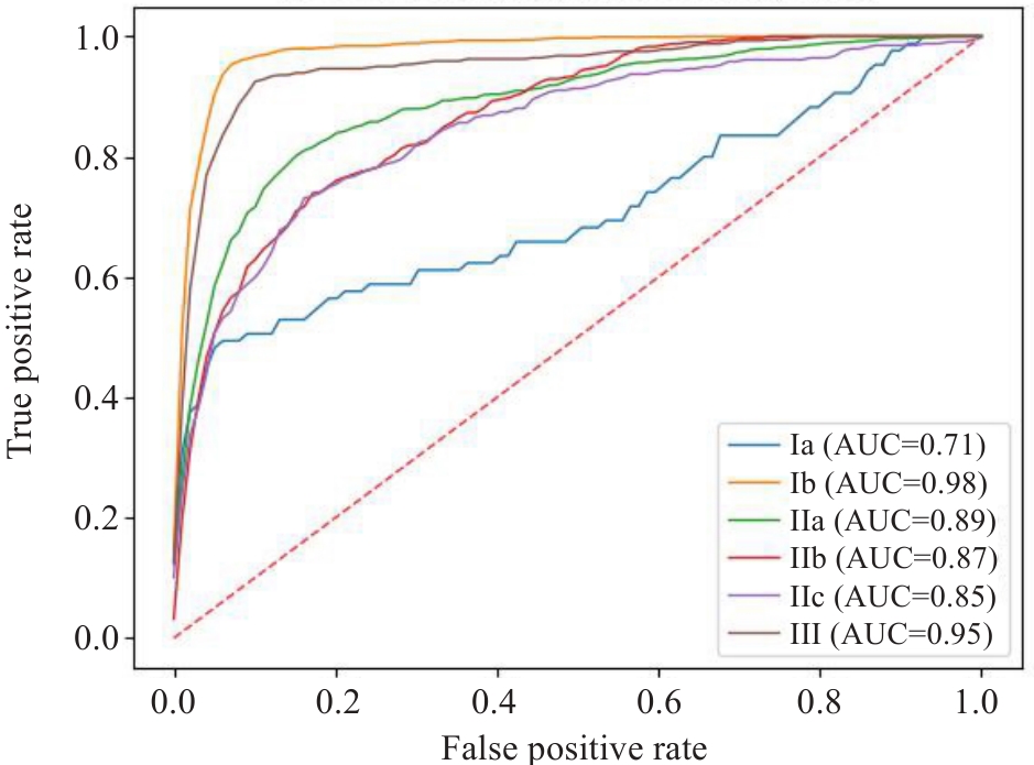
Fig.5 ROC curve of the model.
| Method | Precision (%) | Recall(%) | F1-score (%) | ||||||
|---|---|---|---|---|---|---|---|---|---|
| High Risk | Low Risk | No Therapy | High Risk | Low Risk | No Therapy | High Risk | Low Risk | No Therapy | |
| MFFM | 93.34±0.90 | 81.33±1.50 | 74.24±2.59 | 94.14±0.90 | 81.34±3.00 | 73.19±4.95 | 93.74±0.55 | 81.30±1.41 | 73.59±2.09 |
Tab.5 Performance evaluation of the grading model in simplified ulcer risk stratification
| Method | Precision (%) | Recall(%) | F1-score (%) | ||||||
|---|---|---|---|---|---|---|---|---|---|
| High Risk | Low Risk | No Therapy | High Risk | Low Risk | No Therapy | High Risk | Low Risk | No Therapy | |
| MFFM | 93.34±0.90 | 81.33±1.50 | 74.24±2.59 | 94.14±0.90 | 81.34±3.00 | 73.19±4.95 | 93.74±0.55 | 81.30±1.41 | 73.59±2.09 |
| Subject | Accuracy (%) | Precision (%) | Recall (%) | F1-score (%) |
|---|---|---|---|---|
| Trainee doctor | 59.93 | 71.76 | 59.93 | 58.54 |
| Junior physician | 76.59 | 76.21 | 76.60 | 75.97 |
| Senior physician | 77.02 | 78.12 | 75.48 | 76.34 |
| MFFM | 74.94 | 74.45 | 74.94 | 74.27 |
Tab.6 Comparison of the diagnostic results by the grading model and endoscopists with different levels of experience
| Subject | Accuracy (%) | Precision (%) | Recall (%) | F1-score (%) |
|---|---|---|---|---|
| Trainee doctor | 59.93 | 71.76 | 59.93 | 58.54 |
| Junior physician | 76.59 | 76.21 | 76.60 | 75.97 |
| Senior physician | 77.02 | 78.12 | 75.48 | 76.34 |
| MFFM | 74.94 | 74.45 | 74.94 | 74.27 |
| Subject | F1-score (%) | |||||
|---|---|---|---|---|---|---|
| Ia | Ib | IIa | IIb | IIc | III | |
| Trainee doctor | 59.09 | 56.17 | 55.25 | 53.93 | 33.73 | 79.47 |
| Junior physician | 88.54 | 70.10 | 53.62 | 60.94 | 75.00 | 82.95 |
| Senior physician | 70.30 | 87.00 | 76.00 | 54.03 | 66.15 | 89.33 |
| MFFM | 40.07 | 90.16 | 75.44 | 53.97 | 48.84 | 77.13 |
Tab.7 Comparison of F1 scores for different ulcer rebleeding risk grades between the grading model and endoscopists with different levels of experience
| Subject | F1-score (%) | |||||
|---|---|---|---|---|---|---|
| Ia | Ib | IIa | IIb | IIc | III | |
| Trainee doctor | 59.09 | 56.17 | 55.25 | 53.93 | 33.73 | 79.47 |
| Junior physician | 88.54 | 70.10 | 53.62 | 60.94 | 75.00 | 82.95 |
| Senior physician | 70.30 | 87.00 | 76.00 | 54.03 | 66.15 | 89.33 |
| MFFM | 40.07 | 90.16 | 75.44 | 53.97 | 48.84 | 77.13 |
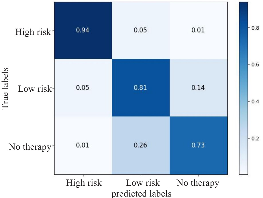
Fig.6 Simplified hierarchical ulcer grading confusion matrix.
| DFM | HFM | FFM | Accuracy (%) | Precision (%) | Recall (%) | F1-score (%) |
|---|---|---|---|---|---|---|
| √ | 69.61±3.22 | 69.84±3.17 | 69.61±3.22 | 68.98±3.07 | ||
| √ | √ | 73.84±1.06 | 73.34±1.39 | 73.84±1.06 | 73.20±1.29 | |
| √ | √ | √ | 74.94±1.10 | 73.88± 1.29 | 74.59±1.10 | 73.83±1.01 |
Tab.8 Hierarchical performance comparison of each feature extraction module in ablation experiments
| DFM | HFM | FFM | Accuracy (%) | Precision (%) | Recall (%) | F1-score (%) |
|---|---|---|---|---|---|---|
| √ | 69.61±3.22 | 69.84±3.17 | 69.61±3.22 | 68.98±3.07 | ||
| √ | √ | 73.84±1.06 | 73.34±1.39 | 73.84±1.06 | 73.20±1.29 | |
| √ | √ | √ | 74.94±1.10 | 73.88± 1.29 | 74.59±1.10 | 73.83±1.01 |
| DFM | HFM | FFM | F1-score (%) | |||||
|---|---|---|---|---|---|---|---|---|
| Ia | Ib | IIa | IIb | IIc | III | |||
| √ | 20.60±13.10 | 86.63±0.87 | 69.09±4.24 | 51.41±5.52 | 38.84±6.88 | 73.11±5.22 | ||
| √ | √ | 36.55±15.75 | 89.22±1.06 | 73.94±0.91 | 56.23±5.05 | 44.84±6.50 | 75.91±3.65 | |
| √ | √ | √ | 40.07±5.43 | 90.16±0.86 | 75.44±1.27 | 53.97±2.81 | 48.84±5.43 | 77.13±1.64 |
Tab.9 Effect of different feature extraction modules on F1 score for different ulcer rebleeding risk grades in ablation experiments
| DFM | HFM | FFM | F1-score (%) | |||||
|---|---|---|---|---|---|---|---|---|
| Ia | Ib | IIa | IIb | IIc | III | |||
| √ | 20.60±13.10 | 86.63±0.87 | 69.09±4.24 | 51.41±5.52 | 38.84±6.88 | 73.11±5.22 | ||
| √ | √ | 36.55±15.75 | 89.22±1.06 | 73.94±0.91 | 56.23±5.05 | 44.84±6.50 | 75.91±3.65 | |
| √ | √ | √ | 40.07±5.43 | 90.16±0.86 | 75.44±1.27 | 53.97±2.81 | 48.84±5.43 | 77.13±1.64 |
| 1 | 陈 丽, 常青霞, 陈少銮. 在内镜下治疗消化性溃疡并发上消化道出血的临床疗效[J]. 中国药物经济学, 2024, 19(6): 67-9, 73. |
| 2 | Wang BL, Yu WT, Zhang ZY, et al. Assessing peptic ulcer risk with the HAMPROW score in the general Chinese population[J]. Sci Rep, 2024, 14(1): 4442. |
| 3 | Soldner T, Bakke K, Savage S. Surgical management of upper gastrointestinal bleeding[J]. Gastrointest Endosc Clin N Am, 2024, 34(2): 301-16. |
| 4 | Vakil N. Endoscopic diagnosis, grading, and treatment of bleeding peptic ulcer disease[J]. Gastrointest Endosc Clin N Am, 2024, 34(2): 217-29. |
| 5 | 李兆申. 消化性溃疡出血的Forrest分级与内镜治疗[J]. 中华消化内镜杂志, 2013, 30(11): 601-3. |
| 6 | 张秀敏, 赵昌东, 李 雪, 等. 基于Forrset分级分层指导内镜下治疗消化性溃疡出血的临床价值[J]. 中国急救复苏与灾害医学杂志, 2023, 18(3): 362-5. |
| 7 | 付 强. 消化性溃疡出血患者进行内镜下Forrest分级的临床意义[J]. 世界最新医学信息文摘, 2017, 17(9): 74. |
| 8 | Chiu PW. Bleeding peptic ulcers: the current management[J]. Dig Endosc, 2010, 22(): S19-21. |
| 9 | 聂玉强, 李瑜元, 吴惠生, 等. 消化性溃疡出血的FORREST分级及其与预后关系[J]. 内镜, 1995, 12(1): 3. |
| 10 | Romstad KK, Detlie TE, Søberg T, et al. Treatment and outcome of gastrointestinal bleeding due to peptic ulcers and erosions - (BLUE study)[J]. Scand J Gastroenterol, 2022, 57(1): 8-15. |
| 11 | Mackiewicz-Pracka A, Nehring P, Przybyłkowski A. Emergency endoscopic interventions in acute upper gastrointestinal bleeding: a cohort study[J]. Diagnostics, 2023, 13(23): 3584. |
| 12 | Yen HH, Wu PY, Wu TL, et al. Forrest classification for bleeding peptic ulcer: a new look at the old endoscopic classification[J]. Diagnostics, 2022, 12(5): 1066. |
| 13 | Klang E, Barash Y, Levartovsky A, et al. Differentiation between malignant and benign endoscopic images of gastric ulcers using deep learning[J]. Clin Exp Gastroenterol, 2021, 14: 155-62. |
| 14 | 王智杰, 高 杰, 孟茜茜, 等. 基于深度学习的人工智能技术在早期胃癌诊断中的应用[J]. 中华消化内镜杂志, 2018, 35(8): 551-6. |
| 15 | 黄 丽, 李艳霞, 吴练练, 等. 基于深度学习的良恶性胃溃疡人工智能辅助诊断系统研究[J]. 中华消化内镜杂志, 2020, 37(7): 476-80. |
| 16 | Yen HH, Wu PY, Su PY, et al. Performance comparison of the deep learning and the human endoscopist for bleeding peptic ulcer disease[J]. J. Med. Biol. Eng, 2021, 41(4): 504-13. |
| 17 | Yen HH, Wu PY, Chen MF, et al. Current status and future perspective of artificial intelligence in the management of peptic ulcer bleeding: a review of recent literature[J]. J Clin Med, 2021, 10(16): 3527. |
| 18 | Afonso J, Saraiva MJM, Ferreira JPS, et al. Development of a convolutional neural network for detection of erosions and ulcers with distinct bleeding potential in capsule endoscopy[J]. echniques and Innovations in Gastrointestinal Endoscopy, 2021, 23(4): 291-6. |
| 19 | He KM, Zhang XY, Ren SQ, et al. Deep residual learning for image recognition[C]//2016 IEEE Conference on Computer Vision and Pattern Recognition (CVPR). June 27-30, 2016. VegasLas, NV, USA. IEEE, 2016: 770-8, . |
| 20 | Sunitha S, Sujatha SS. An improved bleeding detection method for wireless capsule endoscopy (WCE) images based on AlexNet[C]//2021 3rd International Conference on Signal Processing and Communication (ICPSC). May 13-14, 2021. Coimbatore, India. IEEE, 2021: 11-15. |
| 21 | Sainju S, Bui FM, Wahid K. Bleeding detection in wireless capsule endoscopy based on color features from histogram probability[C]//2013 26th IEEE Canadian Conference on Electrical and Computer Engineering (CCECE). May 5-8, 2013. Regina, SK, Canada. IEEE, 2013: 1-4. |
| 22 | Dilna C, Gopi VP. A novel method for bleeding detection in Wireless Capsule Endoscopic images[C]//2015 International Conference on Computing and Network Communications (CoCoNet). December 16-19, 2015. Trivandrum, India. IEEE, 2015: 854-8. |
| 23 | Yuan YX, Li BP, Meng MQH. Bleeding frame and region detection in the wireless capsule endoscopy video[J]. IEEE J Biomed Health Inform, 2016, 20(2): 624-30. |
| 24 | Tuba E, Tomic S, Beko M, et al. Bleeding detection in wireless capsule endoscopy images using texture and color features[C]//2018 26th Telecommunications Forum (TELFOR). November 20-21, 2018. Belgrade. IEEE, 2018: 1-4. |
| 25 | Chen BZ, Li JX, Lu GM, et al. Lesion location attention guided network for multi-label thoracic disease classification in chest X-rays[J]. IEEE J Biomed Health Inform, 2020, 24(7): 2016-27. |
| 26 | Simonyan K, Zisserman A. Very deep convolutional networks for large-scale image recognition[C]// International Conference on Learning Representations (ICLR). May 7-9, 2015. San Diego, CA, USA. ICLR, 2015: 1-14. |
| 27 | Tan M, Le Q. EfficientNet: Rethinking Model Scaling for Convolutional Neural Networks[C]//Proceedings of the 36th International Conference on Machine Learning (ICML). Jun 9-15, 2019. Long Beach, CA, USA. Proceedings of Machine Learning Research, 97: 6105-14. |
| 28 | Cao WZ, Mirjalili V, Raschka S. Rank consistent ordinal regression for neural networks with application to age estimation[J]. Pattern Recognition Letters, 2020, 140: 325-31. |
| 29 | Niu ZX, Zhou M, Wang L, et al. Ordinal regression with multiple output CNN for age estimation[C]//2016 IEEE Conference on Computer Vision and Pattern Recognition (CVPR). June 27-30, 2016. Las Vegas, NV, USA. IEEE, 2016: 4920-8. |
| 30 | Polat G, Ergenc I, Kani HT, et al. Class distance weighted cross-entropy loss for ulcerative colitis severity estimation[C]//Annual Conference on Medical Image Understanding and Analysis. Cham: Springer, 2022: 157-71. |
| 31 | Liu TJ, Xie SN, Yu J, et al. Classification of thyroid nodules in ultrasound images using deep model based transfer learning and hybrid features[C]//2017 IEEE International Conference on Acoustics, Speech and Signal Processing (ICASSP). March 5-9, 2017. New Orleans, LA. IEEE, 2017: 919-23. |
| 32 | Xie JH, Guo LH, Zhao CK, et al. A Hybrid Deep Learning and Handcrafted Features based Approach for Thyroid Nodule Classification in Ultrasound Images[J]. J Physics ConSer, 2020, 1693(1): 012160. |
| No related articles found! |
| Viewed | ||||||
|
Full text |
|
|||||
|
Abstract |
|
|||||