Journal of Southern Medical University ›› 2024, Vol. 44 ›› Issue (11): 2192-2200.doi: 10.12122/j.issn.1673-4254.2024.11.16
Qihui CAI1( ), Haiqiang LAN2, Bojun XIAN1, Lian LIU3, Nan WANG1, Xiaolei HUANG1, Xiaolu NIU1, Xinyu HU1, Chen LI1, Junyi XIE1, Zhaohong LIAO1,2()
Received:2024-05-16
Online:2024-11-20
Published:2024-11-29
Contact:
Zhaohong LIAO
E-mail:m19802076244@163.com;liao1219315353@163.com
Qihui CAI, Haiqiang LAN, Bojun XIAN, Lian LIU, Nan WANG, Xiaolei HUANG, Xiaolu NIU, Xinyu HU, Chen LI, Junyi XIE, Zhaohong LIAO. E2 signaling in myofibers promots macrophage efferocytosis in mouse skeletal muscles with cardiotoxin-induced acute injury[J]. Journal of Southern Medical University, 2024, 44(11): 2192-2200.
Add to citation manager EndNote|Ris|BibTeX
URL: https://www.j-smu.com/EN/10.12122/j.issn.1673-4254.2024.11.16
| Gene | Primer sequence (5'-3') |
|---|---|
| ERα | For:ACTGGCCAATCTTTCTCTGC Rev:CAATTCATCCCCAAAGACATGGAC |
| ERβ | For:TCACTTCTGCGCTGTCTGCAGCG Rev:CCTGGGTCGCTGTGCCAAG |
| GAPDH | For:CAATGTGTCCGTCGTGGATCT Rev:GTCCTCAGTGTAGCCCAAGATG |
Tab.1 Primer sequence for qRT-PCR
| Gene | Primer sequence (5'-3') |
|---|---|
| ERα | For:ACTGGCCAATCTTTCTCTGC Rev:CAATTCATCCCCAAAGACATGGAC |
| ERβ | For:TCACTTCTGCGCTGTCTGCAGCG Rev:CCTGGGTCGCTGTGCCAAG |
| GAPDH | For:CAATGTGTCCGTCGTGGATCT Rev:GTCCTCAGTGTAGCCCAAGATG |
| Gene | Primer sequence(5'-3') |
|---|---|
| IL-1β | For:GCCCATCCTCTGTGACTC Rev:TGTGCCGTCTTTCATTAC |
| IL-10 | For:TTTCAAACAAAGGACCAG Rev:GGATCATTTCCGATAAGG |
| iNOS | For:CTTCCGGGCAGCCTGTGAGACG Rev:ATCCCCAGGTGTTCCCCAGGTAGG |
| TNF-α | For:GCTGTCTCCCCCGAAAGATG Rev:AGGCAGGTGTAGATGTTGTGG |
| Arg1 | For:CTCCAAGCCAAAGTCCTTAGAG Rev:AGGAGCTGTCATTAGGGACA |
| Mrc1 | For:CTCTGTTCAGCTATTGGACGC Rev:TGGCACTCCCAAACATAATTTGA |
| GAPDH | For:CAATGTGTCCGTCGTGGATCT Rev:GTCCTCAGTGTAGCCCAAGATG |
Tab.2 Primer sequence for qRT-PCR
| Gene | Primer sequence(5'-3') |
|---|---|
| IL-1β | For:GCCCATCCTCTGTGACTC Rev:TGTGCCGTCTTTCATTAC |
| IL-10 | For:TTTCAAACAAAGGACCAG Rev:GGATCATTTCCGATAAGG |
| iNOS | For:CTTCCGGGCAGCCTGTGAGACG Rev:ATCCCCAGGTGTTCCCCAGGTAGG |
| TNF-α | For:GCTGTCTCCCCCGAAAGATG Rev:AGGCAGGTGTAGATGTTGTGG |
| Arg1 | For:CTCCAAGCCAAAGTCCTTAGAG Rev:AGGAGCTGTCATTAGGGACA |
| Mrc1 | For:CTCTGTTCAGCTATTGGACGC Rev:TGGCACTCCCAAACATAATTTGA |
| GAPDH | For:CAATGTGTCCGTCGTGGATCT Rev:GTCCTCAGTGTAGCCCAAGATG |
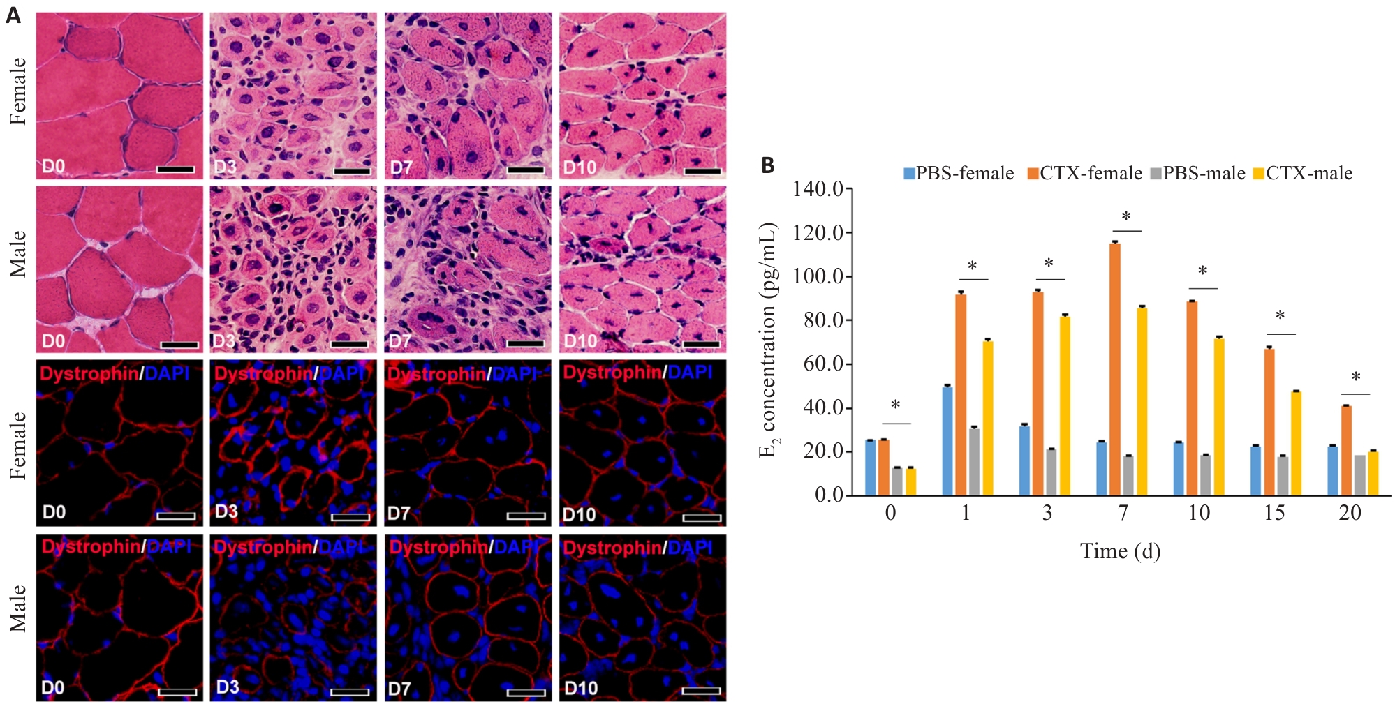
Fig.1 Effect of E2 in myofiber signaling on acute skeletal myositis in mice. A: HE and immunofluorescence staining for detecting inflammation in the tibialis anterior muscle (TA) after CTX injury (Scale bar=50 μm). B: ELISA for detection of serum E2 in male and female mice at different time points after muscle injury (*P<0.05).

Fig.2 Expression of estrogen receptors in acute skeletal myositis. A: Western blotting of ERα and ERβ expression level in mature myotubes derived from C2C12 cells. B: qRT-PCR analysis of ERα and ERβ mRNA levels in the damaged muscle of female mice. C: Immunofluorescence staining of ERβ expression level in injured mouse muscle (Scale bar=50 μm). *P<0.05, **P<0.01.

Fig.3 Effect of E2 signaling in myofibers on exudation of intramuscular mononuclear macrophages after acute skeletal muscle injury. A: ELISA for detecting serum E2 level in female mice after different treatments. B: ELISA for detecting serum E2 levels in female mice at different time points after OVX treatment. C: HE staining for detecting muscular inflammation in female mice with different treatments after CTX injury. D: Immunofluorescence staining for detecting muscular inflammation in female mice after CTX injury with different treatments. *P<0.05, **P<0.01; Scale bar=50 μm.

Fig.4 Effect of E2 signaling in myofibers on phenotype of intramuscular macrophages in mice after acute skeletal muscle injury. A: Flow cytometric analysis of mononuclear macrophages in the damaged muscle of female mice after OVX treatment. B: Flow cytometric analysis of the phenotype of macrophages in the injured muscles of female mice after OVX treatment. *P<0.05, **P<0.01.

Fig.5 Effect of E2 signaling in myofibers on intramuscular inflammatory cytokines after acute skeletal muscle injury. A: qRT-PCR analysis of inflammatory cytokines in female mice after OVX treatment. B: qRT-PCR analysis of inflammatory cytokines in macrophages from the injured muscles isolated by flow cytometry in female mice after OVX treatment. *P<0.05, **P<0.01.

Fig.6 E2 signaling in myofibers drives intramuscular macrophage efferocytosis in acute skeletal myositis. A: Immunofluorescence detection of intramuscular macrophage efferocytosis in the damaged muscle. (scale bar=50 μm). B: Flow cytometry of intramuscular macrophage efferocytosis in the damaged muscle (*P<0.05).

Fig.7 Effect of E2 signaling on macrophage phenotypes and efferocytosis in an in vitro co-culture system. A: Immunofluorescence detection of macrophage phenotypes in the in vitro co-culture system. B: Immunofluorescence detection and flow cytometry analysis of macrophage efferocytosis in the in vitro co-culture system. *P<0.05; Scale bar=50 μm.

Fig.8 E2 signaling in myofibers regulates regeneration and repair of intramuscular myofibers in injured mouse skeletal muscles of female mice after different treatments (Immunofluorescence staining, scale bar=50 μm; *P<0.05).
| 1 | Zhong HH, Sian V, Johari M, et al. Revealing myopathy spectrum: integrating transcriptional and clinical features of human skeletal muscles with varying health conditions[J]. Commun Biol, 2024, 7(1): 438. |
| 2 | Zeller J, Peter K, Eisenhardt SU. Intravital imaging of leukocyte-endothelial interaction in hindlimb ischemia/reperfusion injury by intravital multiphoton microscopy[J]. Methods Mol Biol, 2024, 2711: 89-104. |
| 3 | Zhang H, Qi G, Wang K, et al. Oxidative stress: Roles in skeletal muscle atrophy [J]. Biochem Pharmacol, 2023, 214: 115664-77. |
| 4 | Al-Kharashi L, Attia H, Alsaffi A, et al. Pentoxifylline and thiamine ameliorate rhabdomyolysis-induced acute kidney injury in rats via suppressing TLR4/NF‑κB and NLRP-3/caspase-1/gasdermin mediated-pyroptosis[J]. Toxicol Appl Pharmacol, 2023, 461: 116387. |
| 5 | Boudhabhay I, Poillerat V, Grunenwald A, et al. Complement activation is a crucial driver of acute kidney injury in rhabdomyolysis[J]. Kidney Int, 2021, 99(3): 581-97. |
| 6 | Geraci A, Calvani R, Ferri E, et al. Sarcopenia and menopause: the role of estradiol[J]. Front Endocrinol, 2021, 12: 682012. |
| 7 | Ferreira J, Carneiro A, Vila I, et al. Inflammation and loss of skeletal muscle mass in chronic limb threatening ischemia[J]. Ann Vasc Surg, 2023, 88: 164-73. |
| 8 | Petrocelli JJ, McKenzie AI, de Hart NMMP, et al. Disuse-induced muscle fibrosis, cellular senescence, and senescence-associated secretory phenotype in older adults are alleviated during re-ambulation with metformin pre-treatment[J]. Aging Cell, 2023, 22(11): e13936. |
| 9 | Livshits G, Kalinkovich A. Restoration of epigenetic impairment in the skeletal muscle and chronic inflammation resolution as a therapeutic approach in sarcopenia[J]. Ageing Res Rev, 2024, 96: 102267. |
| 10 | Aluganti Narasimhulu C, Singla DK. Amelioration of diabetes-induced inflammation mediated pyroptosis, sarcopenia, and adverse muscle remodelling by bone morphogenetic protein-7[J]. J Cachexia Sarcopenia Muscle, 2021, 12(2): 403-20. |
| 11 | Antuña E, Cachán-Vega C, Bermejo-Millo JC, et al. Inflammaging: implications in sarcopenia[J]. Int J Mol Sci, 2022, 23(23): 15039. |
| 12 | Ji Y, Lin J, Liu R, et al. Celecoxib attenuates hindlimb unloading-induced muscle atrophy via suppressing inflammation, oxidative stress and ER stress by inhibiting STAT3[J]. Inflammo-pharmacology, 2024, 32(2): 1633-46. |
| 13 | Fischer V, Ragipoglu D, Diedrich J, et al. Mast cells trigger disturbed bone healing in osteoporotic mice[J]. J Bone Miner Res, 2022, 37(1): 137-51. |
| 14 | Zhang Y, Chang YC, Han ZW, et al. Estrogen protects against renal ischemia-reperfusion injury by regulating Th17/treg cell immune balance[J]. Dis Markers, 2022, 2022: 7812099. |
| 15 | Kurmann L, Okoniewski M, Dubey RK. Estradiol inhibits human brain vascular pericyte migration activity: a functional and transcriptomic analysis[J]. Cells, 2021, 10(9): 2314. |
| 16 | Rodríguez-Benítez E, López-García K, Xelhuantzi N, et al. Shift from pro-to anti-inflammatory phase in pelvic floor muscles at postpartum matches histological signs of regeneration in multiparous rabbits[J]. Medicina, 2024, 60(4): 675. |
| 17 | Luo ZW, Qi BJ, Sun YY, et al. Engineering bioactive M2 macrophage-polarized, anti-inflammatory, miRNA-based liposomes for functional muscle repair: from exosomal mechanisms to biomaterials[J]. Small, 2022, 18(34): e2201957. |
| 18 | Guimarães-Pinto K, Maia EP, Ferreira JRM, et al. Efferocytosis in lung mucosae: implications for health and disease[J]. Immunol Lett, 2022, 248: 109-18. |
| 19 | Doran AC, Yurdagul A Jr, Tabas I. Efferocytosis in health and disease[J]. Nat Rev Immunol, 2020, 20(4): 254-67. |
| 20 | 吴泽锴, 黄 涛, 廖钊宏, 等. 肌纤维转化生长因子- β信号激活与急性肌损伤炎症反应的相关性研究[J]. 中华创伤骨科杂志, 2021, 23(3): 254-61. DOI: 10.3760/cma.j.cn115530-20210107-00010 |
| 21 | Xiao JW, Huang JW, Jian XT, et al. IRE1α arm of unfolded protein response in muscle-specific TGF-β signaling-mediated regulation of muscle cell immunological properties[J]. Cell Mol Biol Lett, 2023, 28(1): 15. |
| 22 | Hoffman DB, Raymond-Pope CJ, Sorensen JR, et al. Temporal changes in the muscle extracellular matrix due to volumetric muscle loss injury[J]. Connect Tissue Res, 2022, 63(2): 124-37. |
| 23 | Navi A, Patel H, Xu SW, et al. Role of toll-like receptor 4 in skeletal muscle damage in chronic limb-threatening ischemia[J]. JVS Vasc Sci, 2024, 5: 100194. |
| 24 | Hirtz A, Rech F, Dubois-Pot-Schneider H, et al. Estrogen signaling in healthy and tumor brain[J]. Steroids, 2023, 199: 109285. |
| 25 | Geraci A, Calvani R, Ferri E, et al. Sarcopenia and Menopause: The Role of Estradiol[J]. Front Endocrinol (Lausanne), 2021, 12: 682012-16. |
| 26 | Chaiyasing R, Sugiura A, Ishikawa T, et al. Estrogen modulates the skeletal muscle regeneration process and myotube morphogenesis: morphological analysis in mice with a low estrogen status[J]. J Vet Med Sci, 2021, 83(12): 1812-9. |
| 27 | Lou YY, Fu ZJ, Tian Y, et al. Estrogen-sensitive activation of SGK1 induces M2 macrophages with anti-inflammatory properties and a Th2 response at the maternal-fetal interface[J]. Reprod Biol Endocrinol, 2023, 21(1): 50. |
| 28 | Bauerschmitz G, Hüchel S, Gallwas J, et al. Inhibition of increased invasiveness of breast cancer cells with acquired tamoxifen resistance by suppression of CYR61[J]. Cancer Genomics Proteomics, 2023, 20(6): 531-8. |
| 29 | Valero-Breton M, Tacchi F, Abrigo J, et al. Angiotensin-(1-7) improves skeletal muscle regeneration[J]. Eur J Transl Myol, 2023, 33(4): 12037. |
| 30 | Landeros RV, Jobe SO, Aranda-Pino G, et al. Convergent ERK1/2, p38 and JNK mitogen activated protein kinases (MAPKs) signalling mediate catecholoestradiol-induced proliferation of ovine uterine artery endothelial cells[J]. J Physiol, 2017, 595(14): 4663-76. |
| 31 | Mitchnick KA, Mendell AL, Wideman CE, et al. Dissociable involvement of estrogen receptors in perirhinal cortex-mediated object-place memory in male rats[J]. Psychoneuroendocrinology, 2019, 107: 98-108. |
| [1] | Xianheng ZHANG, Jian LIU, Qi HAN, Yiming CHEN, Xiang DING, Xiaolu CHEN. Huangqin Qingrechubi Capsule alleviates inflammation and uric acid and lipid metabolism imbalance in rats with gouty arthritis by inhibiting the PTEN/PI3K/AKT signaling pathway [J]. Journal of Southern Medical University, 2024, 44(8): 1450-1458. |
| [2] | Yuming ZHANG, Shicheng XIA, Linlin ZHANG, Mengxi CHEN, Xiaojing LIU, Qin GAO, Hongwei YE. Protective effect of Lonicerae japonicae flos extract against doxorubicin-induced liver injury in mice [J]. Journal of Southern Medical University, 2024, 44(8): 1571-1581. |
| [3] | Shan XIANG, Zongxing ZHANG, Lu JIANG, Daozhong LIU, Weiyi LI, Zhuoma BAO, Rui TIAN, Dan CHENG, Lin YUAN. Tujia medicine Toddalia asiatica improves synovial pannus in rats with collagen-induced arthritis through the PI3K/Akt signaling pathway [J]. Journal of Southern Medical University, 2024, 44(8): 1582-1588. |
| [4] | Linyu XIAO, Ting DUAN, Yongsheng XIA, Yue CHEN, Yang SUN, Yibo XU, Lei XU, Xingzhou YAN, Jianguo HU. Linarin inhibits microglia activation-mediated neuroinflammation and neuronal apoptosis in mouse spinal cord injury by inhibiting the TLR4/NF-κB pathway [J]. Journal of Southern Medical University, 2024, 44(8): 1589-1598. |
| [5] | Huaixiang TAO, Jinguang LUO, Zhiyuan WEN, Genming YU, Xiao SU, Xinwei WANG, Han GUAN, Zhijun CHEN. High STING expression exacerbates renal ischemia-reperfusion injury in mice by regulating the TLR4/NF-κB/NLRP3 pathway and promoting inflammation and apoptosis [J]. Journal of Southern Medical University, 2024, 44(7): 1345-1354. |
| [6] | Zhixian REN, Beixian ZHOU, Linxin WANG, Jing LI, Rongping ZHANG, Xiping PAN. Inhibitory effect of 5-hydroxy-6,7-dimethoxyflavone on H1N1 influenza virus-induced ferroptosis and inflammation in A549 cells and its possible mechanisms [J]. Journal of Southern Medical University, 2024, 44(6): 1070-1078. |
| [7] | CAO Jiafan, SUN Yue, DING Xin, LI Shengwen, CHEN Bo, LAN Tian. Arbutin ameliorates liver fibrosis in mice by inhibiting macrophage recruitment and regulating the Akt/NF-κB and Smad signaling pathways [J]. Journal of Southern Medical University, 2024, 44(4): 652-659. |
| [8] | XI Jin, ZHANG Min, ZHANG Yongyu, ZHANG Chen, ZHANG Yulu, WANG Rui, SHEN Lin, LI Jing, SONG Xue. Upregulating KLF11 ameliorates intestinal inflammation in mice with 2, 4, 6-trinitrobenesulfonic acid-induced colitis by inhibiting the JAK2/STAT3 signaling pathway [J]. Journal of Southern Medical University, 2024, 44(4): 765-772. |
| [9] | WU Guangyang, SONG Tianli, TANG Lang, WANG Yiming, LIU Xu, HUANG Sheng. Total saponins of Panax japonicus alleviates CCl4-induced acute liver injury in rats by regulating the PI3K/AktNF-κB signaling pathway [J]. Journal of Southern Medical University, 2024, 44(2): 244-251. |
| [10] | WU Ruojie, LIU Rui, ZHANG Yisu, LI Xiaohong. Parecoxib sodium down-regulates CXCL8-CXCR1/2 to improve inflammatory microenvironment and promote patient recovery following laparoscopic radical resection of rectal cancer [J]. Journal of Southern Medical University, 2024, 44(2): 363-369. |
| [11] | Qinjun YANG, Hui WANG, Shuyu XU, Cheng YANG, Huanzhang DING, Di WU, Jie ZHU, Jiabing TONG, Zegeng LI. Shenqi Tiaoshen Formula alleviates airway inflammation in rats with chronic obstructive pulmonary disease and kidney qi deficiency syndrome by inhibiting ferroptosis via regulating the Nrf2/SLC7A11/GPX4 signaling pathway [J]. Journal of Southern Medical University, 2024, 44(10): 1937-1946. |
| [12] | SUN Xiaopeng, SHI Hang, ZHANG Lei, LIU Zhong, LI Kewei, QIAN Lingling, ZHU Xingyu, YANG Kangjia, FU Qiang, DING Hua. Exosomes from ectoderm mesenchymal stem cells inhibits lipopolysaccharide-induced microglial M1 polarization and promotes survival of H2O2-exposed PC12 cells by suppressing inflammatory response and oxidative stress [J]. Journal of Southern Medical University, 2024, 44(1): 119-128. |
| [13] | XU Guiling, GONG Zhaoqian, WANG Junrao, MA Yanyan, XU Maosheng, CHEN Meijia, HU Dapeng, LIANG Jianpeng, ZHAO Wengqu, ZHAO Haijin. Effects of type 2 inflammation on bronchodilator responsiveness of large and small airways in chronic obstructive pulmonary disease [J]. Journal of Southern Medical University, 2024, 44(1): 93-99. |
| [14] | YU Jiachi, LI Ruibing, XIA Tian, WANG Jianan, JIN Jiacheng, YUAN Manqiu, LI Mianyang. PDCD4 knockdown ameliorates lipopolysaccharide- induced endothelial cell damage by improving mitochondrial dynamics [J]. Journal of Southern Medical University, 2024, 44(1): 25-35. |
| [15] | ZHOU Qiao, LIU Jian, WAN lei, ZHU Yan, QI Yajun, HU Yuedi. Xinfeng Capsule alleviates interleukin-1β-induced chondrocyte inflammation and extracellular matrix degradation by regulating the miR-502-5p/TRAF2/NF-κB axis [J]. Journal of Southern Medical University, 2024, 44(1): 108-118. |
| Viewed | ||||||
|
Full text |
|
|||||
|
Abstract |
|
|||||