
南方医科大学学报 ›› 2026, Vol. 46 ›› Issue (9): 2035-2045.doi: 10.12122/j.issn.1673-4254.2026.09.04
• • 上一篇
徐璐1( ), 潘缘瑾1, 汪雨辰2, 陈振宇1, 蒋伟2, 於江泉1,2(), 郑瑞强1,2()
收稿日期:2026-02-04
出版日期:2026-09-20
发布日期:2026-09-30
通讯作者:
於江泉,郑瑞强
E-mail:sxmuxl@163.com;yujiangquan2021@163.com;zhengruiqiang2021@163.com
作者简介:徐 璐,在读硕士研究生,E-mail: sxmuxl@163.com
基金资助:
Lu XU1(), Yuanjin PAN1, Yuchen WANG2, Zhenyu CHEN1, Wei JIANG2, Jiangquan YU1,2(), Ruiqiang ZHENG1,2()
Received:2026-02-04
Online:2026-09-20
Published:2026-09-30
Contact:
Jiangquan YU, Ruiqiang ZHENG
E-mail:sxmuxl@163.com;yujiangquan2021@163.com;zhengruiqiang2021@163.com
Supported by:摘要:
目的 探讨血必净对脂多糖(LPS)诱导的脓毒症相关急性呼吸窘迫综合征(ARDS)小鼠模型的保护作用及其分子机制。 方法 体内实验:C57BL/6J小鼠随机分为对照组、LPS模型组、LPS+低剂量血必净注射液组(LPS+LXBJ)和LPS+高剂量血必净注射液组(LPS+HXBJ)(n=15)。检测生存率、肺组织病理(HE染色、肺损伤评分、湿/干质量比)、炎症因子[肿瘤坏死因子-α(TNF-α)、白细胞介素-6(IL-6)、白细胞介素-1β(IL-1β)]水平、磷脂转运蛋白(PLTP)表达(转录组测序、实时荧光定量PCR、Western blotting、酶联免疫吸附试验、免疫荧光)。体外实验:RAW264.7巨噬细胞给予LPS与不同浓度血必净处理,检测细胞活力、形态、炎症因子及PLTP表达,并利用过表达PLTP验证其功能。信号通路:检测JNK/c-JUN和NF-κB通路蛋白表达。 结果 与LPS组相比,血必净治疗(尤其是高剂量组)提高脓毒症ARDS小鼠的4 d生存率(P<0.05),降低肺组织病理学损伤评分(P<0.001)和湿/干质量比(P<0.001)。血必净降低了血清及肺泡灌洗液中TNF-α、IL-6和IL-1β的水平:在血清中,低剂量血必净即可降低上述指标(P<0.05),高剂量血必净作用更为显著;在肺泡灌洗液中,低、高剂量血必净均可降低IL-1β水平(P<0.05),高剂量血必净降低TNF-α、IL-6(P<0.05)水平,而低剂量血必净的作用未达到统计学意义(P>0.05)。转录组测序及实验验证显示,LPS下调肺组织PLTP表达(P<0.001),而血必净干预可逆转该趋势(P<0.05)。体外实验显示,血必净呈剂量依赖性地抑制LPS诱导的RAW264.7细胞炎症因子释放(P<0.05),并上调PLTP表达。机制研究显示,血必净通过上调PLTP,抑制了JNK/c-JUN和NF-κB信号通路的磷酸化激活(P<0.05),而过表达PLTP则增强其效果(P<0.05)。 结论 血必净通过上调PLTP表达,抑制JNK/c-JUN和NF-κB信号通路,减轻脓毒症相关ARDS的炎症反应与组织损伤,提示PLTP可能为其作用的关键靶点。
徐璐, 潘缘瑾, 汪雨辰, 陈振宇, 蒋伟, 於江泉, 郑瑞强. 血必净注射液通过上调磷脂转运蛋白调控巨噬细胞JNK/c-JUN通路改善脓毒症相关急性呼吸窘迫综合征[J]. 南方医科大学学报, 2026, 46(9): 2035-2045.
Lu XU, Yuanjin PAN, Yuchen WANG, Zhenyu CHEN, Wei JIANG, Jiangquan YU, Ruiqiang ZHENG. Xuebijing Injection ameliorates sepsis-associated acute respiratory distress syndrome in mice by upregulating phospholipid transfer protein to regulate macrophage JNK/c-JUN pathway[J]. Journal of Southern Medical University, 2026, 46(9): 2035-2045.
| siRNA | Sequence (5'-3') |
|---|---|
| siPLTP-sense | GGUUUCAAAUCUACUCAAATTT |
| siPLTP-antisense | UUUGAGUAGAUUUGAAACCTT |
| siNC-sense | UUCUCCGAACGUGUCACGUTT |
| siNC-antisense | ACGUGACACGUUCGGAGAATT |
表1 siRNA序列
Tab.1 siRNA sequences
| siRNA | Sequence (5'-3') |
|---|---|
| siPLTP-sense | GGUUUCAAAUCUACUCAAATTT |
| siPLTP-antisense | UUUGAGUAGAUUUGAAACCTT |
| siNC-sense | UUCUCCGAACGUGUCACGUTT |
| siNC-antisense | ACGUGACACGUUCGGAGAATT |
| Primer | Sequence (5'-3') |
|---|---|
| PLTP(mouse)-F | TTCCTCCTCAACCAGCAGATCT |
| PLTP(mouse)-R | CAGGAGGGAGTTGAGCAACAC |
| GAPDH(mouse)-F | TGACGTGCCGCCTGGAGAAA |
| GAPDH(mouse)-R | AGTGTAGCCCAAGATGCCCTTCAG |
表2 qPCR引物序列
Tab.2 qPCR primer sequences
| Primer | Sequence (5'-3') |
|---|---|
| PLTP(mouse)-F | TTCCTCCTCAACCAGCAGATCT |
| PLTP(mouse)-R | CAGGAGGGAGTTGAGCAACAC |
| GAPDH(mouse)-F | TGACGTGCCGCCTGGAGAAA |
| GAPDH(mouse)-R | AGTGTAGCCCAAGATGCCCTTCAG |
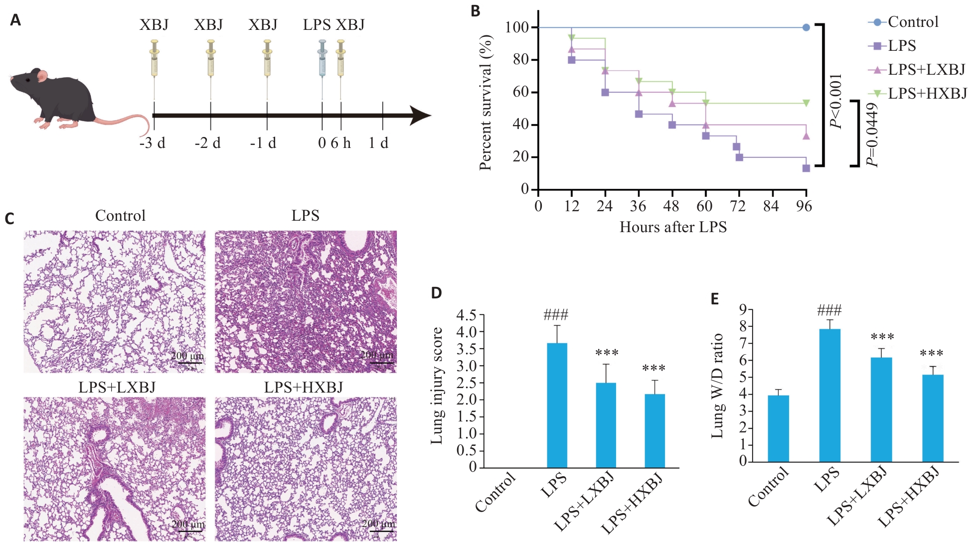
图1 血必净对脓毒症相关ARDS小鼠的保护作用
Fig.1 Protective effect of Xuebijing against sepsis-associated ARDS in mice. A: Schematic diagram of the experimental procedure. B: Four-day survival curves of the mice in each group (n=15). C: HE staining of mouse lung tissues. D: Lung injury scores (n=6). E: Lung wet/dry weight (W/D) ratio (n=10). ###P<0.001 vs Control group; ***P<0.001 vs LPS group.
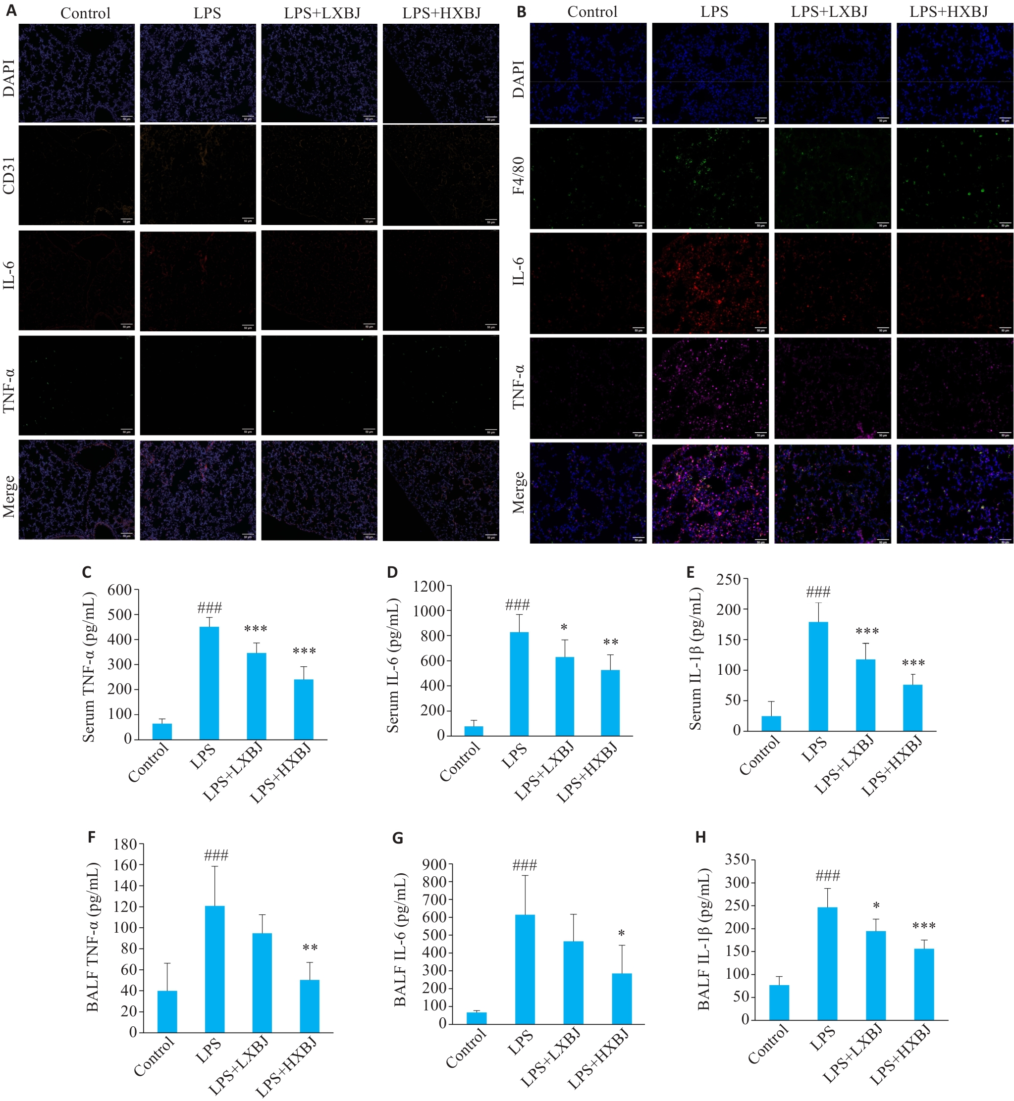
图2 血必净改善脓毒症相关ARDS小鼠的炎症反应
Fig.2 Xuebijing ameliorates inflammatory response in mice with sepsis-associated ARDS. A: Expressions of TNF-α and IL-6 in CD31+ endothelial cells detected by immunofluorescence co-localization (Scale bar=50 μm). B: Expression of TNF‑α and IL-6 in F4/80+ macrophages detected by immunofluorescence co-localization (Scale bar=50 μm). C-H: Results of ELISA for measuring concentrations of TNF-α, IL-6, and IL-1β in serum (C-E) and bronchoalveolar lavage fluid (BALF) (F-H) of the mice (n=6). ###P<0.001 vs Control group; *P<0.05, **P<0.01, ***P<0.001 vs LPS group.
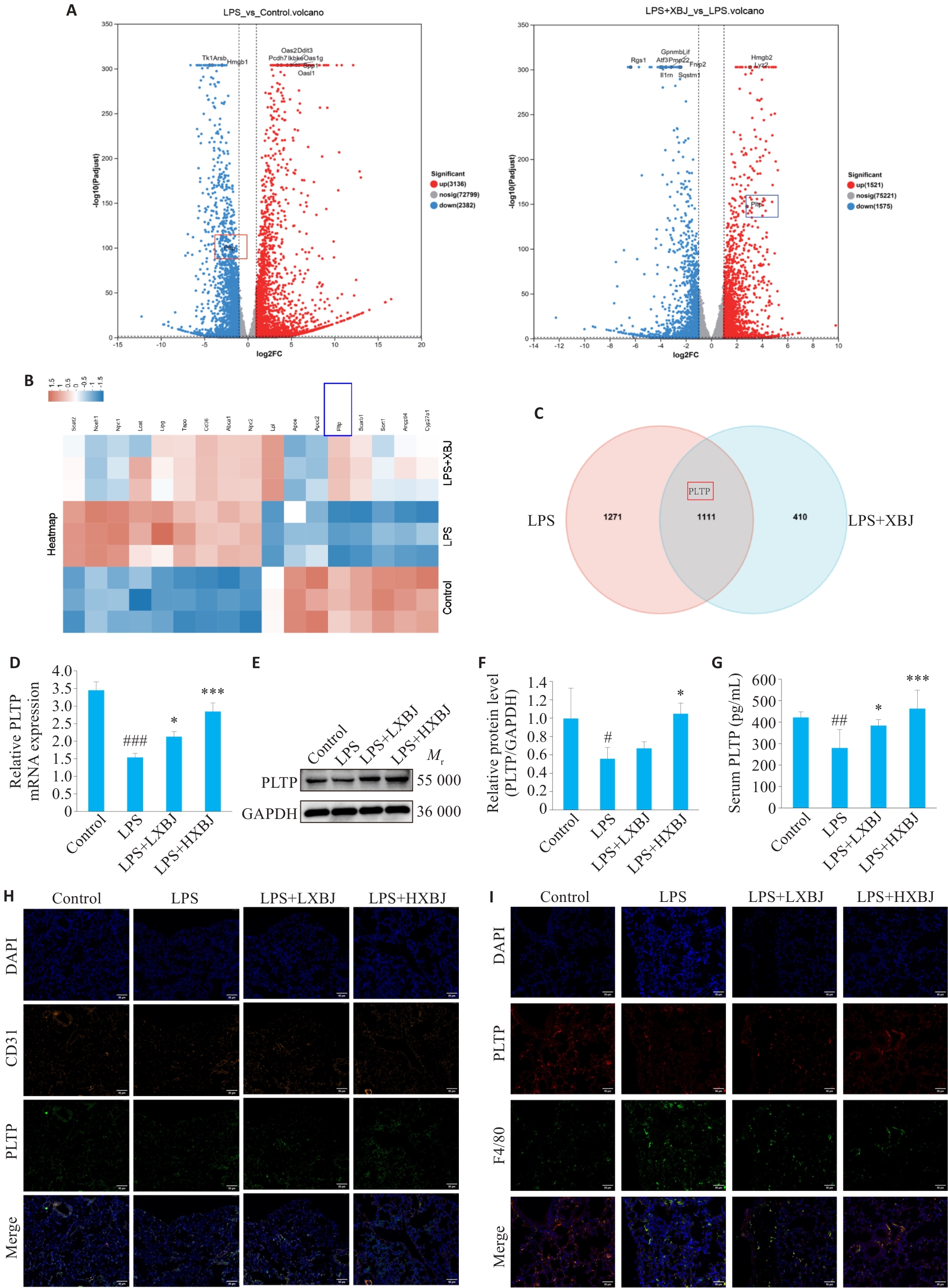
图3 血必净上调脓毒症相关ARDS小鼠的PLTP表达
Fig.3 Xuebijing upregulates PLTP expression in sepsis-associated ARDS mice. A: Transcriptomic sequencing analysis showing downregulation of PLTP by LPS and upregulation by Xuebijing. B: Heatmap of PLTP expression in different groups. C: Venn diagram of differentially expressed genes between LPS and XBJ groups (n=3). D: qRT-PCR analysis of PLTP mRNA levels in mouse lung tissue. E, F: Western blotting for detecting PLTP protein expression in mouse lung tissue. G: ELISA measurement of serum PLTP levels. H: Immunofluorescence co-localization analysis of PLTP (green) with CD31+ endothelial cells (orange) in mouse lung tissue (Scale bar=50 μm). I: Immunofluorescence co-localization analysis of PLTP (red) with F4/80+ macrophages (green) in mouse lung tissue (Scale bar=50 μm). #P<0.05, ##P<0.01, ###P<0.001 vs Control group; *P<0.05, ***P<0.001 vs LPS group.
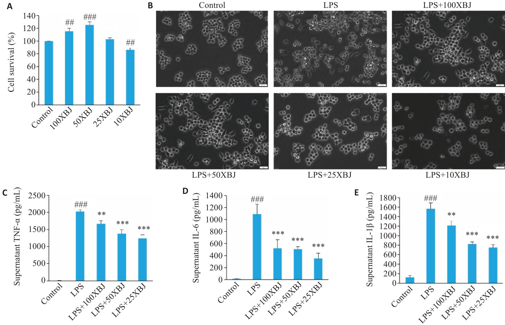
图4 血必净增强RAW264.7细胞活力并抑制LPS诱导的炎症因子释放
Fig.4 Xuebijing enhances viability and suppresses LPS-induced inflammatory cytokine release in RAW264.7 cells. A: Effect of Xuebijing at different dilutions on RAW264.7 cell viability measured by CCK-8 assay (n=3). B: Morphological changes of RAW264.7 cells induced by LPS and pretreatment with different concentrations of Xuebijing observed under optical microscope (Scale bar=100 μm). C-E: ELISA measurement of TNF-α (C), IL-6 (D), and IL-1β (E) levels in the supernatant of RAW264.7 cells pretreated with different concentrations of Xuebijing and stimulated with LPS (n=3). ###P<0.001 vs Control group; **P<0.01,***P<0.001 vs LPS group.
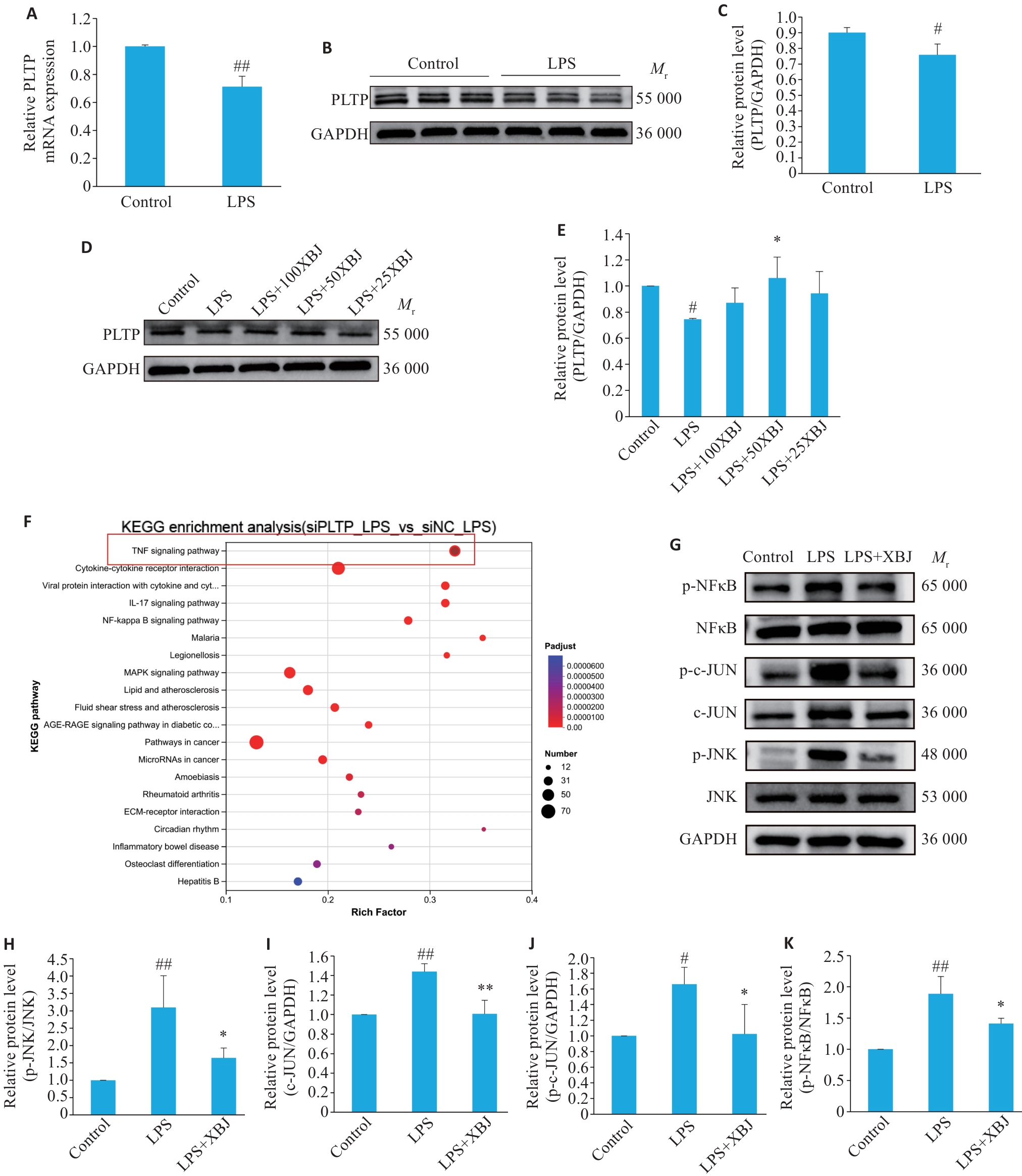
图5 血必净通过调节PLTP介导的JNK/c-JUN和NF-κB通路激活发挥作用
Fig.5 Xuebijing regulates PLTP-mediated activation of JNK/c-JUN and NF-κB pathways. A: qPCR analysis of the effects of LPS and Xuebijing on PLTP mRNA expression in RAW264.7 cells (n=3). B, C: Western blotting for detecting PLTP protein expression in response to LPS stimulation (n=3). D, E: Western blotting for detecting PLTP protein expression following treatment with different concentrations of Xuebijing (n=4). F: KEGG enrichment analysis of differentially expressed genes between PLTP knockdown (siPLTP)+LPS group and control (siNC)+LPS group. G-K: Western blotting for analyzing the effects of LPS and Xuebijing treatment on the expressions of p-JNK, JNK, p-c-JUN, c-JUN, p-NF-κB p65, and NF-κB p65 proteins in RAW264.7 cells (n=3). #P<0.05, ##P<0.01 vs Control group; *P<0.05, **P<0. 01 vs LPS group.
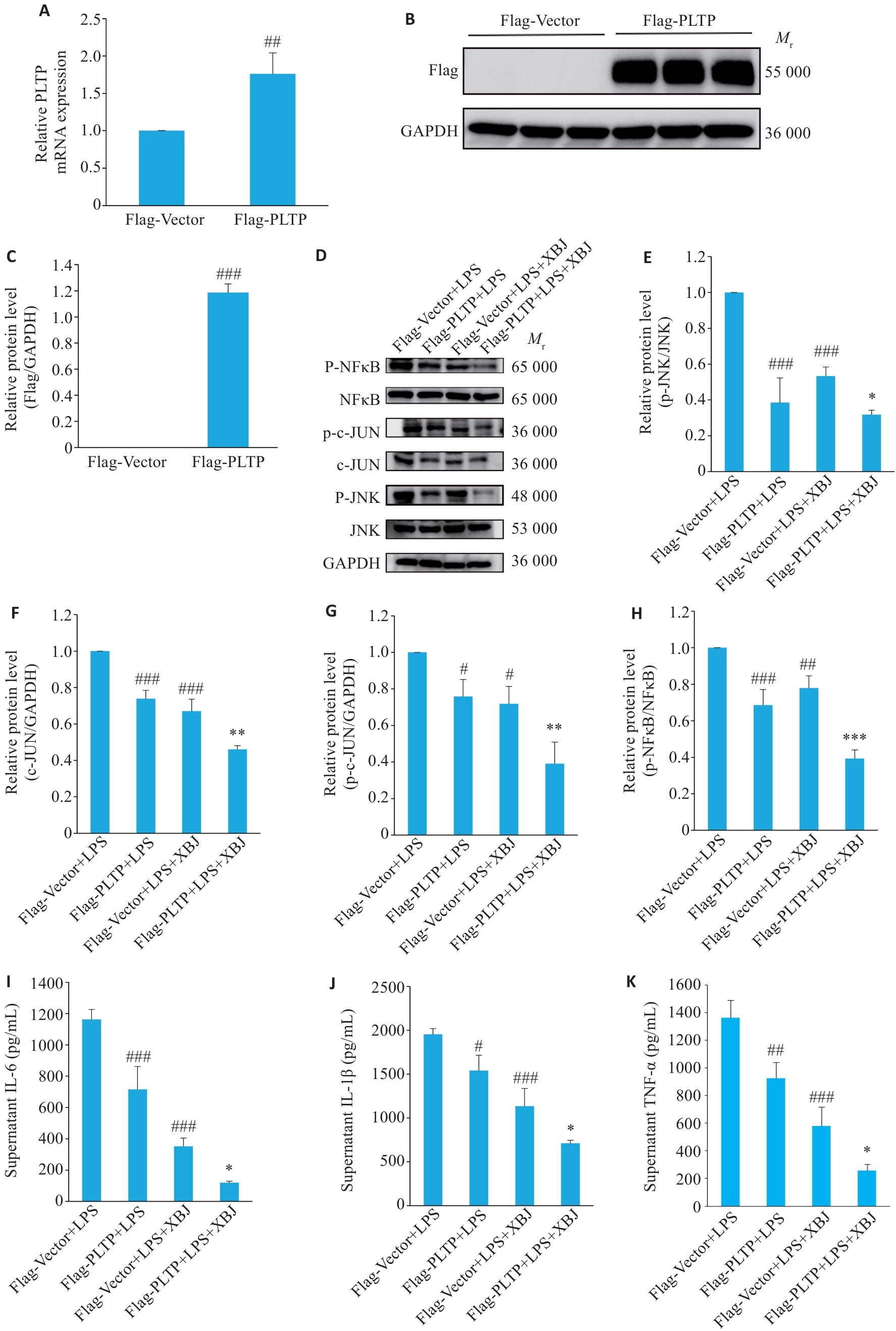
图6 PLTP过表达增强血必净对炎症通路及因子产生的抑制作用
Fig.6 PLTP overexpression enhances the inhibitory effect of Xuebijing on inflammatory pathways and cytokine production. A-C: Validation of PLTP overexpression efficiency in RAW264.7 cells by qPCR (A) and Western blotting (B, C) (n=3; ##P<0.01,###P<0.001 vs Flag-Vector group). D-H: Western blotting for analyzing the effects of PLTP overexpression and Xuebijing treatment on LPS-induced expressions of p-JNK, JNK, p-c-JUN, c-JUN, p-NF-κB p65, and NF-κB p65 proteins (n=3). I-K: Results of ELISA for measurement of the levels of IL-6 (I), IL-1β (J), and TNF‑α (K) in the supernatant of LPS-stimulated RAW264.7 cells with PLTP overexpression and XBJ treatment (n=3). #P<0.05, ##P<0.01, ###P<0.001 vs Flag-Vector+LPS group; *P<0.05, **P<0.01, ***P<0.001 vs Flag-PLTP+LPS group.
| [1] | Abasıyanık MF, Wolfe K, Van Phan H, et al. Ultrasensitive digital quantification of cytokines and bacteria predicts septic shock outcomes[J]. Nat Commun, 2020, 11: 2607. doi:10.1038/s41467-020-16124-9 |
| [2] | Gong HK, Chen Y, Chen ML, et al. Advanced development and mechanism of sepsis-related acute respiratory distress syndrome[J]. Front Med, 2022, 9: 1043859. doi:10.3389/fmed.2022.1043859 |
| [3] | Wick KD, Ware LB, Matthay MA. Acute respiratory distress syndrome[J]. Bmj, 2024, 387: e076612. doi:10.1136/bmj-2023-076612 |
| [4] | Bos LDJ, Ware LB. Acute respiratory distress syndrome: causes, pathophysiology, and phenotypes[J]. Lancet, 2022, 400(10358): 1145-56. doi:10.1016/s0140-6736(22)01485-4 |
| [5] | Flannagan RS, Jaumouillé V, Grinstein S. The cell biology of phagocytosis[J]. Annu Rev Pathol Mech Dis, 2012, 7: 61-98. doi:10.1146/annurev-pathol-011811-132445 |
| [6] | Jones Q, Zheng J, Li ZQ, et al. Effect of phospholipid transfer protein on plasma sphingosine-1-phosphate[J]. J Biol Chem, 2024, 300(11): 107837. doi:10.1016/j.jbc.2024.107837 |
| [7] | Augustin B, Wu DY, Black LP, et al. Multiomic molecular patterns of lipid dysregulation in a subphenotype of sepsis with higher shock incidence and mortality[J]. Crit Care, 2024, 28(1): 431. doi:10.1186/s13054-024-05216-3 |
| [8] | Nguyen M, Alvarez M, Berthoud V, et al. High-density lipoproteins alleviate the endotoxin burden in patients with peritonitis and sepsis: TheLIPSstudy[J]. Eur J Clin Investigation, 2025, 55(12): e70099. doi:10.1111/eci.70099 |
| [9] | Gautier T, Deckert V, Nguyen M, et al. New therapeutic horizons for plasma phospholipid transfer protein (PLTP): Targeting endotoxemia, infection and sepsis[J]. Pharmacol Ther, 2022, 236: 108105. doi:10.1016/j.pharmthera.2021.108105 |
| [10] | Ogier N, Klein A, Deckert V, et al. Cholesterol accumulation is increased in macrophages of phospholipid transfer protein-deficient mice: normalization by dietary alpha-tocopherol supplementation[J]. Arterioscler Thromb Vasc Biol, 2007, 27(11): 2407-12. doi:10.1161/atvbaha.107.151753 |
| [11] | Liao M, Zhang LT, Bai LJ, et al. Xuebijing injection reduces COVID-19 patients' mortality as influenced by the neutrophil to lymphocyte platelet ratio[J]. J Integr Med, 2025, 23(3): 282-8. doi:10.1016/j.joim.2025.04.002 |
| [12] | Li CY, Wang P, Li M, et al. The current evidence for the treatment of sepsis with Xuebijing injection: Bioactive constituents, findings of clinical studies and potential mechanisms[J]. J Ethnopharmacol, 2021, 265: 113301. doi:10.1016/j.jep.2020.113301 |
| [13] | Liu SQ, Yao C, Xie JF, et al. Effect of an herbal-based injection on 28-day mortality in patients with sepsis: the EXIT-SEP randomized clinical trial[J]. JAMA Intern Med, 2023, 183(7): 647. doi:10.1001/jamainternmed.2023.0780 |
| [14] | Kang J, Hu DJ, Li YX, et al. Xuebijing injection reduces 28-day mortality in patients with septic shock[J]. Crit Care, 2025, 29(1): 419. doi:10.1186/s13054-025-05666-3 |
| [15] | Cheng C, Ren C, Li MZ, et al. Pharmacologically significant constituents collectively responsible for anti-sepsis action of XueBiJing, a Chinese herb-based intravenous formulation[J]. Acta Pharmacol Sin, 2024, 45(5): 1077-92. doi:10.1038/s41401-023-01224-1 |
| [16] | Singer M, Deutschman CS, Seymour CW, et al. The third international consensus definitions for sepsis and septic shock (sepsis-3)[J]. Jama, 2016, 315(8): 801. doi:10.1001/jama.2016.0287 |
| [17] | Auriemma CL, Zhuo HJ, Delucchi K, et al. Acute respiratory distress syndrome-attributable mortality in critically ill patients with sepsis[J]. Intensive Care Med, 2020, 46(6): 1222-31. doi:10.1007/s00134-020-06010-9 |
| [18] | Lorencio Cárdenas C, Yébenes JC, Vela E, et al. Trends in mortality in septic patients according to the different organ failure during 15years[J]. Crit Care, 2022, 26(1): 302. doi:10.1186/s13054-022-04176-w |
| [19] | 方邦江, 刘 良. 脓毒症相关急性呼吸窘迫综合征的中西医治疗 [J]. 临床急诊杂志, 2025, 26(01): 79-85. |
| [20] | 丁伟超, 陈 娟, 叶 英, 等. 血必净注射液治疗脓毒症相关急性呼吸窘迫综合征肺热血瘀证患者的临床疗效 [J]. 中草药, 2024, 55(19): 6645-54. |
| [21] | Zhang ZY, Li XY, Zhou JP, et al. Xuebijing injection in the treatment of COVID-19: an update on clinical studies, potentially active metabolites and mechanisms[J]. Front Pharmacol, 2026, 16: 1667022. doi:10.3389/fphar.2025.1667022 |
| [22] | Zhan YX, Wang YB, Zhang BY, et al. Xuebijing injection mitigates instant blood-mediated inflammatory reaction and enhances intrahepatic islet transplantation via target NF-κB pathway[J]. Front Immunol, 2025, 16: 1671966. doi:10.3389/fimmu.2025.1671966 |
| [23] | Ren TY, Chen QH, Xu JZ, et al. Xuebijing alleviates sepsis-induced acute lung injury by inhibiting alveolar epithelial cell apoptosis via cAMP/PKA signaling pathway[J]. J Ethnopharmacol, 2025, 353: 120370. doi:10.1016/j.jep.2025.120370 |
| [24] | Zhang S, Jiang M, Wu C, et al. Modulation of the CEACAM1/TIM3 pathway by traditional Chinese medicine Xuebijing alleviates sepsis-induced acute lung injury through inhibition of NETs formation[J]. J Ethnopharmacol, 2025, 353: 120280. doi:10.1016/j.jep.2025.120280 |
| [25] | Cui JY, Deng YX, Li XC, et al. Herbal-based Xuebijing injection ameliorated vascular endothelial dysfunction via inhibiting ACLY/MYB/RIG-I axis in sepsis-associated lung injury[J]. Phytomedicine, 2025, 140: 156573. doi:10.1016/j.phymed.2025.156573 |
| [26] | Ji HY, Zhang CM, Xu FY, et al. Inhaled pro-efferocytic nanozymes promote resolution of acute lung injury[J]. Adv Sci, 2022, 9(26): 2201696. doi:10.1002/advs.202201696 |
| [27] | Huang WC, Wen LH, Tian H, et al. Self-propelled proteomotors with active cell-free mtDNA clearance for enhanced therapy of sepsis-associated acute lung injury[J]. Adv Sci, 2023, 10(27): 2301635. doi:10.1002/advs.202370184 |
| [28] | Mansuy M, Baille S, Canet G, et al. Deletion of plasmaPhospholipid Transfer Protein (PLTP)increases microglial phagocytosis and reduces cerebral amyloid-β deposition in the J20 mouse model of Alzheimer's disease[J]. Oncotarget, 2018, 9(28): 19688-703. doi:10.18632/oncotarget.24802 |
| [29] | Yazdanyar A, Jiang XC. Liver phospholipid transfer protein (PLTP) expression with a PLTP-null background promotes very low-density lipoprotein production in mice[J]. Hepatology, 2012, 56(2): 576-84. doi:10.1002/hep.25648 |
| [30] | Vergeer M, Boekholdt SM, Sandhu MS, et al. Genetic variation at thePhospholipid transfer ProteinLocus affects its activity and high-density lipoprotein size and is a novel marker of cardiovascular disease susceptibility[J]. Circulation, 2010, 122(5): 470-7. doi:10.1161/circulationaha.109.912519 |
| [31] | Vuletic S, Dong W, Wolfbauer G, et al. PLTP regulates STAT3 and NFκB in differentiated THP1 cells and human monocyte-derived macrophages[J]. Biochim Biophys Acta BBA Mol Cell Res, 2011, 1813(10): 1917-24. doi:10.1016/j.bbamcr.2011.06.013 |
| [32] | Jiang W, Song L, Gong WL, et al. The role of phospholipid transfer protein in sepsis-associated acute kidney injury[J]. Crit Care, 2025, 29(1): 33. doi:10.1186/s13054-025-05253-6 |
| [1] | 于梦洁, 赵安博, 郝艺璇, 王奕人, 刘月娴, 叶红伟, 高琴. 敲除脂质运载蛋白2通过抑制NF-κB通路改善脓毒症急性肺损伤小鼠血管内皮屏障功能[J]. 南方医科大学学报, 2026, 46(8): 1779-1789. |
| [2] | 高建红, 史册, 李蔚然, 尚祥, 王飞, 杨琪琪, 李飞. 电针改善血管性痴呆大鼠认知障碍并抑制TLR4/MyD88/NF-κB通路介导的星形胶质细胞活化[J]. 南方医科大学学报, 2026, 46(8): 1814-1822. |
| [3] | 王伟丽, 高怡琳, 洪馨, 陈义珍, 程梦, 张磊, 戴荣, 王亿平. 清肾颗粒通过调控Akt3介导的M1型巨噬细胞极化改善单侧输尿管梗阻小鼠肾纤维化[J]. 南方医科大学学报, 2026, 46(6): 1216-1227. |
| [4] | 王梓渝, 伍静雯, 王涵冰, 徐艳, 姜春玲, 周莉. 红细胞分布宽度/红细胞压积与脓毒症患者28 d全因死亡率的关系[J]. 南方医科大学学报, 2026, 46(6): 1256-1266. |
| [5] | 杨云恒, 贾雅清, 尹高生, 李双秀, 杨萍. 乳酸通过“心-胃轴”促进巨噬细胞 M1 型极化诱导胃黏膜损伤[J]. 南方医科大学学报, 2026, 46(6): 1267-1277. |
| [6] | 王泽安, 刘丽丽, 施美, 侯勇, 董莉莉, 张国梁. 益气解毒方通过抑制NF-κB通路的过度激活改善小鼠肝癌免疫微环境[J]. 南方医科大学学报, 2026, 46(3): 629-637. |
| [7] | 冯鹏, 王洁, 褚江伟, 王鹤霖, 田永衍, 张杰昌, 王明露, 王宇彤. 石菖蒲挥发油通过抑制TLR4/MyD88/NF-κB通路介导的小胶质细胞极化改善大鼠抽动障碍[J]. 南方医科大学学报, 2026, 46(3): 646-654. |
| [8] | 尹梦霞, 李蕊臣, 杨凯博, 焦伟杰, 刘新光. 利奈唑胺联用血必净通过抑制Hla-NLRP3通路减轻MRSA肺炎小鼠的炎症损伤[J]. 南方医科大学学报, 2026, 46(2): 412-422. |
| [9] | 王喆, 孔柯瑜, 金明昊, 伍信儒, 范文轩, 翟赞京, 胡子豪, 牛琳, 齐岩松, 徐永胜. 叶酸预处理髌下脂肪垫来源间充质干细胞来源的外泌体调控巨噬细胞极化[J]. 南方医科大学学报, 2026, 46(1): 166-174. |
| [10] | 沙桐, 王文研, 宣佳斌, 吴洁, 石能贤, 何劲, 胡鸿彬, 张耀元. 基于Th1/Th2细胞因子检测的脓毒症免疫状态分型及预后分析:一项回顾性研究[J]. 南方医科大学学报, 2026, 46(1): 6-22. |
| [11] | 李楠, 张亮, 郭俏凤, 周越, 刘长江. 有氧运动通过调控miR-221-3p介导的脂肪组织巨噬细胞极化改善小鼠胰岛素抵抗[J]. 南方医科大学学报, 2026, 46(1): 74-82. |
| [12] | 薄海美, 曹新营, 邢平川, 王志军. 外泌体来源的miR-1275通过上调淋巴细胞中IL-38的表达抑制脓毒症心肌细胞凋亡[J]. 南方医科大学学报, 2025, 45(8): 1608-1615. |
| [13] | 汤忠富, 黄传兵, 李明, 程丽丽, 陈君洁, 尚双双, 刘思娣. 芪黄健脾滋肾颗粒通过抑制MyD88/NF-κB通路减轻MRL/lpr小鼠肾损害[J]. 南方医科大学学报, 2025, 45(8): 1625-1632. |
| [14] | 张兆君, 吴琼, 谢苗苗, 叶洳吟, 耿晨晨, 石纪雯, 杨清玲, 王文锐, 石玉荣. 层状双氢氧化物负载si-NEAT1通过miR-133b/PD-L1轴调控乳腺癌紫杉醇耐药及巨噬细胞极化[J]. 南方医科大学学报, 2025, 45(8): 1718-1731. |
| [15] | 涂舒谕, 陈祥宇, 李程辉, 黄丹萍, 张莉. 补阳还五汤通过调控外泌体miR-590-5p介导的巨噬细胞极化延缓大鼠血管衰老[J]. 南方医科大学学报, 2025, 45(6): 1251-1259. |
| 阅读次数 | ||||||
|
全文 |
|
|||||
|
摘要 |
|
|||||
